2483
Development 126, 2483-2494 (1999) Printed in Great Britain © The Company of Biologists Limited 1999 DEV7716
The Caenorhabditis elegans genes egl-27 and egr-1 are similar to MTA1, a member of a chromatin regulatory complex, and are redundantly required for embryonic patterning Florence Solari1,*, Alex Bateman2 and Julie Ahringer1,*,‡ 1University of Cambridge, Department of Genetics, Downing Street, Cambridge CB2 3EH, UK 2The Sanger Centre, Wellcome Trust Genome Campus, Hinxton, Cambridge CB10 1SA, UK
*Present address: Wellcome CRC Institute, Tennis Court Road, Cambridge CB2 1QR, UK ‡Author for correspondence (e-mail:
[email protected])
Accepted 19 March; published on WWW 4 May 1999
SUMMARY We show here that two functionally redundant Caenorhabditis elegans genes, egl-27 and egr-1, have a fundamental role in embryonic patterning. When both are inactivated, cells in essentially all regions of the embryo fail to be properly organised. Tissue determination and differentiation are unaffected and many zygotic patterning genes are expressed normally, including HOX genes. However, hlh-8, a target of the HOX gene mab-5, is not expressed. egl-27 and egr-1 are members of a gene family that includes MTA1, a human gene with elevated
expression in metastatic carcinomas. MTA1 is a component of a protein complex with histone deacetylase and nucleosome remodelling activities. We propose that EGL27 and EGR-1 function as part of a chromatin regulatory complex required for the function of regional patterning genes.
INTRODUCTION
structure (for recent reviews, see Kadonaga, 1998; Struhl, 1998). Here we show that the C. elegans gene egl-27 encodes a protein similar to MTA1, a component of a protein complex with ATP-dependent nucleosome remodelling and histone deacetylation activities (Xue et al., 1998). No previous members of this gene family have been identified by mutation. The C. elegans genome contains a related gene, egr-1 that has a redundant role with egl-27; in embryos where both are inactivated, the pattern of cells in all regions of the embryo is abnormal. Furthermore, although HOX genes have normal expression, a HOX gene target gene fails to be expressed. We suggest that protein complexes containing EGL-27 family members regulate the activity of transcription factors involved in embryonic patterning.
During embryonic development, cells are determined, differentiate and become organised into tissues. These events are regulated by extrinsic and intrinsic signals that are often translated into changes in the expression and/or activity of transcription factors. Therefore, how transcription factor activities are regulated is important for understanding most developmental processes. For example, the HOX genes encode transcription factors that pattern tissues along the anteroposterior axis of most or all animals (reviewed in McGinnis and Krumlauf, 1992). Recently, the identification of co-factors that act with HOX proteins have given insights into the mechanism by which they regulate their targets (for recent reviews, see Fisher and Caudy, 1998; Mann and Affolter, 1998). However, how developmental transcription factors act in the context of chromatin is still largely unknown. Recent studies have revealed that a gene’s transcriptional activity can be regulated through chromatin modifications: hyperacetylated chromatin is generally associated with active genes, whereas hypoacetylated chromatin is associated with repressed genes. Protein complexes that carry out ATPdependant nucleosome remodelling also affect the activity of particular promoters. Multiprotein complexes with the above activities can interact with sequence-specific transcription factors, suggesting a model whereby the transcriptional activity of a promoter is regulated by locally perturbing chromatin
Key words: SANT, ELM1, ELM2, vab-7, NURD, Histone deacetylation, Nucleosome remodelling, Caenorhabditis
MATERIALS AND METHODS Strains and alleles Cultivation of C. elegans was as in Brenner (1974). The following mutant alleles and deficiencies were used: LG II: bli-2(e768), dpy-10(e128), egl-27(e2394), egl-27(n170), egl27(we3), unc-4(e120), mnDf30, mnDf96, mnDf39, mnDf68, mnDf61, mnDf88 and maDf4. LGIII: vab-7(ed6), unc-49(e362) and vab-7(ed6). The following strains were used as markers in expression studies: ayIs7 [hlh-8::gfp] IV (Harfe et al., 1998), eIs24 [vab-7::lacZ] II
2484 F. Solari, A. Bateman and J. Ahringer (Ahringer, 1996), muIs6 [lin-39::lacZ + rol-6(su1006)] (Wang et al, 1993), muIs13 [egl-5::lacZ + rol-6(su1006)] V (kindly provided by C. Kenyon), muIs16 [mab-5::gfp + dpy-20(+)] II; dpy-20(e1282) IV (kindly provided by C. Kenyon), weEx20 [lin-44::gfp + hsp16-2-vab7 + hsp16-41-vab-7 + rol-6(su1006)] (F. S. and J. A., unpublished; lin-44::gfp plasmid (pMHE004) kindly provided by M. Herman), wIs1 [SCM::lacZ + rol-6(su1006)] (Terns et al., 1997), hlh-1::gfp (PD7963; K. Dej, S. Xu and A. Fire, personal communication.). Isolation and mapping of we3 unc-49(e362) vab-7(ed6) animals were mutagenized using EMS as in Wood (1988). 484 F1s were cloned (representing 968 chromosomes) and their progeny scored for stronger posterior defects. 5 enhanced strains were isolated, the strongest of which contained we3. we3 was mapped to position +0.09 on chromosome II: from the strain + we3 + /dpy-10(e128) + unc-4(e120), 23/24 Unc non-Dpy recombinants carried we3 and 1/15 Dpy non-Unc recombinants carried we3. The deficiencies mnDf96 and mnDf30 remove we3; mnDf39, mnDf68, mnDf61, mnDf88 and maDf4 do not remove we3. we3 is maternally rescued: whereas 92% of we3 mutants descended from we3 homozygotes have abnormal body morphology at 15°C, only 4% of homozygotes are abnormal if they inherit wild-type maternal product. When grown as a homozygote, we3 behaves as a strong loss-offunction mutation: we3 mutants are 89.5% lethal at 15°C. we3/mnDf30 animals are 78% lethal at 15˚C (n=118) when they inherit wild-type maternal product, but 88% (n=84) lethal at 15°C when the mother was we3/mnDf30. Unless otherwise stated, all analyses of we3 were done at 15°C. egl-27(we3) enhances the phenotype of vab-7(ed6) zygotically. vab-7(ed6) animals range from having nearly wild-type morphology (31%) to having blunt, round tail regions (16%). Zygotic enhancement was tested using unc-4(e120) as a marker for egl-27(we3): Only 1% of egl-27(we3) unc-4(e120); vab-7(ed6) that come from egl-27(we3) unc-4(e120) /+ +; vab-7(ed6) appear wild type and 50% have blunt tail regions. The progeny of the next generation have slightly stronger defects than egl-27(we3) single mutants (not shown). Only 4% (1/27) of egl-27(we3) unc-4(e120) that come from egl-27(we3) unc-4(e120) /+ + had posterior defects. Complementation tests Phasmid dye filling was assayed in adult hermaphrodites as in Herman and Horvitz (1994). 0% (0/532) of wild-type hermaphrodites have defective phasmid dye filling, whereas 100% of phasmids from egl27(e2394) (39/39) or egl-27(n170) (51/51) fail to fill with dye. At 15°C, phasmids fail to fill in 66% of surviving we3 mutants. Male tail morphology was assayed using DIC optics: 0/20 wild-type, 8/12 egl27(n170) and 3/7 surviving we3 males have abnormal tail morphology. we3 was found to be an allele of egl-27 by complementation tests: egl-27(e2394) or egl-27(n170) hermaphrodites were crossed with we3 unc-4(e120)/++ males and assayed for dye-filling defects (Dyf), abnormal male tail morphology (Mtl) and abnormal body morphology (Mor). In the egl-27(e2394) cross, 9/15 hermaphrodites were Dyf, 16/41 males were Mtl and no Mor animals were found. Progeny testing confirmed that one-half were we3 unc-4(e120)/egl-27(e2394). In the egl-27(n170) cross, 4/8 hermaphrodites were Dyf, 18/39 males were Mtl and no animals were Mor. Therefore, we3 fails to complement egl-27(n170) and egl-27(e2394) for Dyf and Mtl, but not for Mor in this test. However, we3 did fail to complement egl27(n170) for Mor when the mother was egl-27(we3). In the reciprocal cross of we3 unc-4(e120) hermaphrodites with egl-27(n170)/+ males: 21/47 non-unc hermaphrodites were Mor, 5/37 were Dyf and 8/61 males were Mtl. For comparison, in a cross of we3 unc-4(e120) with wild-type males, 24/144 were Mor, 0/80 non-unc hermaphrodites were Dyf and 0/30 males were Mtl. The egl-27(n170) phasmid-filling defect has a maternal effect. Whereas 51/51 of egl-27(n170) homozygotes from homozygous
mother have phasmids that fail to fill with dye, none out of 30 progeny from an egl-27(n170) /+ mother had phasmid dye-filling defects.
egl-27 cloning egl-27 (we3) animals were injected with pools of cosmids covering the region where we3 was mapped: T24B4+F41G3, F41G3+T13C2, T13C2+F31E8, F31E8+C04A2, C04A2+C44B7, C44B7+B0252, B0252+F22D3, F22D3+C15F1 at a concentration of 5 µg/ml with the plasmid pRF4 (100 µg/ml) which confers a dominant Roller phenotype (Mello et al., 1991). Transgenic lines were scored for the rescue of egl-27(we3) embryonic lethality at 15˚C. Two pools containing C04A2 (C04A2+C44B7 and F31E8+C04A2) showed rescue. These cosmids were injected individually into egl-27(we3) and lines tested for phasmid dye filling. Two lines carrying C04A2 showed rescue (phasmids failed to fill in 0% and 20% of rollers compared to 66% of egl-27(we3) mutants). Tissue assays Antibody staining of embryos was essentially as in Albertson (1984). The primary antibodies used are: anti-LIN-26 rabbit polyclonal serum (marks all epidermal and other non-neuronal ectodermal cells; Labouesse et al., 1996) mAb MH27 which recognizes a component of epithelial adherens junctions (Francis and Waterston, 1991), mAb NE2/1B4 against an antigen expressed in seam cells (Schnabel, 1991) and mAb 3NB12, which detects a subset of pharyngeal muscle cells (Priess and Thomson, 1987). For the time course, the development of wild-type and double mutants was followed by DIC microscopy for 8 hours at 1 hour time intervals at 22˚C, the embryos were placed at 15˚C overnight and the terminal phenotype viewed 12 hours later. RNAi Templates for RNA synthesis contained T3 and T7 phage polymerase promoter sites; single strands were synthesized separately and then annealed as described (Fire et al., 1998). Templates were prepared by PCR using the following primers: for egl-27(exon 11), 5′-ATTAACCCTCACTAAAGGGAGACTTCGATGAGCGAGACTCC and 5′AATACGACTCACTATAGGGAGATCGCATTTGATGTTGGAATGC using cosmid C04A2 as a template; for egl-27(exons 1-8), T3 and T7 primers using pJA54 (a RACE clone containing exons 1-8) as a template; egr-1(RNAi), T3 and T7 primers using yk394g5 (an egr-1 cDNA of 436nt covering exons 1, 2 and 199 bp of exon 3) as a template. Double-stranded RNA was injected into the body cavity of adult hermaphrodites at a concentration of 0.5-1 mg/ml. Injected animals were singled on plates for 12 hours, then transferred to new plates every 24 hours. Phenotypes were assayed on the first new plate. Control injections of dsRNA to egl-27(exon 11) into weEx33 (carrying the translational egl-27::gfp reporter gene) resulted in the absence of GFP expression indicating that RNAi removes egl-27 proteins. Isolation and sequencing of cDNA clones and northern blotting For RACE experiments, the template was mixed stage C. elegans cDNA to which 5′ and 3′ anchors were ligated (Clontech, Marathon cDNA Amplification Kit), kindly provided by Howard Baylis. PCR was carried out using a primer in egl-27 (5′ CATGTTGTCAACGTCTGTCG) and a primer to the anchor (5′ CCATCCTAATACGACTCACTATAGGGC). Several products were obtained and cloned. The major product contained exons 1-8; 2/10 clones sequenced had part of SL1 at the 5′ end of exon 1 (one was named pJA54). Several smaller minor products were sequenced; two had part of SL1 (TTTGAG) at the 5′ end of exon 6. Further PCR reactions using wildtype mixed stage cDNA confirmed additional exons. Using JA45 (in exon 8; 5′ TCACATACTTCATTTTCAGCT) and JA42 (in exon 11; 5′ GGACCATGACCATTTGTGAG) a product containing exons 8, 9, 10 and 11 was obtained. PCR reactions, using a primer to SL1 and one in exon 11 (JA42), identified products with SL1 on the 5′ ends of
Global embryonic patterning in C. elegans 2485 exon 11 and of exon 10. egl-27 cDNA clones yk27d3 and yk1e2 (kindly supplied by Yuji Kohara) were completely sequenced and found to begin within exon 10 (yk27d3 at bp 9726 of C04A2 and yk1e2 at bp 9800) and end with poly(A) tails in exon 13 at position 15204 of C04A2. Because the small major RNAs are the most abundant messages (northern blot, Fig. 3), and SL1 is trans-spliced to exons 10 and 11, this suggests that these are the true 5′ ends of the small major messages. The start and stop positions of the egl-27 exons in cosmid C04A2 are as follows: exon 1 (2318-2462), exon 2 (2752-2870), exon 3 (3924-4064), exon 4 (5149-5274), exon 5 (5324-5388), exon 6 (74907652), exon 7 (7748-7927), exon 8 (7980 or 7989-8207), exon 9 (8782-9010), exon 10 (9707-9902), exon 11 (12659-13784), exon 12 (13828-14337) and exon 13 (14388-15203). The northern blots were done as in Ahringer (1996), using approximately 1 µg of mixed-stage wild-type poly(A)+ RNA per lane. RNA markers (Promega) were used as size standards. Construction of egl-27::gfp A genomic fragment covering exons 1-11 (encoding amino acids 1845) of the egl-27 large major transcript were fused in frame to the coding region of GFP: a 13.4 kb SmaI-BamHI fragment from C04A2 (see Fig. 2) was ligated to the promoterless GFP vector pPD95.75 (A. Fire, J. Ahnn, G. Seydoux and S. Xu, personal communication) that had been cut with HindIII, filled in with Klenow, then cut with BamHI. The resulting plasmid is called pJA53. Three independent transgenic lines containing pJA53 (20 µg/ml) and pRF4 (100 µg/ml) were produced as described (Mello et al., 1991): weEx33, weEx34 and weEx35. All three express GFP in essentially all somatic nuclei. Computational analyses Regions of sequence similarity were found using Blast and PSI-Blast (Altschul et al., 1997) using the NCBI server (http://www.ncbi.nlm.nih.gov/BLAST/) and default parameters. The zinc finger was found using ProfileScan (Bairoch and al., 1997; http://www.isrec.isb-sib.ch/software/PFSCAN_form.html). Alignments were done using ClustalW (Thompson et al., 1994) with manual editing.
RESULTS Isolation of egl-27(we3) Strong or null mutations in the C. elegans even-skipped homologue vab-7 cause abnormal patterning of posterior
muscle and epidermal cells, which results in a disorganised posterior end (Ahringer, 1996). To identify additional genes important for embryonic patterning, we screened for genetic enhancers of a weak vab-7 allele. vab-7(ed6) mutants have very mild posterior defects (Fig. 1A). We obtained 5 enhanced strains with stronger posterior defects from 968 chromosomes screened and from the strongest isolated the mutant we3. When separated from vab-7(ed6), we3 mutants are 89.5% lethal and have variable body defects which are most severe in mid- and posterior regions (Fig. 1B; Table 1). These defects are cold sensitive (Table 1) and maternally rescued (see Materials and Methods). we3 retains some zygotic activity, but when grown as a homozygote at 15˚C, it behaves as a strong loss-of-function mutation (see Materials and Methods). We retested the enhancement of vab-7(ed6) by we3 and found that it has a zygotic component. we3 homozygotes derived from we3/+ mothers have essentially wild-type morphology due to maternal rescue. However, we3; vab-7(ed6) homozygotes derived from we3/+; vab-7(ed6) mothers have stronger posterior defects than vab-7(ed6) alone (see Materials and Methods). From complementation tests, we found that we3 is a new allele of the gene egl-27 (see Materials and Methods). The canonical allele of egl-27, n170, has pleiotropic defects shared by we3: hermaphrodites are variably egg-laying defective (Egl), male tails (the copulatory structure) have abnormal morphology and the phasmid sensory neurons, which are normally exposed to the environment, fail to fill with dye (Desai et al., 1988; Garriga et al., 1993; Trent et al., 1983; Herman et al., 1999; Table 1, Materials and methods, and data not shown). However, unlike we3, egl-27(n170) mutants have normal body morphology. Muscles and epidermal cells are disorganised in egl27(we3) mutants To investigate the body morphology defects in egl-27(we3) embryos, we assayed the organization of two tissues where vab-7 mutants are known to have patterning defects: body wall muscles and the epidermis (Ahringer, 1996). In wild-type embryos, muscles are arranged in 4 rows running from anterior to posterior (Fig. 1C,D). In egl-27(we3) mutants, although muscle cell number is normal (Table 2), muscle cells are found
Table 1. egl-27(we3) and RNAi phenotypes Strains wild-type (n=532) egl-27(we3) 15°C (n=456) egl-27(we3) (n=734) egl-27(exon 11)15°C (n=187) egl-27(exon 11) (n=425) egl-27(exons 1-8) (n=510) egr-1(RNAi) (n=671) egr-1(RNAi) +egl-27(exon11) (n=679) egr-1(RNAi) +egl-27(we3) (n=342) egr-1(RNAi) +egl-27(exons 1-8) (n=152) egr-1(RNAi) +egl-27(n170) (n=241)
Embryo-L1 lethal
Viable with body defects
Wild-type morphology
Dye-filling defective*
0.4% 89.5% 7% 96.8% 20.5% 0% 0% 100% 100% 96.7% 100%
0% 8% 42% 0% 61% 0% 3% 0% 0% 0% 0%
99.6% 2.5% 51% 3.2% 18.5% 100% 97% 0% 0% 3.3% 0%
0% (n=450) 66% (n=41) 68% (n=28) nd 100% (n=104) 47% (n=98) 0% (n=109) na na nd na
The first column specifies the strain and the number of individuals scored for the phenotypes in columns 2-4. When no temperature is given, the experiment was done at 22°C. *Dye filling was scored in adult hermaphrodites; n specifies the number of individuals examined; na, not applicable; nd, not done.
2486 F. Solari, A. Bateman and J. Ahringer in clusters and often do not form rows (Fig. 1E,F). This phenotype is similar to, but stronger than that seen in vab-7 null mutants (Ahringer, 1996). These defects are not due to loss of vab-7 protein since vab-7 is expressed normally in egl27(we3) mutants (not shown). In addition, the abnormal pattern of muscle cells in egl-27(we3) mutants is not confined to the posterior as in vab-7 mutants. To analyse epidermal cell patterning, we looked at seam cells, which are lateral epidermal cells. Late stage embryos have 20 seam cells that lie in two rows along the animal (Fig. 1G; Sulston et al., 1983). In egl-27(we3) mutants, the seam is often branched and interrupted (Fig. 1H), indicating that seam cell organization is abnormal. However, seam cells are
Table 2. Muscle and seam cell number in egl-27(we3) and double mutants Strains wild-type egl-27(we3) egl-27(exon 11) + egr-1 (RNAi)
Muscle cell count
Seam cell count
61.2 (n=6; range=55-68) 62.5 (n=6; range=54-70) 59.4 (n=7; range=46-69)
15.8 (n=39, range=10-20) 17.8 (n=56, range=8-20) 4.5* (n=23, range=1-12)
Muscle cell numbers were counted from the expression of hlh-1::GFP (Krause et al., 1994); muscle numbers counted are lower than the 81 expected in wild-type embryos, probably due to mosaic expression of the reporter. Seam cell number was counted using SCM::lacZ (Terns et al., 1997). *Seam cell number appeared normal based on the expression of a seam antigen (i.e., many seam cells not expressing SCM did express the seam antigen in Fig. 7D; data not shown).
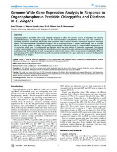
differentiated as shown by their expression of a seam antigen (Fig. 1H), and their number is normal (Table 2), as counted by the nuclear seam cell marker SCM (Terns et al., 1997). These data show that egl-27 is not required for the production or differentiation of muscle or seam epidermal cells, but is needed for their proper positioning in the embryo. As shown previously for vab-7 mutants (Ahringer, 1996), such disorganisation is an indication that patterning within a tissue is defective. Postembryonic defects of egl-27(we3) mutants (in egg-laying, male tail morphology and phasmid dye filling) suggest that egl-27 also has additional developmental roles. Cloning of egl-27 The genetic mapping of egl-27 narrowed the gene to a region of 9 cosmids. We first injected pools of cosmids and then single cosmids into egl-27(we3) mutants and tested for rescue of the mutant phenotype. We found that cosmid C04A2 had rescuing activity (see Materials and Methods). To determine which gene on C04A2 encodes egl-27, we took advantage of RNA-mediated gene inactivation (RNAi; Fire et al., 1998). Injection of double-stranded RNA (dsRNA) corresponding to a gene into a wild-type hermaphrodite will inactivate that gene in her progeny, mimicking a null phenotype (Fire et al., 1998). We found that C04A2.2(RNAi) resulted in progeny Fig. 1. Characterisation of egl-27(we3) and egl-27::gfp expression. (A) vab-7(ed6) L1 hermaphrodite is nearly identical to wild type except for a small bulge at the tip of the tail (arrow), where the wildtype tail is sharply pointed. (B) egl-27(we3) L1 grown at 15°C. (C-F) Muscle patterning in wt (C,D) and egl-27(we3) (E,F) embryos at 1.5-fold stage of development. Muscle cells in D and F are visualised using hlh-1::gfp, a reporter for the C. elegans MyoD homolog (Krause et al., 1990; K. Dej, S. Xu, and A. Fire personal communication); (C,E) DIC images of the same embryos. In D, two of the four muscle rows are visible in this focal plane. Arrow in F points to a cluster of muscle cells. (G,H) Seam patterning in wt (G) and egl-27(we3) (H), visualised using antibody NE2/1B4.14; arrows in H shows forked and disrupted posterior seam. (I) L1 larvae induced by egl-27(exon 11) RNAi. (J,K) egl-27::gfp expression in 1.5-fold embryo (J) and L3 hermaphrodite (K). All somatic cells appear to express the reporter gene. Scale bar in B is for B,I; scale bar in C is for C-H and J.
Global embryonic patterning in C. elegans 2487 1 kb
we3 (Q to STOP)
SmaI
BamHI 1
2
3
45
6
7 8
9
10
11
12
13
C04A2.2 C04A2.3 SL1
4.3kb RNA SL1
2.75kb RNA SL1
2.55kb RNA
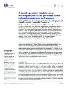
Fig. 2. egl-27 gene structure. The top line shows 15 kb of C04A2, containing all of the egl-27 exons (open boxes). Sizes of introns and exons are to scale. C04A2.2 and C04A2.3 are gene structures predicted (The C. elegans Sequencing Consortium, 1998). Below are our predicted major RNA gene structures, based on the sequences of cDNA and RACE clones and on northern blotting. Excluding a poly(A) tail, the lengths of the predicted major RNAs are 4051 nt, 2664 nt and 2468 nt (including 22 nt from SL1), in good agreement with the sizes of 4.3 kb, 2.75 kb and 2.55 kb from northern blotting (see Fig. 3; Materials and Methods). The SmaI and BamHI sites marked were used to make the egl-27::gfp translational fusion.
with a phenotype very similar to that of egl-27(we3) (Fig. 1I). By sequencing this locus from egl-27(we3) DNA, we found that the we3 mutation introduces a stop codon into the gene (see below), confirming that C04A2.2 corresponds to egl-27. Interestingly, like egl-27(we3), the C04A2.2(RNAi) phenotype is cold-sensitive (Table 1, egl-27(exon 11) rows). The similarity in phenotype between egl-27(we3) and egl27(RNAi) supports the view that egl-27(we3) is a strong lossof-function mutation.
egl-27 encodes multiple transcripts As C04A2.2 was only a gene prediction (The C. elegans Sequencing Consortium, 1998), we sequenced cDNA clones and performed RACE experiments to determine the gene structure. egl-27 extends much farther 5′ than the C04A2.2 prediction (Fig. 2). We found evidence for multiple egl-27 transcripts from our cDNA clones: in different cDNAs, the SL1 trans-spliced leader was found on exon 1, exon 6, exon 10 and exon 11 (see Materials and Methods). Spliced leaders are added to the 5′ ends of 70% of mRNAs in C. elegans (Zorio et al., 1994). We also found micro-heterogeneity in the 3′ splice site for intron 7 (see legend to Fig. 4). To explore further the transcripts produced from the egl-27 locus, we performed northern blots. Fig. 3 shows that at least 7 transcripts are produced: three major transcripts of 4.3 kb, 2.75 kb and 2.55 kb, and four minor ones of 3.5 kb, 3.2 kb, 3.0 kb and 1.5 kb; an exon 11 probe detects all transcripts, but exons 1-8 detect only the 4.3 kb major transcript and some minor transcripts (Fig. 3). The predicted structures of the three major transcripts is shown in Fig. 2, based on the results from northern blotting and cDNA sequencing. We sequenced egl-27 exons from egl-27(we3) DNA and found that we3 introduces a stop codon into exon 11 (Figs 2, 4), so we3 should affect all egl-27 products. By northern blotting, this mutation does not result in the loss of any egl-27
RNAs (Fig. 3). This suggests that egl-27 is not needed for its own expression. An egl-27 reporter gene is expressed ubiquitously in nuclei To learn where egl-27 is expressed, we constructed a translational fusion of the coding region of GFP to exon 11 of egl-27. Since exon 11 is contained in all egl-27 transcripts, we expect this reporter gene to reflect expression of all products made from the locus. egl-27::gfp is expressed in the somatic nuclei of most or all cells from the 50-cell stage of embryogenesis through to adulthood (Fig. 1J,K; additional data
Fig. 3. egl-27 produces multiple transcripts. Northern blots of wild-type (wt) or egl-27(we3) mixed stage poly(A)+ RNA, probed with exons 10-13 (lanes 1 and 2), exons 1-8 (lane 3), or exon 11 (lane 4). Lanes 1 and 2 were run on a different gel from lanes 3 and 4. Sizes are estimated based on standards (not shown).
2488 F. Solari, A. Bateman and J. Ahringer
Fig. 4. EGL-27 sequence. Shown is the translation of the 4.3 kb major egl-27 RNA. The small major RNAs are both predicted to encode a protein that begins at methionine 513 (boxed). The four domains described in the text are underlined: ELM1 with diagonals, ELM2 with verticals, SANT with black, and the GATA-like zinc finger with grey. Shown is the egl-27(we3) mutation, identified in three independent PCR clones, which changes a CAA codon to a TAA (stop) and the position where GFP is fused in the egl-27::gfp reporter gene. Positions of introns are marked with filled triangles. In our RACE clones, either of two 3′ splice sites for intron 7 were used; for the protein shown, the second splice was used as all of our long RACE clones (i.e., those that had ends at or near the beginning of exon 1) used this site. The splice sites differ by 9 nucleotides; splicing at the first one results in a protein with SLQ inserted at the position marked for intron 7.
MSRFDSQCSSEDVNKEDECVPSSSEDSQDGVSSPMENDDEPEFSQKHYDIEPCYYSLTGKSDRNCRGIVY
70
RYRQDSDLKGFQSHDGTLYRLRDSVFVEVSQNEPYVIAAICGFKYTKRDHVVVKLTRYFRADDIPEISLN
140
LMKQERAELEINPHLCPQSLNRELFNSELQITQPVSCLRGKCIVEYVKDVRHARTVADFSLDNDTFFFCL
210
HYNQDSTKLASTHYAIRVGTSFQATLPPMAECSVGDDSDRDELLYRPNSIESGEEEDYIKLARCYRTYTL
280
SGNHMLDSQKNARVSDLLMDEAIIQLHRSGYKIDDALSELNANDIILTTDVDNMTQDDAKKFAKGIKQLG
350
KNFSRIHRELLPHHSREQLVSYYYLWKKTPEATKPKQAARRVNPTSIKRPTKEKVKASRPTSTEYLDFDS
420
ASESDVENNGPSGRACHHCYGAESKDWHHANGLLLCTDCRLHYKKYGQLRQIANRPSQVPACLFKRSNSD
490
EEESGVRTRAGKKEQRRRTPSSMSETPDRRSPSTVSNGAPNLTAEETPTKKLNGSVKRAPKRPLHNGVIN
560
NVEKSNSSEEPASPTTPPPTLTNGLTNGHGPESSTPNGETISKRMKVEPSYDDDDDEEEGKMTIDEGDDD
630
PMPVLNGFKKEESVEEIKLELNGTIKKENGVETDPTTPTCSMEAENEVCETPAVVSVEIRDETNGETNSD
700
LKDDENVEPDSPEDTFELGSNVEFETKNAMFVRSIVRSCGPRCARTDLIFKIKVGGVWEKSIKEKEERKK stop (we3) VHLQNQRIQDSEKVAIQQNQIKKEQQQSQPTPQQIHQQQAQQNAQHLQQLQQAVMLGHLPPEVLRQMMPP GFP QFGVDPTAILMQQMMAGQQSQGVNAAFQHQMALQQQLEAHQVQFQLMMAHQHQQKMIAEQQQQQRHAAAQ
770
QLREREQREQRERERERQHQQQAQQALHQQQQQHAAAAANQLNPAMMQMMALMANSAASQQDIARLMEMA
980
AQQQQQQQQAAQAQAQRDQERERREREAREREAAREREREQAAREAAARDQAAREHAQAVQAAAAAAQQA
1050
QALTPDMQHMHLLQQLMLNPALMMQLQQAQAQQQQQQPQVTNPLQMLQHGMAAQSANQAEMMRRIHPEPA
1120
MRPQHQ*
1126
not shown). egl-27 is likely to be expressed maternally as well, since egl-27(we3) has a maternal effect. This would not have been seen with our reporter construct because transgenes in C. elegans usually do not report germline expression (Kelly et al., 1997). Since the GFP coding sequence introduced did not contain a nuclear localisation signal, nuclear expression of the reporter gene suggests that egl-27 encodes nuclear protein(s). EGL-27 is similar to a component of a chromatin regulatory complex with histone deacetylase and nucleosome remodelling activities Conceptual translation of the major 4.3 kb RNA yields a protein of 1126 amino acids (Fig. 4). The 2.75 kb and 2.55 kb major transcripts would encode an identical protein that begins at methionine 513 of the large protein (boxed in Fig. 4). EGL-27 has highest similarity (by Blast; Altschul et al., 1997) to two human genes and a C. elegans gene: KIAA0458, identified from a human brain cDNA library (Seki et al., 1997), MTA1, which has elevated expression in metastasizing mammary adenocarcinomas (Toh et al., 1995), and T27C4.4, a predicted C. elegans gene (Figs 5, 6). Comparisons between EGL-27, T27C4.4, KIAA0548 and MTA1 showed that EGL27 is most related to KIAA0458 whereas T27C4.4 is most similar to MTA1 (Fig. 5A). MTA1 has recently been shown to be a component of the NURD complex, which has histone deacetylase and ATPdependent nucleosome remodelling activities (Xue et al., 1998). Histone deacetylation is usually associated with transcriptional repression, whereas nucleosome remodelling is usually associated with transcriptional activation. However, neither the in vivo function of the NURD complex nor transcription factors regulated by it are yet known. The finding that EGL-27 is similar to a component of this complex
840 910
argues that EGL-27 has a role in the regulation of chromatin structure. EGL-27 family members are multidomain proteins with a myb-like SANT DNA-binding domain and a zinc finger Sequence analyses of EGL-27 identified four conserved domains (Fig. 5): (1) A SANT domain, which is similar to the DNA-binding domain of myb (Aasland, 1996; Fig. 5B). The SANT domain was first found in SWI3 (a yeast component of the SWI/SNF complex; Peterson and Tamkun 1995), ADA2 (a component of a histone acetylase complex; Horiuchi et al., 1995), N-CoR (a nuclear hormone co-repressor; Horlein et al., 1995) and the B′′ subunit of TFIIIB (a basal pol III transcription factor in yeast; Kassavetis et al., 1995). We propose that this region of EGL-27 has a structure essentially the same as that of myb. (2) A zinc finger related to the GATA family (Fig. 5C). (3) A conserved N terminus that is shared with MTA1 and T27C4.4, which we call the ELM1 (EGL-27 and MTA1 homology) domain. This domain is also found in a number of nuclear proteins, including cytosine-5 methyl transferases, ORC1 (part of the origin recognition complex), the Drosophila absent gene (a trithorax group protein) and several bromodomain-containing proteins (Fig. 5D), but the region of similarity does not cover any previously defined domains of these proteins. (4) A further conserved region (ELM2), also found in additional proteins, many of which contain SANT (Figs 5E, 6). This region is not similar to any domains of known function. Finally, the C terminus of EGL27 is acidic and glutamine rich (Fig. 4). The four named domains are encoded by the large major transcript but not the small ones. Fig. 6 shows a cartoon of the domains that we defined in
Global embryonic patterning in C. elegans 2489 A
Pairwise sequence identities
KIAA0458 T27C4.4 MTA1 B
C
T27C4.4
27% 36%
45%
LGKTRWTREEDEKLKKLVEQNGTDDWKVIANYLPNRTDVQCQHRWQKVLNPE .......HHHHHHHHHHHHH.....HHHHHHH.....HHHHHHHHH...... TDVDNMTQDDAKKFAKGIKQLGKNFSRIHRELLPHHSREQLVSYYYLWKKTP Q09228 LIEKCWTEDEVKRFVKGLRQYGKNFFRIRKELLPNKETGELITFYYYWKKTP g3413878 DQLEEWSTPEMNLFEDALDKVGKDFNEIRAEYLPWKSIRDIVEYYYLMKASN g3165588 DEMEEWSASEANLFEEALEKYGKDFTDIQQDFLPWKSLTSIIEYYYMWKTTD Q13330 QFMNVWTDHEKEIFKDKFIQHPKNFGLIASYLE.RKSVPDCVLYYYLTKKNE g2137603 VETSRWTEEEMEVAKKGLVEHGRNWAAIAKMVG.TKSEAQCKNFYFNYKRRH g2137603 EELSVWTEEECRNFEQGLKAYGKDFHLIQANKVRTRSVGECVAFYYMWKKSE g2529737
GATA-like domain
1gat ss EGL-27/433-480 KIAA0458/236-283 T27C4.4/584-635 MTA1_HUMAN/390-441 D
KIAA0458
SANT domain
1mbe ss EGL-27/329-380 KIAA0458/123-174 T27C4.4/479-530 MTA1_HUMAN/283-334 N-CoR/435-485 N-CoR/622-672 Er1_xenla/272-323
Fig. 5. Alignments of EGL-27 domains. (A) Percent identity between EGL-27, KIAA0458, T27C4.4 and MTA1, summed over the ELM2, GATA and SANT domains. (B-E) Alignments of EGL-27 domains. Conserved residues are highlighted using the belvu program (E. Sonnhammer, personal communication) in default mode. White letters on a black background are the most conserved, white letters on a grey background show intermediate conservation, and black letters on a grey background are the least conserved. The secondary structure is marked in the ss row, with E denoting β-strand and H denoting α-helix. For each sequence, the start and end points follow the identifier of the sequence, with the TrEMBL or SWISSPROT accession number or genbank PID following the alignment. (B) Alignment of myb-like SANT domains with the structurally solved myb domain 1mbe (Ogata et al., 1994). (C) Alignment of the GATA-like domain with the structurally solved gata domain 1gat (Omichinski et al., 1993). (D) Alignment of the ELM1 domain. (E) Alignment of the ELM2 domain. Not all identified matching proteins are included in the alignments.
EGL-27 40% 22% 26%
GTVCSNCQTSTTTLWRRSPM...GDPVCNACGLYYKLHQVNRPLTMRKDGIQ ..............EEEE......EEEEHHHHHHHHHH.....HHH...... GRACHHCYGAESKDWHHAN....GLLLCTDCRLHYKKYGQLRQIANRPSQVP Q09228 GYACRHCFTTTSKDWHHGG..RENILLCTDCRIHFKKYGEL..PPIEKPVDP g3413878 DNPCENCGTLDALNWYQWGGVGDKKVLCSTCWIQWKKFAGLNQKHELERFDK g3165588 GRACESCYTTQSYQWYSWGPPNMQCRLCASCWTYWKKYGGLKMPTRLDGERP Q13330
ELM1 domain
EGL-27/87-187 T27C4.4/13-245 MTA1_HUMAN/4-131 Q43479/26-112 ORC1_HUMAN/45-138 P70049/45-138 YG23_YEAST/368-452 Q18210/734-819 Q90941/954-1038 Q24189/1914-2021 Q18210/942-1026 Q90941/1155-1237 MTDM_ARATH/735-817 Q27746/743-825
TLYRLRDSVFVEVSQN.......EPYVIAAICGFKYTKRDHV.VVKLTRYFRADDIP. VTYAVGDFVYFDDTSA.(24).CVVYLRRRDIPQHLLKIADQAQRRFDNYYEVDKKK. NMYRVGDYVYFENSSS......NPYLIRRIEELNKTANGNV..EAKVVCFYRRRDIS. GVIRPGDSVLMKAPDS.(1)..KPPYVAKIEEIEAAGPRGAN.VKVKVRWYYRPEES. IHIQIGQFVLIEGDDD......ENPYVAKLLELFEDDSDPPPKKRARVQWFVRFCEV. ITVTPGDFVLIEGENE......ERPFVAKLQELYDDGNEKHTSKHAIVQWFLRYEEV. EKYQIGDWVLLHNPND.(1)..NKPIVGQIFRLWSTTDGN...KWLNACWYFRPEQT. TKYVAPCYAYVSRSDE.(2)..TPLHIFRIERTFKDENGE...KALQGHWVYRPEET. SMYHVGDYVYVEPAEA.(1)..LQPHIVCIERLWEDSAGE...KWLYGCWFYRPNET. LQVRQGDAVYVLRDIP.(24).QECDIFRVEHLWKNELGK...RFIFGQHFLRPHET. KFFWLGQCVLVFNNMK.(1)...LCDVMKINKIWREKDGS...EWFSGCWFARPSET. MWLKVGDCVFIKSHGL......VRPRVGRIEKMWVRDGA....AYFFGPIFIHPEET. EMVAVGGAVTLEVDDP.(1)..EMPAIYFVEYMFESTDHC...KMLHGRFLQRGSMT. EKIEIGDCVLIHPDDP.(1)..KPLFMARVIYMWQESQGE...MMFHAQWFVYGSET.
EGL-27/87-187 T27C4.4/13-245 MTA1_HUMAN/4-131 Q43479/26-112 ORC1_HUMAN/45-138 P70049/45-138 YG23_YEAST/368-452 Q18210/734-819 Q90941/954-1038 Q24189/1914-2021 Q18210/942-1026 Q90941/1155-1237 MTDM_ARATH/735-817 MTDM_HUMAN/634-716 Q27746/743-825
.(18)..HLCPQSLNRELFNSE...LQITQPVSCLRGKCIVEYV .(124).DQRLKLRQHEIFMTR...QSEILPAAAIRGKCRVVLL .(35)..KLKHQLRHRELFLSR...QLESLPATHIRGKCSVTLL .(2)...GRRPFHCEKEVFLSD...HQDVQSADTIECKCNVYSF .(6)...LLGRKPGAQEIFWYDYPACDSNINAETIIGLVRVIPL .(6)...LLGREPHQEEIFLYDVPSCENDIDAETIIGSVKVTQL .(2)...RVDRLFYKNEVMKTG...QYRDHPIQDIKGKCYVIHF .(2)...LASRKFMKQEVFLTP...FRDTVLAERLRGRCVVISL .(2)...LATRKFLEKEVFKSD...YYNKVPVSKILGKCVVMFV .(2)...EPSRRFYPNEVVRVS...LYEVVPIELVIGPCWLLDR .(2)...DEGRLFFKNEVIAVYR..NDETRKLCEIQRVCDVMPA .(2)...EPTKMFYKKEVFLSN...LEETCPMSCILGKCAVLSF .......VLGNAANERELFLTN...ECMTTQLKDIKGVASFEIR .......VLGATSDPLELFLVD...ECEDMQLSYIHSKVKVIYK .......VLGETSDPLEVFPID...ECQDTYLGSVNAKCTVIYK
E
Q09228 g3165588 Q13330 Q43479 Q13415 P70049 P53236 Q18210 Q90941 Q24189 Q18210 Q90941 P34881 P26358 Q27746
ELM2 domain
EGL-27/224-281 KIAA0458/16-74 T27C4.4/278-401 MTA1_HUMAN/165-226 KIAA0071/14-75 F10E7.11/81-137 Er1_xenla/169-229 D1014.9/107-167 F53H10.2/501-565
IESG...EEEDYIKLARCYRTYTLS YAIRVGT.SFQATLPPMA.(12).LLYRPNSI GEIRVGP.SHQAKLPDLQ.(14).ELVWMPGVNDC...DLLMYLRAARSMAAFAG. GAIRVGE.KYQAVVDEWM.(78).VWHPHHALTDR...DIDQYMIVARSVGLFARA GEIRVGN.RYQADITDLL.(16).VWEAHNPLTDK...QIDQFLVVARSVGTFARA GGMRVGP.QYQAVVPDFD.(17).VWSPNQNLSEA...KLDEYIAIAKEKHGYNM. RSIRVDPVLFQADVPLFN.(14).LWTIDQTNQPSD.EVIDNYLKDVVGLRKAH.. KEIMVGS.MFQAEIPVGI.(16).LWNPEYVMEER....VIDFLNEASRRTCEERG DEINVGT.EFQAKIADLN.(16).IWNTPETIDDE...KLEAFIRESSDRYLIPI. PHINLGK.NYQARVKKWC.(17).IVFSSEILQDIDPEQITAFELLACSQACPRA.
EGL-27 with other proteins that contain them. T27C4.4 is likely to be the only other C. elegans gene that contains all the identified domains of EGL-27, as >99% of the C. elegans genomic sequence is available (The C. elegans Sequencing Consortium, 1998); we have named this gene egr-1, for egl-27related gene.
Q09228 g3413878 g3165588 Q13330 g505104 g1086837 g2529737 g1256279 Q20733
egr-1 and egl-27 have a redundant embryonic function Because egr-1 shares the domain structure of egl-27, we wondered whether it had a similar function. To investigate this, we used RNAi to inactivate egr-1, alone or in combination with egl-27. egr-1(RNAi) animals are completely viable, with a
2490 F. Solari, A. Bateman and J. Ahringer EGL-27
ELM1
ELM2
SANT GATA
T27C4.4/EGR-1
ELM1
ELM2
SANT GATA
MTA1
ELM1
ELM2
SANT GATA
KIAA0458
ELM2
SANT GATA
Er1
ELM2
SANT
KIAA0071
ELM2
SANT
N-CoR
SANT SANT
F53H10.2
ELM2
SANT
F10E7.11
ELM2
SANT
C26C6.1
ELM1
C14B9.6
SANT
Fig. 6. Proteins with domains similar to those in EGL-27. Proteins shown were found to contain domains similar to EGL-27 using Blastp or PSI-blast (Altschul et al., 1997). Not every protein identified is shown.
small number having mild posterior defects (Table 1; Fig. 7A). However, egr-1(RNAi); egl-27(exon 11) or egr-1(RNAi); egl27(we3) double mutants are 100% embryonic lethal (Table 1), indicating that egl-27 and egr-1 have a redundant function in the embryo. Further RNAi experiments showed that this redundancy is at the level of the large egl-27 RNA. Since exon 11 of egl-27 is contained in all transcripts, egl-27(exon 11) should interfere with all egl-27 transcripts. To interfere with only the large egl27 RNA, we injected dsRNA to exons 1-8 (egl-27(exons 1-8)), which are not contained in the small RNAs. In contrast to egl27(exon 11), egl-27(exons 1-8) animals do not have body morphology defects, but do have dye-filling defects (Table 1), a phenotype similar to that of egl-27(n170) mutants. This is consistent with the findings of Herman et al. (1999), who showed that egl-27(n170) deletes exons in the large transcript, but not the small ones. We next examined egr-1(RNAi); egl27(exons 1-8) and found that these double mutants arrested as embryos with the same phenotype as egr-1(RNAi); egl-27(exon 11) (Table 1 and data not shown). This shows that the large egl-27 RNA, which contains the similarity to egr-1 and MTA1, has a shared function with egr-1. Double mutant embryos are abnormally patterned Double mutant embryos arrest with a striking uniform phenotype: tissues are not properly organised, but they are well
differentiated and morphogenesis fails to occur (Fig. 7B-F). To find out when defects initially occur in double mutants, we compared their development to those of wild-type embryos developing at the same time. Wild-type embryogenesis can be conveniently divided into three phases: first, during the initial cell divisions, the axes of the embryo are established and blastomere fates are determined through the actions of maternal genes (Bowerman, 1998). Second, at the 28-cell stage, gastrulation begins. During this phase, the cells of the body are patterned and most of embryonic cell proliferation occurs. At the end of this second phase, cells lie in precise positions where they will form tissues, but they do not yet express terminal differentiation markers. Disruption of regional patterning during this time causes cells to be abnormally positioned (see e.g., Ahringer, 1996; Chisholm and Horvitz, 1995). During the final stage, morphogenesis causes the organised ball of cells to be transformed into a long thin worm as tissues and organs differentiate and become functional. We found that double mutant embryos develop at the same rate as wild type (Fig. 8). However, cells of the embryo are not properly organised during gastrulation (Fig. 8). For example, intestinal cells in wild type are located in an ordered row but, in double mutants, they are found clustered together (compare Fig. 8C to H). Other cells in double mutants appear similarly disordered (Fig. 7B-F and data not shown). As development proceeds, double mutants do not undergo morphogenesis (compare Fig. 8D,E with I,J), but tissues differentiate normally. For example, intestinal cells in double mutants produce gut granules (a marker of differentiation; Fig. 7O). Muscle cells are found in a wild-type number (Table 2) and are functional, as contractions are evident in double mutants, but they are scattered and are not organised into rows as they are in wild type (compare Fig. 7C to 1E). Pharyngeal tissue is located within a basement membrane and forms a recognizable grinder, but the pharynx is not elongated as in wild type (compare Fig. 7H to 7J). Epidermal cells express the apical membrane antigen MH27 (data not shown), but they do not enclose the embryo (Fig. 7F). Seam cells (lateral epidermal cells) in double mutants are also disorganised but differentiated, as visualised by a seam antigen expressed near the end of embryogenesis (compare Fig. 7D to Fig. 1H). However, most seam cells that express the seam antigen fail to express the nuclear seam cell marker SCM (Table 2 and data not shown). This suggests that SCM may be a target of egl27/egr-1. Muscle, epidermal, pharyngeal, neuronal and intestinal cells appear to be well differentiated and present in the correct number based on antibody staining and visual inspection (Table 2; Fig. 7 and data not shown). The mispositioning of cells in all tissues coupled with normal
Table 3. Patterning gene expression in wild-type and double mutant embryos Strains
vab-7
lin-39
mab-5
egl-5
lin-44
wild-type
21.6 (n=7) range=20-25
12.8 (n=12) range=10-16
17.7 (n=6) range=15-20
20.4 (n=7) range=16-25
4.7 (n=11) range=2-8
double mutant
21.9 (n=15) range=18-31
12.8 (n=10) range=11-16
18.1 (n=9) range=15-21
20.2 (n=16) range=17-25
5.0 (n=8) range=1-8
Double mutants were generated by injecting ds RNA to egr-1(RNAi) + egl-27(exon11) . The number of cells expressing the indicated reporter genes are shown. Counts in wild-type and double mutants were done in embryos of similar ages maintained at 22°C.
Global embryonic patterning in C. elegans 2491
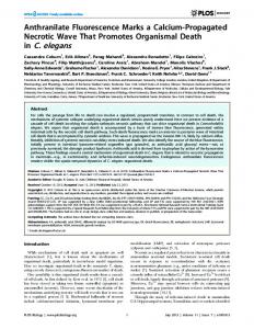
Fig. 7. egl-27 and T27C4.4 have a redundant global patterning function. (A) L2 larvae from an hermaphrodite injected with egr-1 dsRNA; arrow points to slight tail truncation. (B) Double mutant embryo from an hermaphrodite co-injected with egr-1 dsRNA and egl-27 dsRNA; arrow points to a cluster of intestinal cells. (C) hlh-1::gfp expression of (B), showing disorganised muscle cells. (D) Double mutant seam antigen expression; compare to Fig. 1H. (E) LIN-26 expression in a wt threefold embryo in rows of epidermal cells. (F) LIN-26 expression in a double mutant; epidermal cells are clustered on the surface. (G) wt vab-7::lacZ expression. (H) Pharyngeal muscle cells of wt embryo. (I) Double mutant vab-7::lacZ expression. (J) Pharyngeal muscle cells of embryo in I. (K) wt egl-5::gfp expression. (L) Double mutant egl5::gfp expression. (M) wt hlh-8::gfp containing embryo. (N) hlh-8::gfp expression in the M cell of embryo in M. (O) Double mutant hlh-8::gfp containing embryo; arrow points to a round cluster of intestinal cells with visible birefringent gut granules, arrowhead points to the pharyngeal grinder. (P) Weak hlh-8::gfp in the M cell of double mutant embryo in O (arrow). This expression was not visible using the photographic settings for the wild-type expression in N. Scale bar in P is for B-P.
differentiation suggests that egl-27 and egr-1 specifically function in cell patterning in diverse tissue types. A HOX gene target is not expressed in double mutants The global disorganisation of cells of double mutants is similar to that seen regionally in the posterior of vab-7 mutants. Since egl-27/egr-1 are likely to affect transcription, we reasoned that the phenotype could be due to changes in the expression of many regionally acting developmental genes. To test this, we first assayed the expression of vab-7. Double mutants express vab-7 in a normal number of cells, but in a disorganised pattern (Table 3; Fig. 7G,I). Because egl-27 and egr-1 are not required for vab-7 expression, they either act with or downstream of vab-7. Likewise, egl-27::gfp expression is normal in vab-7 mutants suggesting that vab-7 does not regulate egl-27 transcription (not shown). We next asked whether the expression of other regionally acting genes might be affected in double mutants. We assayed
the expression of four zygotic patterning genes that are first expressed in the embryo: three HOX genes (lin-39, mab-5 and egl-5; Costa et al., 1988; Clark et al., 1993; Wang et al., 1993) and lin-44, a wnt homologue required for the correct pattern of cell divisions in the tail (Herman and Horvitz 1994; Herman et al., 1995). Like vab-7, all are expressed in a normal number of cells (Fig. 7K-L; Table 3). This shows that many developmental patterning genes are expressed normally in double mutants. Since EGL-27 and EGR-1 are likely to regulate chromatin structure, thereby affecting the ability of transcription factors to function, one possibility is that they act in conjunction with transcription factors involved in patterning. To test this idea, we assayed the expression of hlh-8, a target of the HOX gene mab-5 (Harfe et al., 1998) in double mutants. So far, this is the only known target of any of the genes assayed above. In wildtype four-fold embryos, where the pharyngeal grinder is well formed, hlh-8::gfp is highly expressed in the M cell (20/20 embryos; Fig. 7N). In contrast, in double mutants with a visible
2492 F. Solari, A. Bateman and J. Ahringer
Fig. 8. Time course of wild-type and double mutant development. The development of wild-type (A-E) and egr-1 (RNAi); egl-27 (exon 11) double mutant (F-J) embryos was followed at 22°C until wild type was undergoing morphogenesis (D); embryos were then placed at 15°C overnight and photographed 12 hours later (E,J). In this figure, the zero time point is at the 50-cell stage (A,F). From this reference, other time points are (B,G) 2 hours; (C,H) 4 hours; (D,I) 8 hours. Cell number in wild-type and double mutant embryos is similar at all time points. (B,G) Wild-type and double mutant embryos are undergoing gastrulation. (C,H) End of gastrulation. Intestinal cells in wild type are arranged in a long anteroposterior tube (arrows in C); in double mutants, intestinal cells form a ball (arrows in H). (D,I) Wild-type embryo undergoing morphogenesis and elongating (D); no elongation occurs in the double mutant embryo (I). (E,J) The wild-type embryo (E) has fully elongated and was moving within the eggshell; the double mutant embryo (J) has not undergone morphogenesis but tissues are well-differentiated (arrow points to pharyngeal tissue, arrowhead to intestinal tissue, muscle contractions and neuronal processes were also evident). Anterior, left.
grinder, hlh-8::gfp is essentially absent (5/10 embryos had no expression and 5/10 had the barely detectable expression shown in Fig. 7P). We believe that double mutants survive long enough for hlh-8 to be expressed, since a late embryonic marker first expressed at this time (the seam antigen in Fig. 7D) is present. This supports the hypothesis that mab-5, and perhaps other zygotic patterning genes such as vab-7, require egl-27 or egr-1 for activity. DISCUSSION egl-27 and egr-1 are C. elegans genes with similarity to MTA1, a protein found in the NURD chromatin remodelling complex. These genes have an important redundant function in the organisation of embryonic cells during the phase of body patterning. Based on our phenotypic analyses and the known role of MTA1 in chromatin remodelling, we propose that EGL27 and EGR-1 are components of a protein complex that specifically functions with sequence-specific transcription factors involved in embryonic patterning. Multiple developmental roles of egl-27 In addition to its redundant function with egr-1, egl-27 also has unique functions. egl-27 mutants have pleiotropic phenotypes, including abnormal body morphology and postembryonic defects in the development of the male tail and the phasmids (Desai et al., 1988; Garriga et al., 1993; Trent et al., 1983; Herman et al., 1999; Table 1, Materials and methods, and data not shown). Our studies focused on the embryonic role of egl27 and showed that it is required for proper pattern of muscle and epidermal cells. Herman et al. (1999), who also cloned egl27, studied other aspects of the egl-27 phenotype. They found that egl-27 mutants have defects in T cell polarity, which has been shown to be controlled by lin-44, a wnt family member (Herman and Horvitz, 1994; Herman et al., 1995). In addition, they showed that egl-27 mutants have defects in cell migrations and divisions, some of which are controlled by HOX genes.
Taken together, these results support the view that there is a widespread requirement for egl-27 function in developmental patterning. Functional redundancy of egl-27 and egr-1 egl-27 has partial functional redundancy with a related C. elegans gene, egr-1. Inhibiting their activities together results in the abnormal positioning of cells without affecting tissue determination or differentiation. To our knowledge, no previously characterised mutants have this phenotype. As we have only studied egr-1 using RNAi, an important goal for the future will be to identify mutations in the gene. Although double mutants have global defects in development, several observations argue that egl-27/egr-1 are not affecting cellular health or gene expression generally. First, tissues in double mutants, though not organised, appear healthy. We did not see increased cell deaths, widespread necrosis or lack of adhesion between cells during the time of normal embryogenesis. Second, cell lineages and the process of gastrulation appear to be normal based on the positions and numbers of cell types. Third, tissues in double mutants are well differentiated and functional where we can assay them (e.g., muscle twitching), and even late differentiation markers are expressed (e.g., the seam antigen in Fig. 7D). Therefore, double mutants are not blocked in development at an early stage and egl-27/egr-1 are not needed for the transcription of terminal differentiation products. Rather egl-27/egr-1 appear to have a specific role in positioning cells in the embryo at the time of major body patterning. This phenotype of double mutants is consistent with a defect in patterning in all regions of the embryo. We therefore investigated whether the expression of a number of regional developmental genes (vab-7, HOX genes and lin-44) was affected in double mutants, but found that these are expressed in a normal number of cells. Although expression is normal, the activity of the HOX gene mab-5, a homeodomain transcription factor (Costa et al., 1988), is apparently altered, as a target (hlh-8) is not expressed in double mutants. This
Global embryonic patterning in C. elegans 2493 suggests that MAB-5 requires EGL-27/EGR-1 in order to activate hlh-8. We propose that EGL-27/EGR-1 affect the ability of many transcription factors involved in patterning to function, and that cells fail to be organised because their patterning is absent. For example, vab-7 patterns muscle and epidermal cells in the posterior of the embryo; in its absence, these cells still differentiate as muscle and epidermal tissue, but they are abnormally positioned resulting in disorganisation of the posterior end (Ahringer, 1996). Likewise, the vab-3 gene, which encodes a Pax6 homologue, patterns anterior epidermal cells; in its absence, the anterior region is disorganised (Chisholm and Horvitz, 1995). We also found that SCM, a nuclear marker of seam cells, fails to be expressed although seam cells are present and differentiated. One possibility is that SCM may be involved in seam cell patterning and may be regulated by EGL-27/EGR-1. Neither the function of SCM nor how SCM expression is controlled are yet known. Studying how hlh-8 and SCM expression are activated should shed light on how egl-27/egr1 and regional patterning genes might cooperate. Histone acetylation and deacetlylation in development Many sequence-specific transcription factors function with multiprotein complexes that alter histone acetylation or remodel nucleosomes (reviewed in Kadonaga, 1998; Struhl, 1998). Histone hyperacetylation is associated with transcriptional activity, whereas histone hypoacetylation is correlated with transcriptionally silent chromatin and heterochromatin. Alterations in chromatin structure by ATPdependent nucleosome remodelling complexes is thought to activate transcription. Recently, functions for histone acetylation (HAT) complexes in development have been identified. For example, dCBP, a Drosophila homologue of the mammalian HAT proteins CBP and p300, is involved in signalling pathways important for pattern formation (Akimaru et al., 1997a,b; Waltzer and Bienz, 1998). In C. elegans, the CBP homologue cbp-1 is required for the development of all non-neuronal tissues: in its absence, no mesodermal, epidermal or intestinal development occurs and most cells appear to differentiate into neurons (Shi and Mello, 1998). Developmental roles for histone deacetylases (HDA) have also been identified. For example, one of a number of C. elegans histone deacetylases, hda-1, antagonizes the effect of cbp-1 on intestinal differentiation (Shi and Mello, 1998). Further, two C. elegans RbAp46/48 homologues, histone-associated proteins which are found in HDA complexes (Taunton et al., 1996) are needed for development past the 100-cell stage, suggesting a shared general cellular function (Shi and Mello, 1998). Recently, the C. elegans gene lin-53 was shown to encode one of these RbAp46/48 homologues (Lu and Horvitz, 1998). LIN-53 is involved in negatively regulating vulval cell fates promoted by the Ras pathway and is likely to act in a multiprotein complex containing the histone deacetlyase HDA1 and the retinoblastoma homolog LIN-35 (Lu and Horvitz, 1998). In summary, chromatin remodelling complexes have important roles in development, but few of their targets are known. Finding these will help our understanding of how transcription factor activities are regulated to promote particular developmental outcomes.
Possible functions for egl-27 and egr-1 Recently, the human protein MTA1, which is similar to EGL27 and EGR-1 was found to be a component of a multiprotein complex called NURD, which has both ATP-dependent nucleosome remodelling and histone deacetylation activities (Xue et al., 1998). A similar complex containing an MTA1 related protein, MTA2, was identified by Zhang et al. (1998). Besides MTA1 the NURD complex contains the histone deacetylases HDAC1 and HDAC2, the two histone-binding proteins RbAp48/46, CHD4 (for chromodomain-helicaseDNA binding), and several unidentified proteins. Based on the similarity between EGL-27/EGR-1 and MTA1, and the phenotype of double mutants, we suggest that EGL-27 and EGR-1 are components of a multi-protein chromatin regulatory complex that is required for the functions of regional patterning proteins. Good candidates for interaction with an EGL-27 complex are VAB-7 and the HOX proteins. In the future, it will be important to identify proteins with which EGL-27 and EGR1 interact, as these should shed light on their biochemical function. How might an EGL-27-containing complex regulate the activity of these proteins? In one simple model, these transcription factors could associate with the complex and bring it to target genes. This could cause chromatin around the target genes to be altered, leading to changes in transcriptional activity. It is not yet known whether the NURD complex has transcriptional repressing or activating activities, or both; experiments in Xue et al. (1998) suggest a repressive function. However, loss of expression of hlh-8 and SCM in double mutants suggests an activating function. Genetic and biochemical studies in C. elegans involving egl-27 and egr-1 should help both to identify further components and to understand the functions of the complex. We thank Jose de Celis, Richard Durbin, Gos Micklem, Jordan Raff, Daniel St. Johnston and members of the laboratory for helpful comments on the manuscript, and Vivian Bardwell, Erik Miska and Tony Kouzarides for useful discussions. We are grateful to Cara Neades for providing excellent technical assistance. We also thank Howard Baylis, Alan Coulson, Andy Fire, Mike Herman, Jonathan Hodgkin, Yuji Kohara, Michel Labouesse and Cynthia Kenyon for strains, reporter constructs, cDNAs and antibodies. M. Herman, Q. Ch’ng, S. Hettenbach, T. R. Ratliff, C. Kenyon, and R. K. Herman kindly communicated results prior to publication. Some strains used in this study were obtained from the CGC, which is supported by the NIH-NCRR. F. S. was supported by an EMBO fellowship and a European Union Marie-Curie postdoctoral fellowship, A. G. B. is supported by Wellcome Trust grant 048880, and J. A. was supported by a Wellcome Trust Career Development Award (No. 045515/Z/95/Z/PMG/AH).
REFERENCES Aasland, R. (1996). The SANT domain: a putative DNA-binding domain in the SWI-SNF and ADA complexes, the transcriptional co-repressor N-CoR and TFIIIB. Trends Biochem. Sci. 21, 87-88. Ahringer, J. (1996). Posterior patterning by the Caenorhabditis elegans evenskipped homolog vab-7. Genes Dev. 10, 1120-1130. Akimaru, H., Chen, Y., Dai, P., Hou, D.-X., Nonaka, M., Smolik, S. M., Armstrong, S., Goodman, R. H. and Ishii, S. (1997a). Drosophila CBP is a co-activator of cubitus interruptus in hedgehog signalling. Nature 386, 735-738. Akimaru, H., Hou, D.-X. and Ishii, S. (1997b). Drosophila CBP is required
2494 F. Solari, A. Bateman and J. Ahringer for dorsal-dependent twist gene expression. Nature Genetics 17, 211214. Albertson, D. G. (1984). Formation of the first cleavage spindle in nematode embryos. Dev. Biol. 101, 61-72. Altschul, S. F., Madden, T. L., Schaffer, A. A., Zhang, J., Zhang, Z., Miller, W. and Lipman, D. J. (1997). Gapped BLAST and PSI-BLAST: a new generation of protein database search programs. Nucleic Acids Res. 25, 3389-3402. Bairoch, A., Bucher, P. and Hofmann, K. (1997). The PROSITE database, its status in 1997. Nucleic Acids Res. 25, 217-221 Bowerman, B. (1998). Maternal control of pattern formation in early Caenorhabditis elegans embryos. Curr. Top. Dev. Biol. 39, 73-117. Brenner, S. (1974). The genetics of Caenorhabditis elegans. Genetics 77, 7194. Chisholm, A. D. and Horvitz, H. R. (1995). Patterning of the Caenorhabditis elegans head region by the Pax-6 family member vab-3. Nature 377, 52-55. Clark, S. G., Chisholm, A. D. and Horvitz, H. R. (1993). Control of cell fates in the central body region of C. elegans by the homeobox gene lin-39. Cell 74, 43-55. Costa, M., Weir, M., Coulson, A., Sulston, J. and Kenyon, C. (1988). Posterior pattern formation in C. elegans involves position-specific expression of a gene containing a homeobox. Cell 55, 747-756. Desai, C., Garriga, G., McIntire, S. L. and Horvitz, H. R. (1988). A genetic pathway for the development of the Caenorhabditis elegans HSN motor neurons. Nature 336, 638-646. Fire, A., Xu, S., Montgomery, M. K., Kostas, S. A., Driver, S. E. and Mello, C. C. (1998). Potent and specific genetic interference by double- stranded RNA in Caenorhabditis elegans. Nature 391, 806-811. Fisher, A. L. and Caudy, M. (1998). Groucho proteins: transcriptional corepressors for specific subsets of DNA-binding transcription factors in vertebrates and invertebrates. Genes Dev. 12, 1931-40. Francis, R. and Waterston, R. H. (1991). Muscle cell attachment in Caenorhabditis elegans. J. Cell Biol. 114, 465-479. Garriga, G., Desai, C. and Horvitz, R. H. (1993). Cell interaction control the direction of outgrowth, branching and fasciculation of the HSN axons of Caenorhabditis elegans. Development 117, 1071-1087. Harfe, B. D., Vaz Gomes, A., Kenyon, C., Liu, J., Krause, M. and Fire, A. (1998). Analysis of a Caenorhabditis elegans Twist homolog identifies conserved and divergent aspects of mesodermal patterning. Genes Dev. 12, 2623-2635. Herman, M. A. and Horvitz, H. R. (1994). The Caenorhabditis elegans gene lin-44 controls the polarity of asymetric cell divisions. Development 120, 1035-1047. Herman, M. A., Vassilieva, L. L., Horvitz, H. R., Shaw, J. E. and Herman, R. K. (1995). The C.elegans gene lin-44, which controls the polarity of certain asymmetric cell divisions, encodes a Wnt protein and acts cell nonautonomously. Cell 83, 101-110. Herman, M. A., Ch’ng, Q., Hettenbach, S. M., Ratliff, T. R., Kenyon, C. and Herman, R. K. (1999). EGL-27 is similar to a metastasis-associated factor and controls cell polarity and cell migration in C. elegans. Development126, 1055-1064. Horiuchi, J., Silverman, N., Marcus, G. A. and Guarente, L. (1995). ADA3, a putative transcriptional adaptor, consists of two separable domains and interacts with ADA2 and GCN5 in a trimeric complex. Mol. Cell. Biol. 15, 1203-1209. Horlein, A. J., Naar, A. M., Heinzel, T., Torchia, J., Gloss, B., Kurokawa, R., Ryan, A., Kamei, Y., Soderstrom, M., Glass, C. K., et al. (1995). Ligand-independent repression by the thyroid hormone receptor mediated by a nuclear receptor co-repressor. Nature 377, 397-404. Kadonaga, J. T. (1998). Eukaryotic transcription: an interlaced network of transcription factors and chromatin modifying enzymes. Cell 92, 307313. Kassavetis, G. A., Nguyen, S. T., Kobayashi, R., Kumar, A., Geiduschek, E. P. and Pisano, M. (1995). Cloning, expression, and function of TFC5, the gene encoding the B′′ component of the Saccharomyces cerevisiae RNA polymerase III transcription factor TFIIIB. Proc. Natl. Acad. Sci. USA 92, 9786-9790. Kelly, W. G., Xu, S., Montgomery, M. K. and Fire, A. (1997). Distinct requirement for somatic and germline expression of a generally expressed Caenorhabditis elegans gene. Genetics 146, 227-238. Krause, M., Fire, A., White Harrison, S., Priess, J. and Weintraub, H. (1990). CeMyoD accumulation defines the body wall muscle cell fate during C. elegans embryogenesis. Cell 63, 907-919. Labouesse, M., Hartwieg, E. and Horvitz, H. R. (1996). The Caenorhabditis
elegans LIN-26 protein is required to specify and/or maintain all non neuronal ectodermal cell fates. Development 122, 2579-2588. Lu, X. and Horvitz, H. R. (1998). lin-35 and lin-53, two genes that antagonize a C.elegans ras pathway, encode proteins similar to Rb and its binding protein RbAp48. Cell 95, 981-991. Mann, R. S. and Affolter, M. (1998). Hox proteins meet more partners. Curr. Opin. Gen Dev. 8, 423-429. McGinnis, W. and Krumlauf, R. (1992). Homeobox genes and axial patterning. Cell 68, 283-302. Mello, C. C., Kramer, J. M., Stinchcomb, D. and Ambros, V. (1991). Efficient gene transfer in C. elegans: extrachromosomal maintenance and integration of transforming sequences. EMBO J. 10, 3959-3970. Ogata, K., Morikawa, S., Nakamura, H., Sekikawa, A., Inoue, T., Kanai, H., Sarai, A., Ishii, S. and Nishimura, Y. (1994). Solution structure of a specific DNA complex of the Myb DNA-binding domain with cooperative recognition helices. Cell 79, 639-648. Omichinski, J. G., Glore, G. M., Schaad, O., Felsenfeld, G., Trainor, C., Apella, E., Stahl, S. J. and Gronenborn, A. M. (1993). NMR structure of a specific DNA complex of Zn-containing DNA binding domain of GATA1. Science 261, 438-446. Peterson, C. L. and Tamkun, J. W. (1995). The SWI-SNF complex: a chromatin remodeling machine? Trends Biochem. Sci. 20, 143-146. Priess, J. R. and Thomson, J. N. (1987). Cellular interactions in early C.elegans embryo. Cell 48, 241-250. Schnabel, R. (1991). Cellular interaction involved in the determination of the early C. elegans embryo. Mech. Dev. 34, 85-99. Seki, N., Ohira, M., Nagase, T., Ishikawa, K., Miyajima, N., Nakajima, D., Nomura, N. and Ohara, O. (1997). Characterization of cDNA clones in size-fractionated cDNA libraries from human brain. DNA Res. 4, 345-349. Shi, Y. and Mello, C. (1998). A CBP/p300 homolog specifies multiple differentiation pathways in Caenorhabditis elegans. Genes Dev. 12, 943955. Struhl, K. (1998). Histone acetylation and transcriptional regulatory mechanisms. Genes Dev. 12, 599-606. Sulston, J. E., Schierenberg, E., White, J. G. and Thomson, J. N. (1983). The embryonic cell lineage of the Nematode Caenorhabditis elegans. Dev. Biol. 100, 64-119. Taunton, J., Hassig, C. A. and Schreiber, S. L. (1996). A mammalian histone deacetylase related to the yeast transcriptional regulator Rpd3. Science 272, 408-411. Terns, R. M., Kroll-Conner, P., Zhu, J., Chung, S. and Rothman, J. H. (1997). A deficiency screen for zygotic loci required for establishment and patterning of the epidermis in Caenorhabditis elegans. Genetics 146, 185206. The C. elegans Sequencing Consortium. (1998). Genome sequence of the nematode C. elegans: a platform for investigating biology. Science 282, 2012-2018. Thompson, J. D., Higgins, D. G. and Gibson, T. J. (1994). CLUSTAL W: improving the sensitivity of progressive multiple sequence alignment through sequence weighting, position-specific gap penalties and weight matrix choice. Nucleic Acids Res. 22, 4673-80. Toh, Y., Pencil, S. D. and Nicolson, G. L. (1995). Analysis of the complete sequence of the novel metastasis-associated candidate gene, mta1, differentially expressed in mammary adenocarcinoma and breast cancer cell lines. Gene 159, 97-104. Trent, C., Tsung, N. and Horvitz, H. R. (1983). Egg-laying defective mutants of the nematode Caenhorhabditis elegans. Genetics 104, 619-647. Waltzer, L. and Bienz, M. (1998). Drosophila CBP represses the transcription factor TCF to antagonize Wingless signaling. Nature 395, 521-525. Wang, B. B., Muller-Immergluck, M. M., Austin, J., Robinson, N. T., Chisholm, A. and Kenyon, C. (1993). A homeotic gene cluster patterns the anteroposterior body axis of C. elegans. Cell 74, 29-42. Wood, W. B., Ed. (1988). The Nematode Caenorhabditis elegans. New York: Cold Spring Harbour Laboratory. Xue, Y., Wong, J., Moreno, G. T., Young, M. K., Cote, J. and Wang, W. (1998). NURD, a novel complex with both ATP-dependent chromatinremodeling and histone deacetylase activities. Molecular Cell 2, 851-861. Zhang, Y., LeRoy, G., Seelig, H.-P., Lane, W. S. and Reinberg, D. (1998). The dermatomyositis-specific autoantigen Mi2 is a component of a protein complex containing histone deacetylase and nucleosome remodeling activities. Cell 95, 279-289. Zorio, D. A., Cheng, N. N., Blumenthal, T. and Spieth, J. (1994). Operons as a common form of chromosomal organization in C. elegans. Nature 372, 270-2.