Apr 15, 2006 - most such segments are stretches of hydrophobic amino acids of sufficient length to .... 1986), and increases the proportion of it found as dimer. (Weiner et al. ..... Samples containing different molar ratios were prepared, and the mixtures ..... face would also include several of the glycine residues present.
Vol. 267, No. 11, Issue of April 15,pp. 7683-7689,1992 Printed in U.S.A.
THEJOURNALOFBIOLOGICAL CHEMISTRY Q 1992 by The American Society for Biochemistry and Molecular Biology, Inc.
Glycophorin A Dimerization Is Driven by Specific Interactions between Transmembrane a-Helices* (Received for publication, October 13,1991)
Mark A. LemmonS, John M. Flanagan, John F. Hunt, Brian D. Adair, Barbara-Jean Bormanng, Christopher E. Dempseyll, and Donald M. Engelman(( From the Department of Molecular Biophysicsand Biochemistry, Yale University, New Haven, Connecticut 06511 and SBoehringerZngelheim Pharmaceuticals, Ridgefield, Connecticut 06877
Specific side-by-side interactions between trans- ices are important in,defining tertiary and quaternary strucmembrane a-helices may be importantin the assembly ture. In the case of bacteriorhodopsin the correctly folded and function of integral membrane proteins. We de- molecule can be regenerated from two chymotryptic fragments scribe a system for the genetic and biophysical analysis containing two and five helical transmembrane segments, of these interactions. The transmembrane a-helical do- respectively (Popot et al., 1987), and further from two synmain of interest is fused to the C-terminus of staphy- thetic helical transmembrane peptides plus the five-helix lococcal nuclease. The resulting chimera can be ex- chymotryptic fragment.’ These results led to the proposal of pressed at high levels in Escherichia coliand is readily a model for the folding of integral membrane proteins in two purified. In our initial application we study the single stages (Popot and Engelman, 1990). In stage I independently transmembrane a-helix of human glycophorin A stable a-helices are established across the lipid bilayer, and (GpA), thought to mediate the SDS-stable dimerization in stage I1 these interact to form functional transmembrane of this protein. The resulting chimera forms a dimer in SDS, which is disrupted upon addition of a peptide structures. Similarly, oligomerization of a number of proteins that corresponding to the transmembrane domain ofGpA. Deletion mutagenesis has been used to delineate the cross the bilayer once is thought to be important in their maybe mediated byspecific minimumtransmembranedomain sufficient for this structureandfunction,and behavior. Site-specific mutagenesis shows that a me- noncovalent interactions between the transmembrane dothionine residue, previously implicatedas a potential mains, in a process analogous to stage I1 of the above model. interfacial residue, canbe replaced with other hydro- A recently demonstrated example is the interaction between T-cell receptor-a and CD36 (Manolios et al., 1990; Cosson et phobic residues without disrupting dimerization. By contrast, rather conservative substitutions at avaline al., 1991) in theassembly of the T-cell receptor complex. Each on a different faceof the a-helix disrupt dimerization, of these proteins has a single predicted transmembrane asuggesting a high degree of specificity in the helix- helix, and ion pairs may form between oppositely charged helix interactions. This approachallows the interface residues in the two transmembrane domains. The context of between interactinghelices to be defined. these charged residues is important in determining theirability to mediate the association, indicating that they may not constitute the sole determinant for this interaction. Association of transmembrane domains which contain no charged Integral membrane proteinscontain one or more memgroups is also seen. The human erythrocyte sialoglycoprotein brane-spanning segments. Excluding those found in a small class of membrane proteins that contain intramembraneous glycophorin A (GpA)’ forms SDS-stable dimers through inp-sheets as theirmajor structural element (Weiss et al., 1991), teractions mediated by its single transmembrane domain, most such segments are stretchesof hydrophobic amino acids which contains no strongly polar residues (Furthmayr and of sufficient length to span the lipid bilayer as a-helices. This Marchesi, 1976). Addition of a synthetic peptide with the characteristic has been exploited in the development of algo- sequence of the GpA transmembrane domain disrupts the rithms for the prediction of membrane protein secondary dimer, resulting in the formation of a peptide-protein heterodimer, whereas addition of a variety of other transmembrane structure (Engelman et al., 1986; Kyte and Doolittle, 1982). In bacteriorhodopsin (Henderson et al., 1990) and thepho- peptides with similar amino acid composition and charge tosynthetic reaction center (Deisenhofer et al., 1985) specific organization has no such effect (Bormann et al., 1989). Carboxymethylation of the single methionine residue (Met-81) interactions between the constituent transmembrane a-helin thetransmembrane domain of GpA prevents dimerization * This work was supported by National Institutes of Health Grant (Silverberg et al., 1976). Thus it has been shown that there 5P01-GM39546, National Science Foundation Grant DMB8805587, are specific structural features of the GpA transmembrane funds from Boehringer Ingelheim Inc., and the National Foundation domain which cause it toform a stable dimer.
for Cancer Research. The costs of publication of this article were defrayed in part by the payment of page charges. This article must therefore be hereby marked “advertisement” in accordance with 18 U.S.C. Section 1734 solelyto indicate this fact. $ A Predoctoral Fellow of the Howard Hughes Medical Institute. 11 Present address: Dept. of Biochemistry, School of Medical Sciences, University Walk, Unversity of Bristol, Bristol, United Kingdom BS8 1TD. 11 To whom correspondence should be addressed Dept. of Molecular Biophysics and Biochemistry, Yale University, 260 Whitney Ave., New Haven, CT 06511.
T. W. Kahn, and D. M. Engelman, submitted for publication. The abbreviations used are: GpA, glycophorin A; SDS, sodium dodecyl sulfate; LB, Luria broth;TB, terrific broth; SN, staphylococcal nuclease; PMSF, phenylmethylsulfonyl fluoride; HPLC, highpressure liquid Chromatography; PAGE, polyacrylamide gel electrophoresis; PCR, polymerase chain reaction; EGFR, epidermal growth factor receptor; HER-2, human version of the neu oncogene; BrA, bacteriorhodopsin helix A TM, transmembrane domain; T(is), GpA transmembrane peptide derived from trypsin cleavage; MOPS, 4morpholinepropanesulfonic acid.
7683
7684
Specific Interactions Transmembrane betweena-Helices
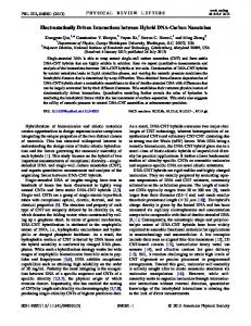
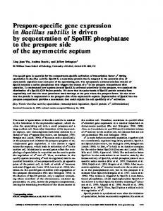
Similar interactions may be important in transmembrane A. signalling by growth factor receptorswith asingle transmembrane a-helix. For a number of these, mostnotably the epidermal growth factor receptor (EGFR), there is considerable evidencethat ligand-induced receptor dimerization is the primary event in signalling (Ullrich and Schlessinger, 1990). Specificinteractionsbetweenintramembraneousdomains may provide part of the energy for this dimerization. Indeed, avaline-to-glutamicacidmutationin the transmembrane domain of the neu oncogene product causes this EGFR-like Rrnlon Enccding molecule to become constitutively active (Bargmann et al., 1986), andincreases the proportionof it found as dimer (Weiner et al., 1989). An analogous mutation has beenreported to enhance the tumorigenicityofligand-stimulated EGFR (Kashles et al., 1988). A sequence motif found in the transmembrane domains of such receptors has been proposed to mediate oligomerization ofthese molecules (Sternberg and Gullick, 1990);this motif also occurs in the GpA transmemB. brane a-helix. DNA Seauence in Linking Region. Here, we report the use of a chimeric protein to study the Apa I Rsl I dimerization of the transmembranedomainofGpA. This li 5 . GCT GAT TCG GGC GRli GTR UVL CTT GCC 3 ’ domain is fused to the C terminus of staphylococcal nuclease Residue in G ; l t R V Q L h via a flexible linker. The resulting artificialmembrane protein can be expressedathigh levels in Escherichia coliand is SNGpA. Nudeax GlycophorinA readily purified exploiting the characteristics of the soluble Amino Acid Seauence of SN/GoA101. domain. We delineate the minimum transmembrane domain sufficient to confer uponthis protein the oligomeric behavior described for GpA. We also present preliminarysite-directed mutagenesis studies, designed to define the specific molecular contacts that stabilize the GpA dimer. FIG. 1. A , schematic representationof the constructs encoding the chimeric protein SN/GpA. From pSN/GpA the chimera $A
l
nurnb3s
EXPERIMENTALPROCEDURES
Construction of the Gene for the Chimeric Protein (SN/GpA)-A plasmid encoding staphylococcal nuclease Q149P (pPCV-I) was kindly supplied by A. Frankel (WistarInstitute). Restriction enzymes, T 4 DNA ligase, and polynucleotide kinase were purchased from New England Biolabs. Taq polymerase was from Perkin-Elmer/Cetus. Molecular biological manipulations were performed according to published protocols (Sambrook et al., 1989). Oligonucleotides were synthesized either by the Protein and Nucleic Acid Chemistry Facility a t Yale, or at Boehringer Ingelheim. All media used contained 200 Fg/ml ampicillin. An RsaI to SphI fragment of the gene for human GpA, containing the transmembrane and cytoplasmic domains, was isolated and ligated along with an ApaI to RsaI linker to replace the ApaI to SphI fragment of pPCV-1, thus generating the plasmid pSN/GpAl, shown in Fig. 1. In this construct the gene is under control of the phoA promoter and has the signal sequence of alkaline phosphatase. Low levels of expression of the chimera were achieved from this construct phosphate starvation in glucose-MOPS medium in E. coli ”294 by containing 0.1 mM phosphate (Neidhardt et al., 1974). Induction of SN/GpA began at around 5 h after inoculation, and was allowed to continue for a further 3 h. Detection was achieved by SDS-PAGE of whole cells, followed by Western blotting, using an affinity-purified rabbit anti-nuclease serum detected using a goat anti-rabbit alkaline phosphatase conjugate (Bio-Rad). Blots were developed with nitro phosphate (Reblue tetrazolium and 5-bromo-4-chloro-3-indolyl search Organics). Also shown in Fig. 1is the T7expression construct used for higher levels of protein production. Nuclease had been subcloned between the NdeI and BamHI sites of p E T l l a m i n dI11 (Studier etal., 1990). The Hind 111to BamHI fragment of this construct was replaced with that of pSN/GpAl, to yield pT7SN/GpA. Mutagenesis of SNIGpA-Mutations in SN/GpA were made either by utilization of PCR (Mullis et al., 1986) for deletions and truncations, or using standard techniques for oligonucleotide-directed mutagenesis (Kunkel et al., 1987; Nakamaye and Eckstein, 1986). In the case of the PCR-mediated deletions, primer 1 (5’) incorporated the ApaI site of SN/GpA and the desired deletion, while primer 2 (3’) incorporated the BamHI site of SN/GpA and a stopcodon to achieve the desired truncation. After amplification, the altered ApaI to EamHI fragment was ligated into thedesired construct.
i
D
AGG
S
145
is expressed at low levels in glucose-MOPS medium (Neidhardt et al., 1974)by phosphate starvation.High levels of expression are obtained from the p E T l l a (Studier et al., 1990) derivative, pT7SN/GpA. The origins of the constructs are described under “Experimental Procedures.” B, the DNA sequence in the region linking the nuclease and GpA domains is shown, depicting the reading frame with respect to the ApaI site into which other transmembrane domains of interest may be cloned. Also shown is the amino acid sequence of SN/GpA101, starting with residue 140 of the nuclease domain. The nuclease sequence is given in Shortle (1983). Upper residue numbers are those for SN/GpA. Lower residue numbers are those corresponding to the GpA sequence. The transmembrane domain is boned. Vertical arrows delineate the sequence of the hydrophobic peptide that may be derived from SN/GpA by trypsin treatment. The sequence taken for the synthetic peptide GpATM is underlined. In SN/GpA131, the Cterminal 30 residues, removal of which has no effect upon the tendency of the chimera to associate are
. . .SPSDVKPLPSPDTDVPLSSVEIENPETSDQ.
For oligonucleotide-directed mutagenesis, the EcoRI to BamHI fragment of pSNIGpA1 was subcloned into the polylinker of the phagemid pBS SK+ (Stratagene). In this construct, SN/GpA retains the phoA promoter and signal sequence. Single-stranded template DNA, containing SN/GpA (-), was prepared from dut- ung- E. coli RZ1032 (Kunkel et al., 1987), and mutagenesis was performed using the Amersham in vitro mutagenesis system (Nakamaye and Eckstein, 1986). For the saturation mutagenesis of residues in SN/GpA corresponding to Met-81 and Val-84 of GpA, sense oligonucleotides with a randomized codon a t the position of interest wereemployed. The oligonucleotides were: for mutagenesis of Met-81 TTTGGGGTGNNNGCTGGTGTT; and for mutagenesis of Val-84 ATGGCTGGTNNNATTGGAACG, where N denotes the use of an equimolar mixture of A, C, G, and T. Mutagenesis efficiencies of up to 90% were obtained. Screening and Characterization of Mutations-The DNA products of in vitro mutagenesis were transformed into E. coli “294, and overnight cultures in Luria broth (LB) were grown. Glucose-MOPS medium cultures containing 0.1 mM phosphate were inoculated from these overnights and grown for 8 h, leading to the production of low levels of the mutated SN/GpA as described above. SDS-PAGE of whole cells was performed using a Pharmacia Phastsystem, and the
Specific Interactions between Transmembrane a-Helices
7685
relative amount of monomer and dimer of SN/GpA produced from rehydrated with sonication in 100 mM NaC1, 50 mM Tris-HCI, pH each colony was estimated by Western blotting as described above. 7.9. The reconstituted peptides were then added to detergent in Having identified mutant colonies, plasmid DNA was prepared from subsequent experiments. Preparation of Transmembrane Peptide by Trypsin Treatment of the LB overnights for double-stranded dideoxy nucleotide sequencing, using the Sequenase system (U. S. Biochemicals) (Sanger et al., 1977). SN/GpA”SN/GpA131 prepared by salt extraction from the pellet In addition, a number of clones in which oligomerization did not after cell lysis was subjected to treatment with trypsin as described appear to be affected were sequenced in an effort to find “silent” for whole GpA by Segrest et al. (1973). The salt extract was dialyzed extensively against 1 M NaC1,50 mM Tris,pH 8.0. L-1-Chloromutations. trypsin Expression, Extraction, and Purification of SN/GpA-For high 3-tosylamido-4-phenylethylchloromethyl ketone-treated levels of SN/GpA production, pT7SN/GpA was transformed into E. (Sigma) was added to a trypsin:SN/GpA ratio of approximately 1:lO coli MGT7 (kindly provided by D. LeMaster), containingthe plasmid w/w, and CaC1, was added to 5 mM. Digestion was allowed to occur pLYS-S. Colonies were taken andgrown to logarithmic phase in LB. for 24 h at 37 “C. HCI wasthen added to bring the pH of the solution These cultures were used to inoculate terrific broth (TB) (Sambrook to 2.0, resulting in the precipitation of the hydrophobic peptide, et al., 1990) at a dilution of 1to 100, and were grown to anA600 of 2.5. termed T(is). This precipitate was collected by centrifugation in a Isopropyl 0-D-thiogalactopyranosidewas then added to 0.8 mM, and JA-20 rotor at 10,000 r.p.m. for 10 min. The pellet was washed twice growth continued for a further 3h. After harvesting by centrifugation, with 50 mM ammonium acetate, pH 5.2. Finally, the pellet was cells were resuspended in 1/20 culture volume of50 mM Tris-HCI, resuspended in 5% SDS, 50 mM NaC03, pH 10.0. The resulting pH 7.9, 5 mM EDTA, 1 mM PMSF, 0.025% NaN3. Cells were lysed solution was loaded onto a semipreparative Vydac C4 reversed-phase by three rounds of freeze-thaw in this suspension, lysis being aided HPLC column, and peptide eluted using a water/acetonitrile/isoproby the low level constitutive expression of T7 lysozyme directed by pyl alcohol gradient. Material from the observed peaks was tested for the pLYS-S plasmid or by addition of hen egg-white lysozyme. CaCL its ability to disrupt dimers of SN/GpA as observed in SDS-PAGE. was added to 10 mM to activate the nuclease moiety of SN/GpA. The Material from those peaks positive in this assay was submitted for resulting cellular DNA hydrolysis was complete after incubation for amino acid analysis at theProtein and Nucleic Acid Facility at Yale. Dansylation of T(is) Peptide-To 1 ml of a solution of the trans15 min on ice. The lysate was clarified by centrifugation, and protein extracted from the resulting pellet at 4 “C (at 1/20 culture volume) membrane (T(is)) peptide at 1.2 mg/ml in 5% SDS was added 0.4 ml in a solution containing 2% Lubrol PX (Sigma), 25 mM Tris-HCI, 1 of a 10 mM dansyl chloride (Sigma) solution in ethanol, giving a 15:l molar ratio of dansyl to peptide. The mixture was incubated at room M NaCl, 1 mM EDTA, 1 mM PMSF, 0.025% NaNa, pH 7.9. Particulates were sedimented by centrifugation in a type Ti-55.2 rotor a t temperature for 24 h. The mixture was dialyzed against 2 liters of 5% 54,000 r.p.m. for 1 h at 4 “C.The supernatantwas then dialyzed into SDS, 50 mM NaC03, pH 10.0. The reaction mixture was then subthe same buffer without NaCl and centrifuged as before. The resulting jected to reversed-phase HPLC, using an acetonitrile/isopropyl alcosupernatant contained SN/GpA at a concentration of between 1and hol/water gradient. Fractions containing fluorescent material were checked for their ability to disrupt the dimer of SN/GpA in SDS5 mg/ml. One-step purification of milligram quantities of SN/GpA from the PAGE. Gel Electrophoresis-SDS-PAGE was performed using mini-gels as extract could be achieved by reversed-phase HPLC utilizing an acetonitrile/isopropyl alcohol/water gradient on a semipreparative Vy- described by Laemmli (1970), with the Mighty Small unit (Hoeffer). dac C4 column. Alternatively, purification of larger quantities was Phastgels were runon the Phastsystem(Pharmacia). Gelswere achieved using two rounds of cation-exchange chromatography. This stained with Coomassie Brilliant Blue. Circular Dichroism Measurements-CD spectra were collected on will be described in detail elsewhere? Briefly, the extracted protein in low salt was purified on a phosphocellulose column as described in an AVIV 60DS spectropolarimeter using a Hellma quartz cuvette of Flanagan et al. (1991), with the addition of 0.5% Lubrol PX to the 0.1-mm path length. Spectra were collected in 10 mM Tris-HCI, pH column buffers. For the second round of cation exchange, during 7.5,lOO mM NaC1,1.5% P-octyl glucoside, with temperature regulated to 20 “C. Data were collected at 0.5-nm intervals, using a 1.5-nm which detergent exchange may be achieved, low salt dialyzed SN/ GpA was loaded on one of a variety of commercial carboxymethyl bandwidth, and a 1-sintegration time. The appropriate buffer blank resins equilibrated with low salt buffer containing the detergent of was subtracted. In order to calculate molecular ellipticity, protein choice. SN/GpA was then eluted with a linear gradient of NaCl. concentration was determined to within 10% by amino acid analysis Detergent exchange for 0-octyl glucoside could be most economically of two aliquots of each sample. achieved by binding the protein purified in Lubrol to a Waters SepPak CM cartridge equilibrated with 50 mM Tris-HCI, 0.1 mM EDTA, RESULTS pH 7.9. After extensive washing with the same buffer containing 1.5% The initial chimera (SN/GpA131) contained 13 residues 8-octylglucoside,the protein was eluted from the column with buffer containing this detergent and 1M NaCl. Since Lubrol PX is no longer from the extracellular domain of GpA, plus the entire cytogenerally available, we have found that it can be replaced in this plasmic domain of 36 amino acids. Residues are numbered procedure with the detergent Thesit, available in purified form from according to their position in thesequence of native GpA (131 Boehringer Mannheim Inc. The chimera SN/GpA131, but not truncated forms, could alter- amino acids). Expression of thisinitial chimera from the plasmid pT7SN/GpA (Fig. 1)in E. coli yielded up to 200 mg natively be extracted by sonication of the pellet after cell lysis in 1M of the protein per liter of culture medium. The protein was NaCl, 50 mM Tris-HC1, 5 mM EDTA, 1 mM PMSF, 0.025% NaNa, pH 7.9, containing no detergent. This extraction was of similar extracted from a pellet of cell lysate with the nonionic deterefficiency to thatwith Lubrol, and was utilized in the preparation of gent Lubrol PX. After cation exchange chromatography on material for generation of transmembrane peptide by trypsin treatphosphocellulose followedby a carboxymethyl column (see ment. Preparation of Synthetic Transmembrane Peptides-Peptides with “Experimental Procedures”),the material is at least 95% pure by SDS-PAGE. SN/GpA prepared by this sequences corresponding to the transmembrane domains of the epi- asestimated dermal growth factor receptor (EGFR-TM), GpA (GpATM), the method or by reversed-phase chromatography can be reconproto neu oncogene (neu-TM), HER-2 (HERB-TM), and helix A of stituted intolipid bilayers at high yield bystandard detergent bacteriorhodopsin (BrA-TM) were synthesized at the Protein and dialysis. Nucleic Acid Chemistry Facility (Yale) or at Boehringer Ingelheim. Large quantities of a tryptic fragment of the protein can be Peptide sequences are given in Fig. 3C. prepared (see “Experimental Procedures”),which includes the Reversed-phase HPLC chromatography utilizing an acetonitrile/ transmembrane domain (T(is)), as has previously been deisopropyl alcohol/water gradient ona semipreparative VydacC4 column was employed in peptide purification. The purified material scribed for whole GpA (Segrest et al., 1973). After 24 h of was quantitated by amino acid analysis at the Yale Protein and trypsin treatment, two hydrophobic peptides, which are sepNucleic Acid Chemistry Facility. Pure peptides were dissolved in arable by reversed-phase HPLC,are obtained in approxitrifluoroethanol to 5 mg/ml together with 20 mg/ml of 1,2-dimyristoyl-glycero-sn-3-phosphocholine (Avanti Polar Lipids). The solvent mately equimolar amounts. These correspond to residues 62was removed under vacuum, and the dried peptide-lipid mixture was 100 and residues 62-101, respectively, of GpA, as confirmed :’ J. F.
Hunt, et al., manuscript in preparation.
by amino acid analysis and N-terminal analysis, as indicated in Fig. 1. Approximately 0.45 mol of pure T(is) are obtained
7686
Interactions Specific Transmembrane betweena-Helices
per mol of SN/GpA. With shorter digestion times, a peptide brane peptideswere added to an SDS solution of SN/GpA101. corresponding to residues 62-131 is also obtained. Samples containing different molar ratios were prepared, and SN/GpA Dimerizes in SDS-SDS gel electrophoresis of the mixtureswere run on 12.5% polyacrylamide gels, as shown purified SN/GpA131 shows twodistinct bands (Fig. 2), which in Fig. 3A. Addition of a n equimolar amount of a peptide with have apparent molecular masses of approximately 44 and 22 sequence correspondingtothetransmembrane domain of kDa, and represent homodimer and monomer of SN/GpA, GpA (GpATM) to SN/GpA101 results in significant disruprespectively. The fact that no smearis seen between the two tion of the homodimer,with the resultant formation of a bands indicates that exchangebetween the two states is slow peptide-protein heterodimer migrating atopositionjust above within the gel on the timescale of these experiments. When that of the monomer. Addition of increasing molar excesses the individual bands were excised from unfixed gels and re- of GpATM results in increased disruption of the homodimer electrophoresed, the two bands were regenerated in both cases, withconcomitantheterodimerformation. To confirmour indicating that the isolated monomer and dimer are capable assignment of the heterodimer band,Fig. 3B shows a compeof interconversion (data not shown).By contrast with native tition experiment inwhich a fluorescently labeled GpA transGpA (Furthmayr and Marchesi, 1976), increased temperature membrane peptide derived from trypsin treatment of SN/ has noeffect upon equilibrationof the two forms of SN/GpA. GpA131 was added a t a 10-fold molar excess to a solution of Staphylococcal nuclease with no C-terminal fusion clearly SN/GpA101. The new band that appears upon addition of runs as a monomer of 17 kDa (Fig. 2, lane 3 ) . Thus, we may peptide is clearly fluorescent (Fig. 3B, lane 3 ) and stainswith attribute the appearanceof dimer entirely to the transmem- Coomassie Blue (lane 2). The diffuse fluorescent band at the brane domain in the chimeric protein. When loaded in 2% bottom of lune 3 corresponds to free peptide. The very faint SDS solution, at a concentration of 15 p ~ up, to 90% of the bands above and below the heterodimer correspond to dimer SN/GpA131 is seen to run as the dimer form (Fig. 2, lane 1). and heterodimerformed by a small populationof peptide that of GpA. A truncated form of this chimera, in which a stop codon was also contains the C-terminal tail By contrast with the results seen with GpATM addition, introduced afterlysine 101 (SN/GpA101),behaves identically (Fig. 2, lane 2), indicating that thecytosolic portion of GpA no effect is detectable when peptides corresponding to the is not important for the oligomerization of this protein. Some transmembrane domains of HER-2(HER2-TM),EGFR instability was observed inthepreparation of theinitial chimera, SN/GpA131, which was attributed to proteolysis of A 1 2 3 4 5 6 7 8 the C-terminal tail. This problem was not experienced with the truncated form, which was therefore used in all further experiments. limer Fig. 2 shows adilution series forSN/GpA101. It is apparent that the ratio of dimer to monomer falls with decreasing concentration, demonstrating the expected reversible nature lonmer of this association. Protein loaded at 24 p~ shows more than 90% homodimer (Fig. 2, lane 4 ) . Lanes 5-7 were loaded with a n equal volume of solution, with SN/GpA101 at concentra1 2 3 B tions of 12, 6, and 3 PM, respectively. Upon dilution, the intensity of the monomer band remains constant, whereas the intensity of the dimer band falls rapidly, such that at 3 PM the dimer to monomer ratio is only approximately 2:l. We assume that therelative intensity of the two bandsin the gel reflects themonomer-dimer equilibrium in theloading buffer. A similar disruptionof the dimeris observed upon progressive increase in the concentrationof SDS in thegel loading buffer (not shown). s ~ ~ ~l>r~s\,lllllclic ~ c ~PWIKIC,. I ~ c ~ C Demonstration of the Specificity of the Interaction-In order %\a PEITLIIFG~GVIGTILLISYGIRRLI t o assess the specificity of the interaction, competition experiments were performed in which purifiedsynthetic transmem\rlyL\l -SPVT~IlATWGVLLFLILVVWGlLIKRRRYK LGEKlll
1 2 3 4 5 6 7
It
-
8
RI\SPLTSIIVSAVVGILLVL~W~G~=~~~RYQK
"-
KIPSIATGHVGAILLLLWALGIGL~~RRRYIVRKR
GRPEYIWLALGTALMGLGTLPFLVKGHGVSDPDAKKF
Dimer "
Monomer 0 /
FIG. 2. A 12.5% polyacrylamide SDS mini-gel demonstrating the dimerization of SN/GpA. In lane 1, 10 pl of SN/GpA was loaded a t a concentration of 15 p ~Lane . 2 shows an identical load of SN/GpA101. Lane 3 showsstaphylococcalnucleaseloaded at the sameconcentration. Lanes 4-7 show 10 p l of SN/GpA loaded a t concentrations of 24, 12, 6, and 3 p ~ respectively. , Lane 8 shows low molecular mass markers (Bio-Rad)of 97.4,66.2,45,31,21.5, and 14.4 kDa in order of decreasing molecular mass. All samples were loaded in 2% SDS,and were boiled for 5minbefore loading. Positions corresponding to dimer and monomerof SN/GpA101 are marked.
PIG. 3. A, a 12.5% polyacrylamide SDS Phastgel (Pharmacia) the peptide competition experiment. Lane 1, 1 pl of SN/GpA101 15 p ~ Lanes . 2-4 also include a 1-, 5-, and 10-fold molar excess GpATM, respectively. Lanes 5-8 contain a 10-fold molar excess
of at of of synthetic peptides with sequences corresponding to the transmembrane domain of Neu, EGFR, HER-2, and BrA, respectively. Dimer and monomer positions aremarked. I?, a 12.5% polyacrylamide SDS mini-gelshowinga competitionexperiment performed with GpA transmembrane peptidederived by trypsin treatment of SN/GpA131 and labeled with dansyl a t amino groups. Lane I shows SN/GpA101 loaded a t a concentration of 15 p ~Lane . 2 is identical to lune 1 except for the additionof an approximate10-fold molarexcess of dansylated T(is) peptide. Lane 3 is as lune 2 but was not Coomassie-stained and is photographed underUV illumination. The diffuse fluorescent band at the bottom of the gel corresponds to free dansylated peptide. C, the sequences of the synthetic transmembrane peptides used in the competition experiments.
Specific Interactions between Transmembrane a-Helices (EGFR-TM), the neu oncogene product (mu-TM), or helix A of bacteriorhodopsin (BrA-TM) are added a t a 10-foldmolar excess. Each of these peptides has a similar charge organization andamino acid composition to thatof GpATM, differing only in the sequence of the hydrophobic transmembrane domain (Fig. 3C). Our interpretation of these results is that disruption of the SN/GpA homodimer and peptide-protein heterodimer formation requires sequence-specific association between the hydrophobic a-helical regions of SN/GpA and peptide, this being satisfied only in thecase of addition of GpATM. Delineation of the Minimal Transmembrane Domain Necessary for Dimerization-SNIGpAl31 includes 13 amino acid residues from the extracellular domain of GpA, and the cytoplasmic tail of 36 residues. We have removed these segments from the original product, in order to identify the minimum sequence that is required for oligomerization of the chimeric molecule. The manipulations were performed by PCR, and the results are summarized in Fig.4. Truncation of the C terminus, as mentioned above, such that thecytoplasmic tail from GpA is removed, has no discernible effect upon the association. Indeed, insertion of a stop codon after Lys-101 or Ile-99 gives rise to a product that is a little shorter than the original, but associates to the same extent. Substitution of a stop codon for Ile-95, however, completely abolishes the association (not shown). Progressive deletion of 11 of the13 GpA extracellular residues was performed upon a C-terminally truncated form (SN/GpA99), as shown in Fig. 4. Beginning at the most Cterminal residue of nuclease, deletion of 3,5, or 7 residues has no discernible effect upon the association (Fig. 4, lanes 3-5). Deletion of 9 residues results in a significant disruption (lane A C-Terminal T r u n c a i m I
SNGpAlOl N I I C L B A S E - P E R V Q ~ H H F S E P E ~ G ~ V I ~ R U L I K K
2
SNCpA99
Lk!&ms
NUCLEASE-PERVQLAHHFSEPElTLlI~G~GVIGTI~RRLl
of Extramembranenus GpLSqwns
3
SNCpAA3
NUCLEASC-P"-QLAIIHFSEPEJTLIIFGVXA~X~~Ll
4
SNGpAAS
NUCLEASE-P-----AHHFSEPE~LIIFGWUGVIGTILLISY~RRLI
5
SNCpAA7
NUCLE~\SC-P-------HFSEPEZTLIlP~IGTlLL~EY~RULl
6
SNCpAAR
NUCI.EASE-P---------SEPE~F-~G~~LI
7
SNCpAAII N U C L E A S E - P - - - - - - - - - - - P E L ' & & I ~ F G W V ~ R R L I
8
A9A2
NUCLEASE-P-------AASEPEX.T&UFGVXAGVIGTILLISYC%ZRRLI
9
AllA4
NUCLE~\SE-P-------~APEZTLIIF~GVIGTILLIE~G~RRLI
7687
6). Deletion of 11, which gives a construct (SN/GpAA11) in which a sequence corresponding to GpATM (29 amino acids) is fused directly to nuclease, shows only about 40% dimer (lane 7). At first, this seems to indicate a role for the tetrapeptide HFSE in dimerization of SN/GpA. However, the reduced dimerization of these deletants may simply reflect the removal of a flexible linker between the two domains, rather than thatof amino acid residues specifically involved in the interaction. If the two a-helices interact in a parallel fashion, this may result in steric or electrostatic repulsion between the two nuclease moieties of a homodimer. This hypothesis was tested by the replacement of the deleted residues with alanines. Insertion of 4 alanine residues between the nuclease domain and GpA domain of A l l , to yield AllA4, restored the propensity of this moleculefor dimerization as is seen in Fig. 4, lane 9, such that it appears indistinguishable from 67. Thus the tetrapeptide HFSE can be replaced with AAAA with no effect upon the association, and we may conclude that the minimal membrane-spanning domain, as used for the synthetic transmembrane peptide, is sufficient for conferring upon nuclease the propensity for dimerization, when attached via a flexible linker. The Cterminal 7 residues of nuclease, which are disordered in the crystal structure (Loll and Lattman, 1989; Hynes and Fox, 1991) may constitute part of this linker. We consider that this requirement for a flexible linker supports the view that the interacting a-helicesare parallel. In the cases of 89, A l l , and A9A2 (Fig. 4, lanes 6, 7, and 8), where dimer formation is reduced compared with other constructs, a smearis seen in the gel between the positions of monomer and dimer. This indicates that thereis an increasing rate of exchange between the monomer and dimer states in these cases compared with other constructs. Soluble and Transmembrane Domains of SN/GpA Are Independent Structural Units-To determine whether our view of the chimeric protein as consisting of separate soluble and transmembrane domains is realistic, we have performed measurements of the circular dichroism (CD) of SN/GpA99, staphylococcal nuclease, and GpATM in the nonionic detergent /3octylglucoside.The CD spectra of these molecules are shown in Fig. 5 in units of molecular ellipticity. The CD spectrum of SN/GpA99 shows the characteristics of that seen for nuclease,
I
0 '4
I
B "E
2
1 2 3 4 5 6 7 8 9
-0-
--
-
.
-
I
'?
I
-
-
'1 '.'. : ,._........ .
Dimer
Monomer /
, '
,,
" 0 I
FIG.4. A, the deletions and truncations made in the original construct. B, a 12.5% polyacrylamide SDS mini-gel of the constructs depicted in A, showing their relative tendency to oligomerize. 10-pl samples were loaded a t a protein concentration of 15 p ~ Lane . numbers correspond to theconstruct numbers given in A. The rightmost lane shows low molecularweight markers-(Bio-Rad).
200
220
240
260
280
Wavelength (nm)
FIG. 5. Circular dichroism spectra of SN/GpA99, GpATM, and nuclease solubilized in j3-octylglucoside. A difference spectrum is also shown, obtained by subtraction of the spectrum obtained for nuclease from that seen forSN/GpA99.
Specific Interactions between Transmembrane a-Helices
7688
with significant additional a-helical character. A difference spectrum reinforces this conclusion since it is similar to that of the synthetic 29-mer (GpATM), which is shown for comparison. With the assumption that the 11-residue linker in SN/GpA99 has significant a-helicalcharacter,and that changes occur in the C-terminal portion of nuclease, we may conclude reasonably that CD of the chimera reflects correctly folded nuclease to which has been added a largely a-helical C-terminal domain. GpATM is also highly a-helical in SDS solution, as seen by CD (Welsh et al., 1985), as are the other transmembrane peptides utilized in the competition experiments described above (data not shown). Saturation Mutagenesis for the Analysis of Sequence Specificity-In order to analyze further thespecificity of interaction between the transmembrane domains, we have performed saturation mutagenesis at two sites in the transmembrane ahelix of SN/GpA, the results of which are summarized in Table I. Met-81 of GpA was previously implicated as being importantinthe association since its carboxymethylation abrogates the tendency to dimerize (Silverberg et al., 1976), so we chose to begin with this residue. Using an oligonucleotide with a random codon at this position, we have generated a set of mutations with every natural amino acid substituted for Met-81, except for phenylalanine, tyrosine, and aspartic acid. Substitution of this methionine with any highly polar residue (E, Q, N, K, R, H) results in the complete disruption of the dimer in SDS-PAGE. Serine and threonine substitutions result in a relatively small degree of disruption, as does glycine. These effects may arise from the introduction of polar groups (via backbone exposure in the case of glycine) which could affect the natureof the association of the helix with the SDS micelle. The effect of glycine may alternatively result from helix distortion. Indeed, substitution with proline results in complete disruption, perhaps as a result of helix distortion, which could create an unsatisfied main chain hydrogen bond acceptor. By contrast, substitutions which conserve hydrophobicity (C, A, W, L, I, V) have no discernible effect upon the association. The latterresult would be somewhat unlikely if Met-81 were intimately involved in theinteraction between the a-helices, so we interpret this result as suggesting that
-.
Met-81 is not at thehelix-helix interface. We have performed a similar analysis for Val-84, which would be on a different face of the helix from that of Met-81, assuming canonical a-helical geometry. The effect of equivalent mutations at this position is rather different. We have isolated mutations that contain every residue at this position except for phenylalanine andproline. Again, substitution with highly polar residues (E, Q, D, N, R, K, H) disrupts the dimer, and serine and threonine aremildly disruptive. Replacement of Val-84 with glycine is much more disruptive than is the same substitution at Met-81. Most notably, the set of hydrophobic substitutions at Val-84 shows three distinct subsets. Substitution with residues of a size similar to or smaller than valine (M, A, C) is only slightly disruptive. Substitution with residues with larger side chains (W, Y), however, appears to disrupt to an extent that is commensurate with side-chain volume. Of most interest are those residues of size similar to valine. While isoleucine is accommodated in place of Val-84 with relatively little effect upon dimerization, substitution with leucine results in a significant disruption of the dimer, reducing the proportion of dimer from about 90 to just 40%. We interpret these results as suggesting that Val-84 is intimately involved in the interface, and thatclose packing of the side-chains on adjacent helices is important in their association. In the assembly of the T-cell receptor complex, intramembraneous ion pairs may be important, stabilizing interactions between the transmembrane domains of T-cell receptor-a (with 2 basic residues) and CD3b (with one acidic residue) (Cosson et al., 1991). In order to test whether similar interactions couldbe seen with glycophorin in SDS, chimeric proteins containing oppositely charged residues at either position Met-81 or Val-84 were mixedtogether and themixtures run on SDS gels. In no case was restoration of dimerization observed upon mixing mutants containingpolar substitutions of opposite charge. It is not clear whether this result reflects the requirement for a specific context for the charged residues as is seen in the case of the T-cell receptor, or alterations in the association of the mutated chimerae with the SDS micelle such that charge pair formation is not favored. The latter possibility i s suggested by the altered mobility that isobserved TABLEI for these mutated proteins in SDS-PAGE. Monomers with Summary of the effectsof point mutations polar substitutions migrate with lower mobility than those upon dimerization of SN/GpA with hydrophobic substitutions, indicating that theirSDS Effects upon dimerization were placed into one of four categories: binding properties are altered.
no dimer: +. detectable dimer; ++, significant dimer, butless than wild type; +++, as wild type. Amino acid substitution
SubstitutionatMet-81SubstitutionatVal-84
A% LY5 His
ASP
Glu Asn Gln Pro Ser GlY Thr Ala CYS
Val Leu Ile Met TY Phe Trp a NF. mutation not found.
-
+ ++ ++ +++ +++ +++ +++ +++ +++ NF NF
+++
+ + + + NF
++ + ++ ++ ++ +++ + ++ ++ -
NF
-
DISCUSSION
It has previously been shown that whole GpA forms stable dimers in SDS (Furthmayr andMarchesi, 1976).These dimers could be dissociated by carboxymethylation of the single methionine residue in thetransmembrane domain (Silverberg et al., 1976), or by addition of a molar excess of a synthetic peptide with sequence corresponding to thatof the transmembrane domain (Bormann et al., 1989),which forms a complex with GpA. From these studies, it has been deduced that the transmembrane domain is responsible for mediating the association of this molecule. However, until now, studies of the importance of the transmembrane sequence have not been possible with whole GpA primarily because of the difficulties experienced in the expression of recombinant forms. We report here the use of a chimeric protein to show that the presence of just thetransmembrane domain of GpA, fused to a normally monomeric soluble protein, is sufficient to mediate the dimerization of this artificial membrane protein -in SDS. Purification is straightforward, and the chimera can be reconstituted into lipid bilayers. These manipulations are
Specific Interactions between. Transmembrane &-Helices problematic for GpA itself, which tends to be heterogeneous in glycosylation, and has a tendency to aggregate irreversibly during biochemical manipulation. Through measurement of the CD of the chimera we show that the soluble domain is folded into a structure that is not greatly different from that adopted by staphylococcal nuclease to which no C-terminal fusion has been made. We also show, through preliminary mutational analysis, that the dimerization is exquisitely sequence specific. We find that the methionine residue previously implicated as important in the dimerization of GpA can be replaced with any nonpolar amino acid with little effect upon the association of the chimera (Table I). Substitution with hydrophilic residues at this position is disruptive, and we suggest that this results from an alteration of the association of the transmembrane domain with the detergent micelle. It is likely that this is theexplanation for the observed effect of carboxymethylation of Met-81 (Silverberg et al., 1976), and that thisresidue is not part of the dimer interface. Substitution of hydrophilic residues at Val-84 are similarly disruptive, probably for the same reason. However, at thisposition nonpolar residues with bulky side chains disrupt formation of the dimer. Mutation of the valine to leucine is somewhat disruptive, whereas mutation to isoleucine has littleeffect. We interpret thisresult as being suggestive of the importance of intimate packing of residues at theinterface of the helix dimer. Mutation of Val84 totryptophan, tyrosine, and leucine may be disruptive as a result of steric hindrance to helix packing. The minor disruption seen for other conservative substitutions may be a result of reduced van der Waals interactions between the helices. Thus, the simplest interpretation is that the face of the helix involved in the association includes Val-84. This face would also include several of the glycine residues present in the transmembrane domain, and studies are under way to analyze the role of these and other residues in theassociation. With further mutational analysis, combined with an array of biophysical approaches, it may be possible to obtain a good structural and thermodynamic understanding of the nature of this association. The ease with which the transmembrane peptide fragment can be prepared from SN/GpA with trypsin treatment will permit the preparation of a number of variants of this domain, suitable for spectroscopic, calorimetric, or other biophysical study in detergent micelles and in lipid bilayers. The results presented here may indicate that interhelical interactions are important inmany transmembrane proteins, including those mentioned in the introduction. One particularly intriguing example, which we are currently testingusing the chimeric protein technique described here, is the transmembrane domain of the amyloid precursor protein. Substitution of a valine within this domain with isoleucine (Goate et al., 1991), phenylalanine (Murrell et al., 1991), or glycine (Chartier-Harlin et al., 1991) has recently been found to segregate with familial early onset Alzheimer's disease. One possible explanation for how such conservative mutations could be responsible for the disease is that they may alter the hetero- or homo-oligomeric association of this protein.
7689
Acknowledgments-M. A. L. designed and performed the experiments reported in this paper. However, the initial stages of the project involved significant contributions from several individuals. J. M. F. and J. F. H. proposed the original idea of using a chimeric protein to study transmembrane helix interactions. J. M. F. supervised and participatedin the initial molecular biology, and, with M. A. L., developed the strategy for the mutational analysis. J. F. H. worked out the initial purification protocol for the chimeric protein in detergent. B. D. A. developed conditions for production, purification, and derivatization of transmembrane peptides from SN/GpA. C. E. D. worked out the reversed-phase HPLC purification of SN/GpA. We thank the laboratory of Prof. R. Macnab for advice and use of facilities, Jian Zhang for technical assistance, and Kevin MacKenzie and Ted Kahn for valuable discussions. We would also like to thank A. Frankel for supplying the vector pPCV-1, F. W. Studier for supplying the T7 expression plasmids, D. LeMaster for supplying E. coli MGT7, and the Protein and Nucleic Acid Chemistry Facility at Yale for peptide and oligonucleotide synthesis and amino acid analyses. REFERENCES Bargmann, C. I., Hung, M.-C., and Weinherg, R. A. (1986) Nature 3 1 9 , 226230 Bormann, B.-J., Knowles, W. J., and Marchesi, V. T. (1989) J. Biol. Chem. 264,4033-4037 Chartier-Harlin. M.-C., Crawford, F., Houlden, H., Warren, A., Hughes, D., Fidani, L., Goate, A., Rossor, M., Roques, P., Hardy, J., and Mullan, M. (1991) Nature 353,844-846 Cosson, P., Lankford, S. P., Bonifacino, J. S., andKlausner, R. D. (1991) Nature 351,414-416 Deisenhofer, J., Epp, O., Miki, K., Huber, R., and Michel, H. (1985) Nature 318,618-624 Engelman, D. M., Steitz, T. A., and Goldman, A. (1986) Annu. Reu. Biophys. Biophys. Chem. 15,321-353 Flanagan, J. M., Kataoka, M. Shortle, D., and Engelman, D. M. (1992) Proc. Natl. Acad. Sci. U. S. A. 89: 748-753 Furthmayr, H., and Marchesi, V. T. (1976) Biochemistry 15,1137-1144 Goate, A., Chartier-Harlin, M.-C., Mullan, M., Brown, J., Crawford, F., Fidani, L., Giuffra, L., Haynes, A,, Irving, N., James, L., Mant, R., Newton, P., Rooke, K., Roques, P., Talbot, C., Pericak-Vance, M., Roses, A., Williamson, R., Rossor, M., Owen, M., and Hardy, J. (1991) Nature 349,704-706 Henderson, R., Baldwin, J. M., Ceska, T. A., Zemlin, F., Beckmann, E., and Downing, K. H. (1990) J. Mol. Biol. 2 1 3 , 899-929 Hynes, T.R. and Fox, R. 0. (1991) Protem Struct. Funct.Genet. 10,92-105 Kashles, O., Szapary, D., Bellot, F., Ullrich, A,, Schlessinger, J., and Schmidt, A. (1988) Proc. Natl. Acad. Sci. U. S. A. 85,9567-9571 Kunkel, T. A., Roberts, J. D., and Zakour, R. A. (1987) Methods Enzymol. 154,
,".
t.e. .I..
and Doolit.tle. R. F. (19821 J. Mol. Bid. 157. 10.5-132
Millis, K., Faloona, F., Scharf, S., Saiki, R., Horn, G., and Erlich, H. (1986) Cold Spring HarborSymp. Quant. Biol. 5 1 , 263-273 Murrell, J., Farlow, M., Ghetti, B., and Benson,M. D. (1991) Science 254,9799
Nakamaye, K. L., and Eckstein, F. (1986) Nucleic Acids. Res. 14,9679-9698 Neidhardt, F. C., Bloch, P. L., and Smith, D. F. (1974) J. Bacteriol. 1 1 9 , 736747 Popot, J.-L., and Engelman,D. M. (1990) Biochemistry 29,4031-4037 Popot, J.-L., Gerchman, S.-E., and Engelman, D. M. (1987) J. Mol. Biol. 198, 655-676 Samhrook, J., Fritsch, E. F., and Maniatis, T. (1989) Molecular Cloning: A Laboratory Manual, 2nd Ed., Cold Spring Harbor Laboratory Press, Cold Spring Harbor, NY Sanger, F., Nicklen, S., and Coulson, A. R. (1977) Proc. Natl. Acad. Sci. U. S. A. 74,5463-5467 Segrest, J. P., Kahane, I., Jackson, R. L., and Marchesi, V. T. (1973) Arch. Biochern. Biophys. 1 5 5 , 167-183 Shortle, D. (1983) Gene (Amst.) 2 2 , 181-189 Silverberg, M., Furthmayr, H., and Marchesi, V. T. (1976) Biochemistry 15, 1448-1454 Sternherg, M. J. E., and Gullick, W. J. (1990) Protein Eng. 3,245-248 Studier, F. W., Rosenherg, A. H., Dunn, J. J., and Dubendorff, J. W. (1990) Methods Enzymol. 185,60-89 Ullrich, A., and Schlessinger, J. (1990) Cell 6 1 , 203-212 Weiner, D. B., Liu, J., Cohen, J. A,, Williams, D. V., and Greene, M. I. (1989) Nature 339,230-231 Weiss, M. S., Wacker, T., Weckesser, J., Welte, W., and Schulz, G. E. (1990) FEBS Lett. 267,268-272 Welsh, E. J., Thom, D., Morris, E. R., and Rees, D. A. (1985) Biopolymers 2 4 , 2301-2332