Tom Stückemann and Albert Thommen not only for working hard to produce ...... Smed are also well-suited to study scalable patterning during growth and regeneration. ...... [38] Cochet-Escartin, O., Mickolajczyk, K.J. & Collins, E.M.S. (2015).
Max-Planck-Institut f¨ ur
arXiv:1608.06918v1 [physics.bio-ph] 19 Aug 2016
Physik komplexer Systeme
Growth and Scaling during Development and Regeneration Dissertation zur Erlangung des akademischen Grades
Doctor rerum naturalium
Institut f¨ ur Theoretische Physik
Fakult¨at f¨ ur Mathematik und Naturwissenschaften Technische Universit¨at Dresden
vorgelegt von
Steffen Werner geboren am 3. September 1984 in Bad Mergentheim
Dresden, 2016
Eingereicht am 04. Dezember 2015 Promotionskommission: Prof. Dr. D. Inosov (Vorsitz) Prof. Dr. F. J¨ ulicher (Gutachter) Prof. Dr. St. Grill (Gutachter) Prof. Dr. N. Barkai (Gutachter) Prof. Dr. G. Pospiech Verteidigt am 17. Juni 2016
Abstract in English Life presents fascinating examples of self-organization and emergent phenomena. In multi-cellular organisms, a multitude of cells interact to form and maintain highly complex body plans. This requires reliable communication between cells on various length scales. First, there has to be the right number of cells to preserve the integrity of the body and its size. Second, there have to be the right types of cells at the right positions to result in a functional body layout. In this thesis, we investigate theoretical feedback mechanisms for both self-organized body plan patterning and size control. The thesis is inspired by the astonishing scaling and regeneration abilities of flatworms. These worms can perfectly regrow their entire body plan even from tiny amputation fragments like the tip of the tail. Moreover, they can grow and actively de-grow by more than a factor of 40 in length depending on feeding conditions, scaling up and down all body parts while maintaining their functionality. These capabilities prompt for remarkable physical mechanisms of pattern formation. First, we explore pattern scaling in mechanisms previously proposed to describe biological pattern formation. We systematically extract requirements for scaling and highlight the limitations of these previous models in their ability to account for growth and regeneration in flatworms. In particular, we discuss a prominent model for the spontaneous formation of biological patterns introduced by Alan Turing. We characterize the hierarchy of steady states of such a Turing mechanism and demonstrate that Turing patterns do not naturally scale. Second, we present a novel class of patterning mechanisms yielding entirely selforganized and self-scaling patterns. Our framework combines a Turing system with our derived principles of pattern scaling and thus captures essential features of body plan regeneration and scaling in flatworms. We deduce general signatures of pattern scaling using dynamical systems theory. These signatures are discussed in the context of experimental data. Next, we analyze shape and motility of flatworms. By monitoring worm motility, we can identify movement phenotypes upon gene knockout, reporting on patterning defects in the locomotory system. Furthermore, we adapt shape mode analysis to study 2D body deformations of wildtype worms, which enables us to characterize two main motility modes: a smooth gliding mode due to the beating of their cilia and an inchworming behavior based on muscle contractions. Additionally, we apply this technique to investigate shape variations between different flatworm species. With this approach, we aim at relating form and function in flatworms. Finally, we investigate the metabolic control of cell turnover and growth. We establish a protocol for accurate measurements of growth dynamics in flatworms. We discern three mechanisms of metabolic energy storage; theoretical descriptions thereof can explain the observed organism growth by rules on the cellular scale. From this, we derive specific predictions to be tested in future experiments. In a close collaboration with experimental biologists, we combine minimal theoretical descriptions with state-of-the-art experiments and data analysis. This allows us to identify generic principles of scalable body plan patterning and growth control in flatworms.
i
Zusammenfassung auf Deutsch Die belebte Natur bietet uns zahlreiche faszinierende Beispiele fu anomene ¨ r die Ph¨ von Selbstorganisation und Emergenz. In Vielzellern interagieren Millionen von Zellen miteinander und sind dadurch in der Lage komplexe K¨orperformen auszubilden und zu unterhalten. Dies verlangt noch einer zuverl¨assigen Kommunikation zwischen den Zellen auf verschiedenen L¨ angenskalen. Einerseits ist stets eine bestimmte Zellanzahl erforderlich, sodass der K¨ orper intakt bleibt und seine Gr¨osse erh¨alt. Anderseits muss f¨ ur einen funktionst¨ uchtigen K¨ orper aber auch der richtige Zelltyp an der richtigen Stelle zu finden sein. In der vorliegenden Dissertation untersuchen wir beide Aspekte, die Kontrolle von Wachstum sowie die selbstorganisierte Ausbildung des K¨ orperbaus. Die Dissertation ist inspiriert von den erstaunlichen Skalierungs- und Regenerationsf¨ ahigkeiten von Plattwu urmer k¨onnen ihren K¨orper selbst aus ¨ rmern. Diese W¨ winzigen abgetrennten Fragmenten – wie etwa der Schwanzspitze – komplett regenerieren. Dar¨ uberhinaus k¨ onnen sie auch, je nach F¨ utterungsbedingung, um mehr als das 40fache in der L¨ ange wachsen oder schrumpfen und passen dabei alle K¨orperteile entsprechend an, wobei deren Funktionalit¨ at erhalten bleibt. Diese F¨ahigkeiten verlangen nach bemerkenswerten physikalischen Musterbildungsmechanismen. Zun¨ achst untersuchen wir das Skalierungsverhalten von fru atzen zur ¨ heren Ans¨ Beschreibung biologischer Musterbildung. Wir leiten daraus Voraussetzung f¨ ur das Skalieren ab und zeigen auf, dass die bekannten Modelle nur begrenzt auf Wachstum und Regeneration von Plattw¨ urmern angewendet werden k¨onnen. Insbesondere diskutieren wir ein wichtiges Modell f¨ ur die spontane Entstehung von biologischen Strukturen, das von Alan Turing vorgeschlagen wurde. Wir charakterisieren die Hierarchie von station¨aren Zust¨anden solcher Turing Mechanismen und veranschaulichen, dass diese Turingmuster nicht ohne weiteres skalieren. Daraufhin pr¨ asentieren wir eine neuartige Klasse von Musterbildungsmechanismen, die vollst¨ andig selbstorgansierte und selbstskalierende Muster erzeugen. Unser Ansatz vereint ein Turing System mit den zuvor hergeleiteten Prinzipien f¨ ur das Skalieren von Mustern und beschreibt dadurch wesentliche Aspekte der Regeneration und Skalierung von Plattw¨ urmern. Mit Hilfe der Theorie dynamischer Systeme leiten wir allgemeine Merkmale von skalierenden Mustern ab, die wir im Hinblick auf experimentelle Daten diskutieren. Als n¨ achstes analysieren wir Form und Fortbewegung der Wu ¨ rmer. Die Auswertung des Bewegungsverhaltens, nachdem einzelne Gene ausgeschaltet wurden, erm¨oglicht R¨ uckschl¨ ussse auf die Bedeutung dieser Gene f¨ ur den Bewegungsapparat. Dar¨ uber hinaus wenden wir eine Hauptkomponentenanalyse auf die Verformungen des zweidimensionalen Wurmk¨orpers w¨ ahrend der nat¨ urlichen Fortbewegung an. Damit sind wir in der Lage, zwei wichtige Fortbewegungsstrategien der W¨ urmer zu charakterisieren: eine durch den Zilienschlag angetriebene gleichm¨ assige Gleitbewegung und eine raupenartige Bewegung, die auf Muskelkontraktionen beruht. Zus¨ atzlich wenden wir diese Analysetechnik auch an, um Unterschiede in der Gestalt von verschiedenen Plattwurmarten zu untersuchen. Grunds¨atzlich zielen alle diese Ans¨atze darauf ab, das Aussehen der Plattw¨ urmer mit den damit verbundenen Funktionen verschiedener K¨ orperteile in Beziehung zu setzen.
ii
Schlussendlich erforschen wir den Einfluss des Stoffwechsels auf den Zellaustausch und das Wachstum. Dazu etablieren wir Messungen der Wachstumsdynamik in Plattw¨ urmern. Wir unterscheiden drei Mechanismen f¨ ur das Speichern von Stoffwechselenergie, deren theoretische Beschreibung es uns erm¨ oglicht, das beobachtete makroskopische Wachstum des Organismus mit dem Verhalten der einzelnen Zellen zu erkl¨aren. Basierend darauf leiten wir Vorhersagen ab, die nun experimentell getestet werden. In enger Zusammenarbeit mit Kollegen aus der experimentellen Biologie fu ¨ hren wir minimale theoretische Beschreibungen mit modernsten Experimenten und Analysetechniken zusammen. Dadurch sind wir in der Lage, Grundlagen sowohl der skalierbaren Ausbildung des K¨ orperbaus als auch der Wachstumskontrolle bei Plattw¨ urmern herauszuarbeiten.
iii
Acknowledgements
I would like to acknowledge all the people who supported me during my PhD. My special thanks go to: My supervisors Frank Ju ¨ licher und Benjamin Friedrich for their great support and assistance and for teaching me their inspiring way of doing science. Thanks to Frank J¨ ulicher for his enthusiasm about my projects, for always making sure to be available for insightful discussions and advice and generally for all the stimulating input. Thanks to Benjamin Friedrich for his valuable and highly appreciated guidance, for encouraging me to pursue my own ideas and for always being there to help structuring my work, to make sure I stay on track and to provide new food for thoughts. Jochen Rink for being the driving force in this exciting and fruitful collaboration, for his open-mindedness towards our theoretical ideas and for sharing his visionary thoughts on the flatworm project with us. Thanks also for creating such an enjoyable and inspiring atmosphere in the lab. Tom Stu ¨ ckemann and Albert Thommen not only for working hard to produce some amazing data but also for sharing this data with me, which triggered major parts of this thesis. Thanks for all the motivating discussions and the productive team spirit. Nicole Alt, Johanna Richter and many further student helpers for putting a lot of effort and dedication in taming the beasts under the macroscope, generating beautiful movies for me to analyze. Sarah Mansour, Olga Frank, James Cleland and Shang-Yun Liu for the exciting times we are having discussing science and beyond. All the other members of the Rink lab for creating such a fun atmosphere to work in. Lutz Brusch and Michael Ku ¨ cken for many fruitful discussions during the theory gatherings.
iv
Daniel Aguilar-Hidalgo for being a motivating sparring partner with respect to the scaling project and for the great time we had developing theories. The summer interns Manuel Beir´ an-Amigo and Yihui Quek for the intruiging questions they asked and for their enthusiasm about the flatworm project creating an extra thrust pushing it forward. Ulrike Burkert and many other members of the administration and the IT department for making everything run so smoothly and for the quick and non-bureaucratic help with no matter what question. Vivien Scherr and her colleagues from the library for the great efforts to track down any exotic piece of literature I was asking for. My colleagues and friends for contributing in many ways to provide a great working environment and to make my time at the PKS really enjoyable. Thanks for all the discussions and the advice about science and life in general, all the coffee breaks, kicker sessions, whisky tastings, tango lessons, movie nights, sciency pub outings ... My parents and my brother Jochen for their enduring support during my whole life and for the great feeling that I always can rely on them. Ruth for all her patience and support, for her understanding of my late night working sessions and for giving me great backup during the final stretch as well as for the proofreading of my thesis. I also gratefully acknowledge the funding by the Max Planck Society and the German Federal Ministry of Education and Research (BMBF).
v
Contents
1. Introduction
1
1.1. Development, growth and regeneration . . . . . . . . . . . . . . . . . . . 1.1.1. From cells to tissues to organisms
1
. . . . . . . . . . . . . . . . .
2
1.1.2. Cellular communication and chemical signals . . . . . . . . . . .
4
1.1.3. From signals to genes and back . . . . . . . . . . . . . . . . . . .
6
1.1.4. Gene expression can be modified in experiments . . . . . . . . .
8
1.2. Flatworms as a model organism for scaling, growth and regeneration . .
8
1.3. Theories of body plan patterning by morphogens . . . . . . . . . . . . .
13
1.4. Turing mechanism yields self-organized patterns
. . . . . . . . . . . . .
16
1.5. Open questions in growth control and scalable body patterning . . . . .
19
1.6. Organization of the thesis . . . . . . . . . . . . . . . . . . . . . . . . . .
21
2. Scaling in morphogen systems
22
2.1. Scaling of biological patterns . . . . . . . . . . . . . . . . . . . . . . . .
22
2.2. Morphogen dynamics and concentration profiles . . . . . . . . . . . . . .
23
2.3. Scaling of concentration profiles with system size . . . . . . . . . . . . .
24
2.4. Scaling of morphogen profiles in pre-patterned systems . . . . . . . . . .
27
2.4.1. The concept of an expander as a chemical size reporter . . . . .
27
2.4.2. A simple mechanism of gradient scaling: The expander-dilution model . . . . . . . . . . . . . . . . . . . .
28
2.4.3. Perfect shape scaling vs. approximate scaling: The expansion-repression model . . . . . . . . . . . . . . . . . .
29
2.4.4. Other feedback schemes for self-organized gradient scaling . . . .
32
2.4.5. Conclusions from our analysis of expander models . . . . . . . .
32
2.5. Revisiting the absence of scaling in Turing patterns . . . . . . . . . . . .
34
2.5.1. A hierarchy of steady state solutions . . . . . . . . . . . . . . . .
35
2.5.2. Higher order patterns form in larger systems . . . . . . . . . . .
38
2.6. Summary . . . . . . . . . . . . . . . . . . . . . . . . . . . . . . . . . . .
41
vi
Contents
3. Scaling and regeneration of self-organized patterns
42
3.1. Self-scaling and self-organization . . . . . . . . . . . . . . . . . . . . . .
42
3.2. A minimal model for self-organized pattern scaling . . . . . . . . . . . .
43
3.3. Numeric solution shows scaling and pattern regeneration . . . . . . . . .
44
3.4. Dynamical systems analysis of scaling and regeneration . . . . . . . . .
44
3.5. As single source pattern is attractive for a large region of the phase space 50 3.6. Structural robustness for pattern scaling . . . . . . . . . . . . . . . . . .
51
3.7. Signatures of self-scaling patterns . . . . . . . . . . . . . . . . . . . . . .
53
3.8. Comparison to experiments in flatworms . . . . . . . . . . . . . . . . . .
54
3.9. Summary and discussion . . . . . . . . . . . . . . . . . . . . . . . . . . .
56
4. Flatworm shape dynamics and motility
58
4.1. Modes, movement and morphology . . . . . . . . . . . . . . . . . . . . .
58
4.2. Worm motility reports on patterning defects upon gene knockout . . . .
60
4.3. Shape mode analysis of 2D worm outlines . . . . . . . . . . . . . . . . .
63
4.4. Shape dynamics during crawling and inchworming . . . . . . . . . . . .
65
4.4.1. A bending mode and two width-changing modes . . . . . . . . .
65
4.4.2. The second and third modes characterize inch-worming . . . . .
66
4.5. Discriminating flatworm species by shape . . . . . . . . . . . . . . . . .
66
4.6. Discussion . . . . . . . . . . . . . . . . . . . . . . . . . . . . . . . . . . .
68
5. Quantitative study of flatworm growth and cell turnover 5.1. Homeostasis is a dynamic steady state . . . . . . . . . . . . . . . . 5.2. Size-dependent growth and degrowth dynamics in flatworms . . . . 5.2.1. Allometric scaling laws . . . . . . . . . . . . . . . . . . . . . 5.2.2. Characterizing the immediate growth response upon feeding 5.2.3. Small worms grow and degrow faster than large worms . . . 5.3. Theoretical descriptions of cell turnover dynamics and energy flux 5.3.1. Model 1: Dynamic energy storage . . . . . . . . . . . . . . 5.3.2. Model 2: Fixed proportion energy storage . . . . . . . . . . 5.3.3. Model 3: Size-dependent energy storage . . . . . . . . . . . 5.3.4. Discussion of the turnover models . . . . . . . . . . . . . . 5.4. Control logic for cell turnover and growth . . . . . . . . . . . . . . 5.4.1. Measuring cell turnover on various scales . . . . . . . . . . 5.4.2. Analyzing turnover of the epidermis as an example tissue . 5.5. Summary . . . . . . . . . . . . . . . . . . . . . . . . . . . . . . . . 6. Summary and outlook
70 . . . . . . . . . . . . . .
. . . . . . . . . . . . . .
. . . . . . . . . . . . . .
70 72 72 77 79 83 85 87 89 89 91 91 92 97 98
vii
Contents
A. Reaction-diffusion systems: fixed points and scaling
102
A.1. Morphogen dynamics with linear degradation . . . . . . . . . . . . . . . 102 A.1.1. Reaction, diffusion, advection and dilution . . . . . . . . . . . . . 102 A.1.2. Steady state solution neglecting tissue growth . . . . . . . . . . . 102 A.1.3. Relaxation to the steady state . . . . . . . . . . . . . . . . . . . 103 A.1.4. Steady state without morphogen degradation in the source . . . 104 A.2. Gradient scaling with expander . . . . . . . . . . . . . . . . . . . . . . . 104 A.2.1. On the scaling with an autonomously controlled expander . . . . 104 A.2.2. Scaling by expander feedback with a switch-like production . . . 105 A.2.3. Scaling by expander feedback with a graded production . . . . . 106 A.3. Linear stability analysis of a Turing system . . . . . . . . . . . . . . . . 107 A.3.1. Eigenvalues of the linearized reaction-diffusion matrix . . . . . . 107 A.3.2. The principle of local activation and lateral inhibition . . . . . . 109 A.4. Motivation for the Hill function . . . . . . . . . . . . . . . . . . . . . . . 110 A.5. Homogeneous steady state of our Turing system . . . . . . . . . . . . . . 112 A.6. Inhomogenous steady states of our Turing system . . . . . . . . . . . . . 114 A.6.1. First order steady state solution . . . . . . . . . . . . . . . . . . 114 A.6.2. Source size of the first order steady state . . . . . . . . . . . . . 115 A.6.3. Hierarchy of higher order steady states . . . . . . . . . . . . . . . 117 A.6.4. Stability of the inhomogeneous steady state of our Turing system 117 A.7. On our scalable Turing system . . . . . . . . . . . . . . . . . . . . . . . 120 A.7.1. A homogeneous dynamic state for low expander levels . . . . . . 120 A.7.2. Generalized scalable Turing system . . . . . . . . . . . . . . . . . 121 A.7.3. Scaling of downstream targets with a constant amplitude . . . . 121 A.7.4. On knockout experiments in scalable Turing systems . . . . . . . 122 B. On the numerical solution of reaction-diffusion equations
125
B.1. Euler method and Courant criterion . . . . . . . . . . . . . . . . . . . . 125 B.2. Algorithmic speed-up using a convolution with a Gauss kernel . . . . . . 126 C. Worm handling and measurements of size and shape
129
C.1. Worm handling . . . . . . . . . . . . . . . . . . . . . . . . . . . . . . . . 129 C.2. Image acquisition . . . . . . . . . . . . . . . . . . . . . . . . . . . . . . . 129 C.3. Extracting size and shape from worm movies . . . . . . . . . . . . . . . 130 D. Shape reconstruction and worm bending
132
D.1. On the reconstruction of a closed worm outline . . . . . . . . . . . . . . 132 D.2. On the reconstruction of head shapes . . . . . . . . . . . . . . . . . . . . 132
viii
Contents
E. On growth and cell turnover E.1. Signatures of aging in flatworms . . . . . . . . . . . . . . . . . . . . . . E.2. Additional size measurements and growth dynamics . . . . . . . . . . . E.3. Measuring cell cycle times . . . . . . . . . . . . . . . . . . . . . . . . . . E.4. Measuring cell turnover dynamics on the organism level . . . . . . . . . E.4.1. Measurement of C14 reveals dynamics of neurogenesis in humans E.4.2. Adapting the C14 technique to cell turnover in flatworms . . . . E.4.3. Label dynamics after a single feeding event . . . . . . . . . . . . E.4.4. Challenges and advantages of the application to flatworms . . . .
133 133 134 137 138 138 140 142 144
List of Figures
145
List of Variables
147
References
150
ix
1. Introduction
1.1. Development, growth and regeneration The world around us is populated by a great variety of organisms of very different shapes, sizes and levels of complexity. Many of the most complex organisms, including humans, develop from a single fertilized egg cell, see Fig. 1.1(a). The egg divides multiple times to give rise to the many cells that form the different tissues of the adult organism (68, 244). This embryonic development results in a welldefined body plan of the organism, which eventu-
“Cell and tissue, shell and bone, leaf and flower, ... Their problems of form are in the first instance mathematical problems, their problems of growth are essentially physical problems, and the morphologist is, ipso facto, a student of physical science.” — D’Arcy W. Thompson, On Growth and Form, 1945 (217)
ally can reproduce again. One important aspect of development is growth, i.e. the increase in organism size. The growth at the scale of the organism follows from processes at the cellular scale: (i) an increase in cell number by cell division, (ii) an increase in cell size by cellular growth and (iii) an increase in the extra-cellular material (244). During most of its lifetime, an organism maintains shape and function of its body, despite the fact that cells continuously become damaged and get lost (68, 158, 244). This homeostasis requires the sustained addition of new cells by cell division as well as mechanisms of controlled cell death such as apoptosis. Importantly, the turnover processes have to be well orchestrated at the cell, tissue and organism level. Imperfect homeostasis results in aging of the organism (158). Furthermore, many organisms can regenerate after injury to some extent, see Fig. 1.1(b) (68, 184, 244). Regeneration refers to de-novo formation of large parts of tissues and organs that have been damaged or lost. In contrast to embryonic development, which comprises a fixed sequence of morphogenetic events starting from the fertilized egg as a well-defined initial condition, the starting point of regeneration strongly depends on the injury and is thus variable. It is a major question to what extent both processes are guided by the same principles and depend on the same mechanisms (184). The ability to reproduce and the permanent struggle against decay are important characteristics of life in general (192). We are only beginning to understand the respective
1
1. INTRODUCTION
Figure 1.1.: (a) Human development starts from a single fertilized egg cell, from which the body plan emerges and grows to its final size. (b) Many multi-cellular organisms like lizards and salamanders can regenerate major parts of their body.
processes of development, growth and regeneration in simple model systems with the help of modern molecular biology. In this thesis, we combine minimal theoretical descriptions with the analysis of biological data in flatworms as model systems in order to extract fundamental physical principles for body plan patterning and growth control in multicellular organisms.
1.1.1. From cells to tissues to organisms The question of how an organism forms has puzzled natural philosophers and researchers for more than two millennia. It was Aristotle who performed one of the earliest documented experiments in developmental biology in 350 BC (68, 216, 244). He opened chicken eggs at various time intervals after fertilization and observed that the embryo gradually resembles a chicken. The gradual formation of an organism, called epigenesis, was confronted with the alternative hypothesis of preformation (68, 244). The latter assumes a completely pre-formed miniature body, which then only grows. Despite the work of Aristotle, the theory of preformation was still prevalent in Europe until the 18th century and is embodied in the idea of the “homunculus”, the tiny version
2
1.1 Development, growth and regeneration
of a person encapsulated in the sperm cell (68, 228, 244). Later a related discussion arose about the concepts of pre-encoded and emergent complexity, as will be mentioned below (188). The basic building blocks of higher order organisms are the cells, which come at very different shapes and sizes (68, 244). For example, a muscle cell and a blood cell have a completely different appearance and very different properties but they both originate from a single egg cell, which has divided many times to give rise to all the cells of the body (68, 244). The cells become committed to fulfill specific tasks and change their properties during the process of differentiation. Uncommitted cells that can differentiate into other cell types are called stem cells. Often, there are also tissuespecific stem cells that are already partially committed and can only turn into a subset of cell types. In some organisms, differentiated cells are able to de-differentiate into less committed cells (15, 214). During embryonic development and regeneration, cells differentiate and organize in a position dependent manner to form a well-defined body plan. Most modern animals are Bilaterians, characterized by three main body axis, see Fig. 1.2(a): the anteriorposterior axis from tail to head, the dorso-ventral axis from the front to the backside and a mirror symmetric medio-lateral axis (141). Yet, how is the cellular behavior choreographed with respect to this internal coordinate system? With the advancements in light microscopy and the ability to observe microscopic structures, biologists could address this question and perform experiments, in which they selectively perturbed a specific part of an organism in order to reveal its functions in body plan patterning (42, 68, 108, 125, 128, 244). As a result, biological research changed from a descriptive to an experimental science. Here, we will highlight three early experiments by Chabry, Driesch and Morgan to discuss important concepts of morphogenesis. Chabry selectively killed individual cells in the early embryo of the marine invertebrate Tunicate after the first or second cell division with a needle. In consequence, only certain parts of the organism developed, depending on which cells he had destroyed (68, 188). The results were later confirmed by completely removing the two muscle precursor cells of the 8-cell embryo (68, 241). These separated cells became muscle cells by themselves, while the remaining embryo was lacking the muscles. Driesch performed a similar experiment in sea-urchins but obtained completely opposite results (42). He separated the two cells after the first round of cell division and observed that a single cell can develop into a perfectly patterned organism, just of a smaller size.
3
1. INTRODUCTION
(a)
D P L
M
(b)
L
pre-encoded body plan
?
A self-organized
V
body plan
?
Figure 1.2.: (a) The bilaterian body plan, for example of a frog, is characterized by three perpendicular axes in anterior-posterior (AP), dorso-ventral (DV) and mediolateral (ML) direction. (b) Two concepts of morphogenesis: mosaic development based on pre-encoded structures (exemplified by purple cells forming the head), self-organized formation of a body plan as an emergent phenomenon based on mutual interactions between cells.
At the same time, Morgan was performing various regeneration experiments especially with the freshwater polyp Hydra and flatworms, and he reported that these animals could re-grow perfectly patterned heads, tails and other body parts after amputation (128). These observations can be discussed in the light of two fundamental concepts of morphogenesis, see Fig. 1.2(b) (68, 188): First, the theory of self-differentiation (or mosaic development) builds on the idea of a pre-encoded (hidden) complexity in the early partitioning of the tissue that then only enfolds. Second, the converse theory (sometimes called conditional specification) considers complexity of the body plan as an emergent phenomenon by the interaction of different parts. Today, we begin to appreciate that embryonic development combines both paradigms. The second one has the appeal to account for regeneration in a natural fashion and will be studied in this thesis in the context of self-organized body plan patterning.
1.1.2. Cellular communication and chemical signals At the end of the 19th and the beginning of the 20th century, the existence of “formative substances” was postulated to control cell fate during development and regeneration (128, 129, 174, 188). It was proposed that these substances are found in graded abundance originating from the animal poles as the organizing centers and that they determine polarity and growth of an organism by controlling the cellular behavior (128, 129, 174). Related concepts were built on a “physiological gradient”, inspired for example by the fact that the regenerative capability in some flatworm species varies along the body axis from head to tail (36).
4
1.1 Development, growth and regeneration
These considerations lead to the notion of morphogens as specific signaling molecules that are secreted in distinct source regions and spread in the tissue. The morphogen concentrations provide chemical cues that control division and differentiation of cells (68, 79, 138, 244). The term was originally coined by Turing, who proposed a purely theoretical mechanism for the spontaneous emergence of chemical patterns as a template for the body plan layout (221). As a complementary theoretical approach, it was discussed how a pre-existing organizing region can instruct tissue patterning by secretion of morphogens (174, 243). It was proposed that graded concentration profiles provide cells with the information about their position within the tissue. We will discuss a well-known illustration of this idea, the French flag model, in Section 1.3. Next, we provide biological examples for organizing regions and concentration gradients of signaling molecules. Early experiments by Mangold and Spemann found evidence for an organizing region that instructs body plan patterning in the embryo of the frog Xenopus laevis (204). When transplanting the now so-called Spemann organizer into another frog embryo, the latter developed a second perfectly patterned head. Furthermore, experimental evidence for “organizing substances” at the animal poles was found in leaf hoppers (100, 186, 187). After splitting the embryo in a head and a tail fragment, in most cases neither part developed normally. Yet, if substances from the tail tip were moved to the head fragment, the head fragment developed into a complete embryo. Interestingly, also the tail fragment developed further, indicating a concentration-dependent effect of these tail substances. Pioneering work by N¨ usslein-Volhard and colleagues could identify the protein Bicoid as a signaling molecule in the embryo of the fruit fly Drosophila melanogaster and demonstrated its instructive role in tissue patterning, see Fig. 1.3(a) (45, 52, 56). They also visualized its graded concentration profile decreasing from the anterior tip, which could be fitted by an exponential function (43, 44). Additionally, they showed that Bicoid influences cellular behavior in a concentration-dependent manner (44). There are several key signaling systems for patterning and growth control that are widespread across the animal kingdom. Prominent examples belong to the Transforming growth factor β (TGF-β) superfamily and to the Wnt family. TGF-β proteins can be found in a wide range of organisms from simple worms to mammals. They control growth, patterning, tissue homeostasis and even the immune system (88). In this thesis, we will encounter four examples of these proteins: Activin in the clawed frog Xenopus (72, 75, 80) and Decapentaplegic (Dpp) in the fruit fly (5, 109, 205, 234), see Fig. 1.3(b),
5
1. INTRODUCTION
(a)
Stage 3 (1.5 h)
Stage 1 (0 h)
(b)
Stage 4 (2 h, surface)
Stage 4 (2 h, midplane)
Relative fluorescence
100 μm 1
0.75 0.5
0.25 0 -10 0 10 20 30 40 50 Distance to source boundary [μm]
Figure 1.3.: (a) Bicoid protein gradient (black) in the embryo of the fruit fly Drosophila melanogaster at different stages of development (anterior side at the left, modified with permission from (43), scale bar and approximate timing added by the author) (b) Decapentaplegic protein in the imaginal wing disc of the fruit fly labeled by GFP and quantification by GFP fluorescence (green) and GFP immunostaining (red) (modified with permission from (109)).
as well as Bone morphogenetic protein (Bmp) and Anti-dorsalizing morphogenic protein (Admp) in flatworms (4, 61). Also Wnt family members are found in organisms from invertebrates to humans (9, 40, 107, 141, 144, 164). The name is a portmanteau of Wingless (the corresponding protein in the fruit fly), and Integration 1 (the homolog originally identified in mammal cancer research). These proteins control the division, differentiation and migration of cells as well as the specification of the main body axes. In this thesis, we especially consider the head-tail polarity in flatworms associated with Wnts.
1.1.3. From signals to genes and back The function of a cell is largely determined by the proteins inside (244). There are several classes of proteins. Housekeeping proteins for the maintenance of the basic cellular functions such as protein synthesis, structural support and cell metabolism are present in all cells under physiological conditions. Other proteins are only found in certain types of cells and are involved in specific tasks such as division, developmental signaling, force generation, sensory perception or immune responses. The blueprint for all proteins is chemically encoded in the genome. The genome consists of Deoxyribonucleic acid (DNA) macromolecules, in which characteristic base pair sequences (genes) represent the proteins. This genetic information can be read out from the DNA strands by a gene expression pathway as depicted in Fig. 1.4(a). First, the DNA unfolds at the respective gene site and the gene sequence is successively copied
6
1.1 Development, growth and regeneration
(a)
(b)
Wnt
Frizzled LRP5/6 GSK3
DNA
transcription
Axin β-catenin APC
nucleus
CK1α
β-catenin β-catenin
mRNA
ribosome
β-catenin β-catenin
protein translation
on pti . cri rs ns sso crip tra pre trans ors t re fac
target gene
Wnt
Dishevelled
Axin
nin ate β-c scrip. tran ctors fa
target gene
target gene
Figure 1.4.: (a) Gene expression: proteins are synthesized by transcribing the genetic information saved in the DNA to mRNA molecules, which then act as a blueprint for the protein. (b) Gene expression can be activated or de-activated by signaling molecules (here exemplified for canonical Wnt signaling). In the absence of Wnt molecules, β-catenin is tagged for degradation by a destruction complex, which includes Axin, Adenomatosis polyposis coli (APC), Glycogen synthase kinase 3 (GSK3) and Casein kinase 1α (CK1α). If Wnt is bound to the Frizzled receptors and the Low density lipoprotein receptor-related protein 5 or 6 (LRP5/6), the formation of the destruction complex is suppressed and β-catenin can translocate to the nucleus to act as a co-activator of various genes (68, 143, 244).
to a messenger ribonucleic acid strand (mRNA) during a process called transcription. Again a sequence of nitrogenous bases encodes the specific protein. Second, during translation, the mRNA acts as a template for protein synthesis with the help of ribosomes (i.e. large complexes consisting of proteins and RNA strands). All somatic cells (with a few exceptions) are genetically equivalent because they all stem from the same initial egg cell. During cell division, the DNA becomes duplicated and one copy remains in each daughter cell (68, 244). The cells acquire different fates if different genes are activated and thus different proteins are present in the cells. This activation of genes is in turn also controlled by signaling molecules. Fig. 1.4(b) illustrates the signaling cascade of canonical Wnts as an example. In the absence of Wnt molecules, β-catenin is tagged for degradation by a destruction complex involving several molecules such as Adenomatous poluposis coli (APC) and Axin. Upon binding of Wnt molecules to the Frizzled receptors and co-receptors (LRP5/6), Axin is recruited to the membrane and the formation of the destruction complex is suppressed. Thus, β-catenin can accumulated and reach the nucleus, where it acts as a transcription co-activator for specific target genes. In effect, Wnt has an activating effect on the expression of these target genes. The resulting proteins can fulfill certain tasks for the cell in which they have been
7
1. INTRODUCTION
synthesized, yet they can also be released and act as morphogens to activate or deactivate parts of the genome in other cells. This realizes positive and negative feedback loops, from which complex cellular signaling networks are built.
1.1.4. Gene expression can be modified in experiments Experimentalists can interfere with the synthesis of specific proteins by exploiting the control and error correction machinery of the cells, which ensures robustness of the important processes of DNA duplication, transcription and translation and modifies their outcome (68). One such mechanism is RNA interference (RNAi), for which small interfering RNA (siRNA) pieces are used by the cell to target specific mRNA strands, mainly for destruction (6). For example, this can be an immune response against exogenous RNA introduced by viruses. Similarly, experimentalists can artificially suppress a protein of choice by introducing a RNA sequence for this protein in the cell. Such RNAi techniques are applied to obtain some of the data presented in this thesis.
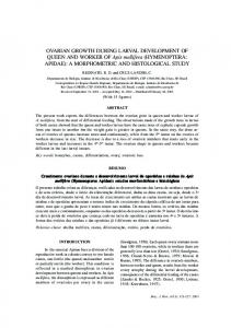
1.2. Flatworms as a model organism for scaling, growth and regeneration Classic experiments on flatworm regeneration already inspired the idea of morphogenetic gradients (36, 128, 129). In recent years, the flatworm Schmidtea mediterranea (Smed ) has become increasingly popular as a model organism to study regeneration and growth, aging, and even behavior (63, 140, 151, 158, 162, 179, 183, 198, 212). There are many different flatworm species populating very diverse habitats all around the world. They are found in saltwater, in freshwater and in the soil; some live more than 1000 m under the sea and some parasitic species (like flukes and tapeworms) in the body of other organisms (151, 170, 179, 202). Smed is a non-parasitic flatworm living in freshwater. Such free-living species are sometimes also referred to as “planarians” (50). Flatworms (Greek: Platyhelminthes) bridge the gap between other model organisms of lower complexity such as the freshwater polyp Hydra and the C. elegans worm and those of higher complexity, such as fruit fly, clawed frog, axolotl, zebrafish and mouse (184, 244), see Fig. 1.5. Many members of the flatworm phylum seem to represent the most evolved organisms that are still able to regenerate any part of their body (50, 128, 140, 184). For example, Smed can restore its complete body plan from amputation
8
1.2 Flatworms as a model organism for scaling, growth and regeneration
regenerates
Hydra: even from cell aggregates
Hydra
C. elegans: no regeneration, rigid lineage
Regeneration capability
Flatworm
Axolotl
Flatworm:
regeneration of all body parts very limited
Fruit fly: regeneration
Zebrafish
in the embryo regeneration
Clawed frog: very limited and only in tadpoles
Axolotl:
Fruit fly
Clawed frog Mouse
regeneration of major body parts like limbs, heart, spinal cord, brain regeneration of
Zebrafish: major body parts like fin, heart, spinal cord regeneration
Mouse: limited to some
C. elegans
tissues like liver
“Complexity”
Figure 1.5.: Flatworms are the most complex model organisms that can still regenerate every tissue. This regeneration capability is shared with simpler organisms such as Hydra. At the same time, flatworms possess organ systems like a centralized nervous system and two distinct brain lobes, which are characteristic for the most complex organisms (50, 68, 128, 140, 143, 184, 244).
fragments as tiny as the very tip of the tail with only about 104 cells (126). By re-growing missing body parts and re-modeling oversized organs, they recover their normal shape scaled to the size of the amputation fragment within 1-2 weeks (4, 170). This astonishing regeneration capability is comparable to the much simpler Hydra and distinguishes flatworms from other model organisms with much less body plan plasticity (158, 184). Thereby, the body plan of Smed shows already key characteristics that are usually associated with higher order organisms (140), see Fig. 1.6. The bilaterally symmetric Smed possess a central nervous system with a distinct bilobed brain and two ventral nerve cords connected by commissural neurons (69, 95, 165, 170, 171, 183). The sensory system processes information from chemo-, rheo- and photorecetors leading to a complex behavioral repertoire (69, 95, 153, 171). Their usual mode of motility is a gliding motion on a secreted layer of mucus being propelled by the beating of numerous short flagella (or cilia) that project from their multi-ciliated ventral epithelium (8, 169, 179, 180). However the worms also use the numerous muscles situated along their inner body wall for (i) steering, (ii) quick escape responses, (ii) exploratory head motion and (iv) a back-up movement strategy (152, 153, 169, 180, 220).
9
1. INTRODUCTION
(a) locomotory and sensory system eye spots
brain lobes
(b) digestive and excretory system protonephridia
(c) hermaphrodite reproductive system ovaries
gut testes
ventral nerve cords pharynx (ventral)
copulatory organs
cilia (mostly ventral)
Figure 1.6.: The body plan of the flatworm Schmidtea mediterranea (Smed ) shows already key characteristics that are usually associated with higher order organisms (37, 51, 171, 173, 231, 245).
Smed belong to the taxon Tricladida, which is reflected by the fact that their highly ramified gut splits in three main branches (69, 140, 165, 170), see Fig. 1.6(b). During feeding, the carnivorous worms suck in food through their extensible pharynx opening. After digestion, the pharynx also functions as an anus for excretion. Protonephridia constitute a further part of the excretory system, which performs similar functions as the kidneys in humans (171). Turnover, growth and regneration completely relies on a pool of stem cells called neoblasts. The fraction of neoblasts among all cells had been estimated to be as large as 25 − 35% (15, 16, 17, 18). If the worms are depleted of all neoblasts by γ-irradiation, they will show a regression of the body starting from the head and finally they fall apart (15, 46, 159, 170). The time scale until the decay sets in varies between species in the range from several days to a few weeks (46, 159). This indicates that there is no de-differentiation of cells to restore the stem cell pool. A subpopulation of neoblasts is pluripotent (and maybe even totipotent) and can develop into every other cell type (15, 63, 84, 140, 158, 170). Wagner et al. have shown that irradiated worms can be rescued by transplanting a single pluripotent neoblast from an intact worm (229). Even though neoblasts are well defined by their progression through the cell cycle resulting in cell division, there is increasing evidence that the neoblast population is not
10
1.2 Flatworms as a model organism for scaling, growth and regeneration
homogeneous (85, 132, 194, 225). Most likely, they also comprise lineage-restricted subpopulations and transiently amplifying cells that go through a few more rounds of cell division during differentiation. Smed show both sexual and asexual reproduction (48, 140, 170, 183). Some strains are hermaphrodites, which develop testes and ovaries symmetrically along their body axis as well as copulatory organs for cross-fertilization, see Fig. 1.6(c). In contrast, asexual strains do not possess a reproductive system and reproduce by fissioning: the worm attaches its tail to the substrate and glides on until the body is ripped into two or more pieces, which develop into new worms after regeneration (140). Fissioning depends strongly on the environmental conditions, in particular on temperature, feeding, light and
“Wenn einem beim Duell ein Ohr oder sonst ein Glied abgeschlagen wurde, so wuchs innerhalb von acht Tagen erstens ein neues Ohr an den Menschen und zweitens ein neuer Mensch an das Ohr. ... Wer sich vermehren wollte, schnitt sich zum Beispiel einen oder zwei oder zehn Finger ab und wartete acht Tage lang.” — Joachim Ringelnatz, Abseits der Geographie, 1924 (168)
worm density (48, 131, 140, 161). For example, fissioning frequency is reduced in crowded environments. Like regeneration abilities, reproduction strategies also vary widely among flatworm species (50). In particular, one observes various approaches to asexual reproduction. Some species like Smed first split and regenerate afterwards, others already grow the respective organs of the new body plan before fissioning. The second strategy comes in two forms: as paratomy with the new body aligned to the old axis and budding with a non-aligned outgrowth at the side or pointing backward. Smed are well-suited model organisms to study growth and cell turnover. First, all somatic cells are continuously replaced by cell turnover at a time scale of weeks (158, 170). Second, growth and cell turnover rely completely on the division of a large population of neoblasts, some of which are pluripotent (15, 63, 85, 140, 158, 170). Third, depending on feeding conditions, the worms can grow and actively shrink (usually referred to as “degrow”), while scaling their body plan over more than one order of magnitude in length in the range of 0.5 mm to 2 cm (140, 151, 170), see Fig. 1.7(a). Active degrowth enables the worms to survive long starvation periods of several months. It has been suggested that worms recycle apoptotic material during degrowth to fuel the metabolism of the remaining cells (69, 70). Smed are also well-suited to study scalable patterning during growth and regeneration. Being able to reversibly grow by a factor of 40 in length and perfectly regenerate even from scrambled body fragments prompts for patterning mechanisms that are not only highly robust and self-organizing but also functional across a wide range in sizes,
11
1. INTRODUCTION
(a) Growth and Degrowth
(b) Regeneration 1mm
Figure 1.7.: Growth and regeneration in flatworms: (a) Schmidtea mediterranea (Smed ) can reversibly grow over a 40fold range depending on feeding conditions (Images taken by Nicole Alt under the supervision of the author). (b) Amputation fragments regenerate to form a perfectly shaped worm within 2 weeks (green lines mark the cuts, white tissue parts indicate the regeneration site before all pigments have been reestablished, modified with permission from (116)).
see Fig. 1.7. For example, regeneration comprises a tightly controlled sequence of responses (4, 10, 13, 14, 69, 81, 117, 128, 158, 165, 170, 237). After an immediate muscle contraction to close the wound, a peak in cell division together with the migration of stem cells within the first 12 h generates an outgrowth of undifferentiated tissue at the wound site, the blastema. In a second step, the now oversized, remaining body parts are re-patterned to match the size of the amputation fragment. The corresponding process is called morphollaxis and comprises a more sustained proliferation and cell death response during the following days. All this pattern formation is guided by an internal coordinate system that is re-established as one of the earliest cues in the remaining worm fragment independently of cell differentiation (81, 141). Our work is especially focussed on the anterior-posterior (AP) axis specifying head and tail position. The Wnt/β-catenin system is a conserved pathway for such AP patterning in many animals and it is also key for the AP polarity in flatworms (4, 9, 54, 81, 82, 141, 160). There are 9 Wnt genes in flatworms. In particular the canonical Wnts (activating β-catenin signaling) are highly expressed in the tail. Inhibition of the Wnt/β-catenin system leads to formation of additional heads, while overexpression induces tail identity everywhere in the worm (3, 82, 82, 160). Remarkably, reducing the level of β-catenin is sufficient to induce full regenerative power to some flatworm species that are usually deficient in head regeneration (116, 200, 222). Taken together, the experiments suggest that β-catenin is instructive for positional information along the AP axis in a concentration-dependent manner.
12
1.3 Theories of body plan patterning by morphogens
Perpendicular to the AP axis, the dorsal-ventral (DV) axis and the medial-lateral (ML) axis are formed (54, 117). For example, the DV axis is specified by the interplay between the TGF-β family members Bmp and Admp. Together, all three body axes are characterized by gradients in abundance and expression of specific molecules, in neoblast mitotic activity as well as in membrane potential (4, 9, 23, 62, 81, 149, 201) and it is a major open question how these gradients robustly guide body plan patterning irrespective of the size of the organism.
1.3. Theories of body plan patterning by morphogens Here, we will discuss two ideas of pattern formation based on cell-cell communication via morphogens. The first type of mechanisms describes how a pre-existing structure can act as an organizing center for the development of further patterns. Such theories are built on the idea of a well-defined, sequential developmental program where existing patterns determine the formation of new patterns, see Fig. 1.8(a). Maternal patterning cues provide a pre-pattern for the embryonic tissues forming from an egg cell and the layout in the embryo determines the future body plan of the animal (2, 57, 172). For example the localization of bicoid mRNA to the anterior pole of the fly embryo is a maternal effect. However, the impressive capabilities of flatworms to regenerate from almost arbitrary initial fragments challenge these concepts, see Fig. 1.8(b). In contrast to the sequential patterning from predefined cues, Alan Turing introduced a second class of mechanisms for self-organized pattern formation (221). We will discuss both approaches and assess them from a biological perspective. Body plan patterning requires that cell fates are assigned depending on the relative position of the cells in the tissue or the organism (68, 244). In order to obtain the information about their spatial position, cells sense their environment and communicate with each other. For that, cells respond to mechanical cues as well as chemical signals such as morphogens (89, 112, 147, 174, 224). Signaling molecules often establish long-range, graded concentration profiles, which can be accounted for by the interplay of transport and degradation (76, 223, 232). Cells respond to the concentration of these molecules in their local environment. Thus, a graded morphogen concentration can provide cells with the information about its spatial distance from the morphogen source (243), see Fig. 1.9(a). This idea forms the basis for the French flag model, which draws a simplified picture of how body plan patterning might be guided by graded morphogen profiles. For example, a stripe pattern can be
13
1. INTRODUCTION
(a)
(b)
Figure 1.8.: (a) Idea of sequential development: The emerging tissues in the fertilized egg determine the future body plan of the juvenile chicken and later of the adult hen. Finally maternal signals break again the spatial symmetry in the new egg and guide development of another chicken. (b) Regeneration in flatworms: Diverse initial fragments are repatterned to form the same body plan scaled to fragment size.
generated if cellular differentiation depends on discrete, genetically encoded threshold levels. In Fig. 1.9(a), cells turn blue if the morphogen concentration is above the first threshold level and red if it is below the second threshold level. Several intersecting gradients can generate more complex patterns. Dose-dependent responses have indeed been observed in experiments, yet in a more complex way than given by the simplified French flag model (72, 74, 75, 80, 104, 191, 242). For example, cells of the frog Xenopus show a distinct differentiation response to at least five concentration thresholds of Activin (72, 75). However, the early response of the individual cells is less specific and very inhomogeneous (73, 242). It appears that only the interaction between cells leads to the well-defined behavior of the cell aggregate. Furthermore, the cells in the tissue are reported to respond in a ratchet-like manner to the highest level of Activin to which they were exposed (80). Similar dosedependent responses have also been recently reported for Wnts in the frog embryo with respect to the AP axis patterning (104). Thus, although the simple concept of a direct threshold-comparison has been questioned in some organisms, the French flag model continues to provide a pictorial representation of the case where a feedback on the concentration gradient by the responding cells can be neglected. As an extension, it also has been proposed that the cells might compare spatial and temporal differences in morphogen concentration and correspondingly adjust cell division, differentiation and motility (135, 167, 175, 233, 234, 235). The described mechanisms can explain patterning, given a pre-defined source region where morphogens are produced locally. Yet, how is this source region established in the first place? One possibility is that another graded morphogen profile specified
14
1.3 Theories of body plan patterning by morphogens
morphogen
cells
(b) Self-organized: morphogen 1
morphogen 2
morphogen concentrations
morphogen concentration
(a) Pre-patterned:
threshold level 1 threshold level 2
Figure 1.9.: (a) The French flag model substantiates the idea that a graded morphogen profile provides positional information for cells in a tissue. Specifically, cells adopt distinct cell fates (sketched blue, white, red) by responding to the local morphogen concentration depending on whether a certain threshold is met. The model assumes a pre-defined source region (grey), which secretes the morphogens. (b) In a Turing system, at least two chemical species (morphogen 1 and 2, red and blue) interact. Source regions (gray) of the morphogens are established in a self-organized way (here: a source forms where the concentration of the first morphogen is larger than the concentration of the second morphogen).
this source by a threshold rule and a sequential developmental program establishes one pattern from an already existing one, see Fig. 1.8(a). An alternative explanation dates back to Alan Turing. In a seminal paper from 1952, he proposed a general framework for the spontaneous formation of biological patterns, independent of pre-patterning cues (221). Turing’s framework was later further explored by Meinhardt and Gierer (64, 65, 66, 106, 120). Turing demonstrated how the interaction of at least two diffusing molecular species can result in chemical patterns in a completely self-organized manner, see Fig. 1.9(b). Thereby, these (often periodic) patterns specify their own production regions. It is an appealing idea that the chemical patterns layout the body plan of an animal. Yet for the following 50 years, there was only little experimental evidence for Turing’s ideas and the focus of developmental biology was shifted to other patterning mechanisms like the French flag model (74). Certainly this was partly due to the fact that it is generally difficult to demonstrate experimentally that a specific pattern is generated by a Turing mechanism. While a unidirectional relationship like in the French flag model (where a concentration of one molecule has a particular effect on other molecules or the cells) is straight forward to analyze, this is less so for Turing patterning which includes cross-reaction terms. The behaviour of a Turing system is often counterintuitive and its analysis might easily yield misleading results. For example, the concentration of some
15
1. INTRODUCTION
Turing molecules peak at the maximum concentration of their repressors, compare to Section 1.4. Furthermore, if there are more than two chemical species involved, the concentration of one of them might decrease upon knockout of its direct activator because of additional indirect effects, see Appendix A.7.4 for an example. Despite the challenging task of revealing the existence of Turing patterns and identifying the involved molecules, recently more and more evidence has been accumulated that Turing’s ideas might be the guiding principles for the formation of a wide range of biological patterns, ranging from the formation of digits in vertebrate limbs to the emergence of left-right asymmetry (49, 119, 134, 137, 164, 191, 197). Some of these examples combine Turing patterning with a French flag model. In fact, in many biological systems, pattern formation might result from a combination of both concepts (74). The feedback loop of a Turing mechanism ensures high robustness and possesses the ability to generate chemical patterns from random fluctuations. In particular, Turing systems can spontaneously generate graded concentration profiles like required for the French flag model. In turn, biological patterns hardly emerge in a completely homogeneous environment without any pre-patterning cues, as Turing already remarked (221). During development as well as during regeneration, pre-existing morphogen profiles and tissue structures can guide the formation of chemical patterns. Thus, Turing mechanisms might often be found downstream of morphogen profiles that modify the respective patterns (74, 164).
1.4. Turing mechanism yields self-organized patterns In 1952, Alan Turing introduced a generic framework for the self-organized formation of chemical patterns in biology (221). He asked the very general question whether there are conditions, under which a system of two or more diffusing and reacting chemical species possesses a homogeneous steady state, which is linearly unstable with respect to inhomogeneous perturbations, such that inhomogeneous patterns form spontaneously. Very importantly, in consequence, any model for self-organized patterning based on diffusing and cross-reacting molecules can be understood within the Turing framework (64, 65, 120, 147, 195, 221). Recently, there are attempts to further generalize the Turing mechanism to generic interactions between two players without a diffusion term (136, 236). In the following we show that the conditions for a Turing instability can be derived from the linear stability analysis of the homogeneous steady state. For this, we briefly
16
1.4 Turing mechanism yields self-organized patterns
recall the basic Turing model comprising two chemical species with concentrations A and B. Details on the derivations are provided in Appendix A.3 or in the literature (64, 65, 146, 195, 221). The general reaction-diffusion system for two molecular species in one space dimension is ∂t A = RA (A, B) + DA ∂x2 A ∂t B = RB (A, B) + DB ∂x2 B ,
(1.1)
with diffusion coefficients DA and DB and two generic functions RA and RB describing the reactions between the different molecules and the effects of each molecular species on itself. In order to spontaneously form stable patterns from random fluctuations, these reaction functions have to fulfill two requirements: the homogenous steady state (A∗h , Bh∗ ), defined by RA (A∗h , Bh∗ ) = 0 and RB (A∗h , Bh∗ ) = 0, has to be (i) stable with respect to homogeneous perturbations (to avoid a diverging behavior), yet (ii) unstable with respect to inhomogeneous perturbations. It is possible to derive a set of necessary conditions for spontaneous pattern formation from these two conditions above by applying linear stability analysis. For this, we consider a small perturbation (a, b) about the homogeneous steady state: A = A∗h + a and B = Bh∗ + b. The linearized form of Eq. 1.1 can be written as
∂t
as
!
bs
=
Ms
as
! .
(1.2)
bs
Here, the perturbation modes as (t) and bs (t) with wavenumber s represent the spatial Fourier transform of a(t, x) and b(t, x), respectively: Z Z as (t) = a(x, t) e−2πsx/L dx , bs (t) = b(x, t) e−2πsx/L dx
(1.3)
The matrix Ms is given by
Ms =
∂A RA − DA (2πs/L)2
∂B RA
∂A RB
∂B RB − DB (2πs/L)2
! ,
(1.4)
where derivatives are evaluated at A = A∗h , B = Bh∗ . In order to fulfill the two conditions on the stability above, (i) the real parts of both eigenvalues qsI and qsII of the matrix Ms have to be negative for s = 0 and (ii) at least one has to be positive for s 6= 0. As shown in Appendix A.3, this results in the following conditions for the trace Tr and the determinant Det of the matrix Ms : Tr[M0 ] < 0 ,
Det[M0 ] > 0
and ∃ s > 0 with Det[Ms ] < 0 .
17
(1.5)
1. INTRODUCTION
(b)
Position
Feedback 2
source of A
Morphogen concentration
Feedback 1
source of A & B
Morphogen concentration
(a)
Position
Figure 1.10.: There are two possible feedback topologies that lead to pattern formation in a Turing system with two molecular species (A and B): (a) an activatorinhibitor scheme with activator A and inhibitor B, (b) a second topology where both chemical species have activating and inhibiting effects. We show two examples of typical concentration profiles corresponding to the two feedback topologies. The selforganized source regions are indicated in gray.
In consequence, we obtain several constraints on the reaction design, which have been summarized by the principle of local activation and lateral inhibition (64, 65, 120, 146, 147), see Appendix A.3 for details and Fig. 1.10 for illustration of the allowed feedback topologies. The necessary conditions for spontaneous pattern formation are: 1. One molecule has to be self-activating and the other self-inhibiting. In the following, we choose A to be the self-activator and B the self-inhibitor: ∂A RA > 0
and ∂B RB < 0 .
(1.6)
2. There have to be cross-reaction terms of opposing sign: � �� � ∂A RB ∂B RA < 0
(1.7)
3. The diffusion coefficient of the self-activator has to be sufficiently smaller than the diffusion coefficient of the self-inhibitor: DA < DB .
(1.8)
As a result for a Turing system with two chemical species, there are two possible network topologies as depicted in Fig. 1.10. In Turing feedback 1, the concentration A has only
18
1.5 Open questions in growth control and scalable body patterning
activating effects and B has only inhibiting effects both on itself and the respective other player. In this case, we can shortly refer to A and B as the concentrations of activator and inhibitor instead of self-activator and self-inhibitor. In Turing feedback 2, both molecular species have activating and inhibiting effects. This includes depletion models, where, for example, binding of both molecular species enhances the production of A but consumes B (66). Note that there is a formal correspondence between the two Turing topologies by the replacement B → Bc − B with a constant parameter Bc . How the principle of local activation and lateral inhibition leads to spontaneous pattern formation can be nicely demonstrated for the Turing feedback 1 of Fig. 1.10(a). At the source, the activator dominates because the inhibitor is diffusing away more quickly. Consequently, the source region stabilizes itself and initially tends to expand. In contrast, some distance away from the source, the inhibitor dominates due to its fast diffusion. This creates at least two distinct regions, which already comprises a simple pattern. In contrast to other systems where diffusion homogenizes a pattern, here diffusive spreading in combination with specific reactions enhances small inhomogeneities in the concentrations. Therefore, it is sometimes also referred to as a diffusion-driven instability (145, 146, 181).
1.5. Open questions in the study of growth control and scalable body patterning During development, growth and regeneration, cells communicate with each other and mutually influence their behavior, especially by changing the expression status of their genes. This information exchange orchestrates cell division, cell death and differentiation. It can also guide cell migration and can elicit the release of further signals, e.g. the secretion of morphogens. As a common theme of this thesis, we devise theoretical descriptions of how cell fate decisions based on local rules on the microscopic scale result in the formation and maintenance of a macroscopic body plan, drawing inspiration from flatworms as a specific model organism. In particular, we address the following questions: How is the turnover of cells regulated during growth and homeostasis and how are fluxes in cell number related to fluxes of metabolic energy? Organisms have to control the number of cells to ensure homeostasis and growth in a well-defined manner. As changes in cell number depend on the balance between cell proliferation and cell loss, there needs to be a communication between dividing and differentiated
19
1. INTRODUCTION
cells (158). Furthermore, the environment and in particular the availability of food provides an external stimulus to influence cell behavior, such as proliferation and cell death (113, 244). What are minimal requirements for self-organized patterns that scale with organism size? Organisms also have to control the type and position of cells for a reliable body plan patterning. Again, this requires a communication between cells, ranging from direct neighbor-neighbor interactions to long range signaling via mobile molecules such as morphogens (54, 68, 244). Importantly, pattern formation can be observed on all length scales from the development of a fertilized egg to growth and regeneration of large scale tissues in mature organisms. Yet, patterning mechanisms often possess fixed characteristic length scales defined by the intrinsic physical properties of the system (76, 223, 232), which is challenged by cases of biological pattern scaling like regeneration in flatworms. We aim to understand scalable body plan patterning and cell turnover dynamics by building on the framework of dynamical systems theory and birth-death processes. Biological systems add a new perspective to those classical concepts. They usually operate far from equilibrium and form patterns with unconventional properties (39, 87, 120). Development and regeneration are subject to a high level of noise ranging from external perturbation like a variable environment to the intrinsically stochastic nature of gene expression (77, 218, 219). Thus, robust growth control and body plan patterning require reliable sensing, transmitting and processing of noisy information (47, 218). We address the question of robustness with respect to initial conditions and physical parameters as well as the structural robustness of the models. This thesis is mainly inspired by the astonishing scaling and regeneration capabilities of flatworms, yet the results obtained here are likely also to be relevant for other biological organisms as well as related questions in biological physics, nonlinear dynamics and pattern formation.
20
1.6 Organization of the thesis
1.6. Organization of the thesis In this thesis, we investigate biological shape and size control on various levels, for which we combine theoretical descriptions and state-of-the-art analyses of experimental data in flatworms. First, we explore mechanisms for self-organized and self-scaling pattern formation (Chapter 2). We discuss to what extent previously proposed theories can account for the scaling of morphogen profiles. For this, we adhere to a strict mathematical definition of scaling and illustrate the difference between perfect scaling and approximate scaling with system size. Furthermore, we demonstrate the absence of scaling in classical Turing pattering, going beyond linear stability analysis. Second, flatworms like Smed challenge existing theories on body plan patterning, which typically can at most only explain one of both: either scaling or self-organization. Here, we introduce and characterize a novel class of mechanisms that combine both features (Chapter 3). The developed theory can act as a framework to understand robust body plan scaling during growth and regeneration in flatworms. Next, worm shape variations cannot only be observed between different stages of development, growth and regeneration. Individual worms also show large body deformation during movement, driven by muscle contractions. We apply a method based on “Principal component analysis” to characterize shape dynamics and analyze movement patterns. A similar shape mode analysis also enables us to compare shapes of different related species. Analyzing motility patterns and comparing shape variations between worm species is the first step towards relating form and function (Chapter 4). Finally, we analyze growth and degrowth dynamics and their dependence on feeding conditions. We investigate mechanisms for size control by metabolic energy balances to explain the macroscopic growth and degrowth behavior in terms of microscopic cell turnover dynamics. Our theory makes testable predictions for the ongoing experiments. Beyond, we develop the theoretical basis for additional measurements on various scales to reveal further details about the control logic of cell turnover (Chapter 5).
21
2. Scaling in morphogen systems
2.1. Scaling of biological patterns The development of multi-cellular organisms with a well-defined, complex body plan is one of the most fascinating processes in nature. A series of patterning events take place “The continuous change in form that takes place from hour to hour puzzles us by its very simplicity. The geometric patterns that present themselves at every turn invite mathematical analysis.” — Experimental embryology, Thomas H. Morgan, 1927 (130)
across various length scales leading to a distinct layout, which is scaled to match the size of the organism. The scaling of body plan patterns becomes especially apparent if a juvenile organism already resembles its adult counterpart or if individuals of different, yet related species look very
much alike besides their great differences in size (27, 76, 223). A third, more subtle example is the robust formation of proportionate patterns in the same organism during development despite size variations that arise from varying environmental conditions or stochastic fluctuations of growth rates (24, 76). In this chapter, we analyze to what extent previously proposed theories can account for pattern scaling. First, we introduce a mathematical definition of gradient scaling. Based on these considerations, we revisit and assess scaling mechanisms proposed for morphogen gradients in pre-patterned systems and extract the main principles underlying scaling. This will later allow us to point out the important differences between scaling mechanisms for pre-patterned and self-organized systems in Chapter 3. Furthermore, we discuss the absence of scaling in self-organized Turing systems beyond the classical approach of linear stability analysis. The latter has been published in Werner et al. (239).
22
2.2 Morphogen dynamics and concentration profiles
2.2. Morphogen dynamics and concentration profiles One of the most simple morphogen systems that result in graded concentration profiles draws on diffusion from a localized source with an effective diffusion coefficient D and a linear degradation with rate β (232). It has been frequently applied to describe morphogen gradients in the fruit fly (24, 44, 76, 78, 234). Note that effective diffusion might result from a wide range of underlying undirected processes, such as active transport or even signaling between neighboring cells without secretion of motile molecules. For simplicity, we consider a one-dimensional system of size L with reflecting boundary conditions. The corresponding time evolution of the morphogen concentration C = C(t, x) is given by ∂t C = D ∂x2 C − β C + ν
(2.1)
∂x C|x=0 = ∂x C|x=L = 0 .
(2.2)
Here, ν(x) = α Θ(w − x) describes localized morphogen production with rate α in a source of width w. Θ denotes the Heaviside step function. Eq. 2.1 also holds in two and three dimensions, if the system is symmetric with respect to the other dimensions and, thus, the morphogen concentration still only depends on x, see Appendix A.1.1. If the tissue grows at a time scale comparable to the time scale of the morphogen dynamics, one has to consider additional terms for advection and dilution. The steady state solution to Eq. 2.1-2.2 is computed in Appendix A.1.2. Here, we are mainly concerned with the steady state concentration outside the source region, which is given by C ∗ (x) = C0
cosh(L/λ − x/λ) cosh(L/λ)
for w ≤ x ≤ L
(2.3)
with amplitude C0 =
α sinh(w/λ) β tanh(L/λ)
(2.4)
p D/β .
(2.5)
and a characteristic length scale λ=
Within the course of a characteristic time scale (which is of the order of 1/β in our example, see Appendix A.1.3), the morphogen will relax towards this steady state profile. Analyzing the steady state is well justified in a case of separated time scales, where other dynamics (e.g. growth and differentiation) are slow in comparison to the relaxation time of the morphogen profile. In the following, we will often assume such
23
2. SCALING IN MORPHOGEN SYSTEMS
a quasi steady state. One should keep in mind that this is a simplifying assumption which is not always fulfilled. In fact, it has been argued that many gradients might be already read out before the steady state is reached as a means to increase robustness (20, 29, 55, 167). Yet, even in these cases, the steady state is the reference state and provides a first order approximation to the concentration the cells actually respond to.
2.3. Scaling of concentration profiles with system size Profiles of signaling molecules have been measured for example in the fruit fly Drosophila melanogaster, which is an important model organism to study biological pattern formation (123, 206). Quantifications of morphogen concentrations in the developing wing and eye of the fly at different stages of development revealed that the concentration profiles maintain an approximately constant shape relative to the changing size of the growing tissue (26, 83, 234, 235). This biological observation of profiles that scale with system size inspired the development of a number of theoretical mechanisms that can account for pattern scaling (11, 24, 26, 27, 96, 135, 148, 154, 223, 233, 234). In this section, we discuss examples, which represent two major classes of these mechanisms. We define a mathematical property of perfect shape scaling and analyze approximations of this property. We define perfect shape scaling as the ideal case of strictly proportional scaling of a concentration profile with system size. In this case, the concentration at steady state can be written as C(x; L) = C0 (L) · Z(x/L) ,
(2.6)
where C0 (L) denotes a possibly size-dependent amplitude and Z(x/L) defines the shape of the profile, which only depends on the relative spatial coordinate x/L. As a result, scaling profiles from systems of different sizes perfectly collapse onto a single master curve if plotted as a function of relative coordinates and normalized by their amplitude, see Fig. 2.1(a). For the steady state solution in Eq. 2.3, perfect shape scaling arises if the characteristic gradient range λ is not constant but strictly proportional to the length of the system L: λ ∝ L.
(2.7)
In a more general case, if the length scale λ increases with L in a monotonic, yet nonlinear fashion, we will classify this as approximate scaling, see Fig. 2.1(b): ∂L λ > 0 .
24
(2.8)
2.3 Scaling of concentration profiles with system size
Rel. Concentration
Rel. Concentration
(a) Perfect shape scaling:
Relative position
Position
Rel. Concentration
Rel. Concentration
(b) Approximate scaling:
Relative position
Position
Figure 2.1.: Illustration of the mathematical concept of perfect shape scaling and approximate scaling: (a) Perfect shape scaling: normalized concentration profiles collapse onto a master curve when plotted as a function of the relative position. (b) In a more general case, concentration profiles might expand with system size, though the collapse onto a single master curve is only approximate. We classify this case as approximate scaling.
In the following, we will strictly distinguish between the two notions of approximate scaling and perfect shape scaling. This slightly academic distinction will prove useful in the subsequent mathematical analysis. Nonetheless, we want to emphasize that approximate scaling can be virtually indistinguishable from the ideal case of perfect shape scaling, both in measurements and simulations. In contrast to our theoretical description, real systems are generally prone to intrinsic fluctuations and measurement inaccuracies. Furthermore, the separation of time scales between system growth and morphogen dynamics assumed here will always be an approximation, such that the morphogen concentration never exactly corresponds to the theoretically computed steady state profile. Therefore, even if the underlying mechanism is in principle able to generate profiles that perfectly scale like in Fig. 2.1, we will never observe a perfect collapse of the measured concentration profiles. On the contrary, mechanisms of approximate scaling that cannot yield perfect shape scaling in the sense of Eq. 2.6, may still fully account
25
2. SCALING IN MORPHOGEN SYSTEMS
Relative Concentration Absolute Position
Concentration
Absolute Position
Absolute Position
Relative Position Relative Concentration
Concentration Concentration
Adjusting Diffusion
Concentration
(i) Fixed Source (ii) Scaling Source
Adjusting Degradation
Absolute Position
Relative Position
Figure 2.2.: Size-dependent amplitudes of scaling profiles: Models for the scaling of morphogen profiles often assume the adjustment of degradation rate β or diffusion coefficient D with system size L. As a result, the amplitude of the morphogen concentration might change depending on whether the morphogen source size (gray) is fixed (upper row) or scales with L (lower row). The reference profile (red) is the same in all plots (Eq. 2.3, α/β = 10, L/λ = 2, L/w = 5). The other profiles correspond to either half (blue) or twice the system size (green) with constant ratio L/λ and either scaling or non-scaling source size w.
for gradient scaling as observed in biological systems. As an example for a mechanism that yields approximate scaling, we discuss a simple realization of a prominent scaling mechanism, the expansion-repression model. We emphasize that this mechanism has been successfully applied to analyse profile scaling in fly wing development (24, 26, 27). According to the definition of λ in Eq, 2.5, scaling can be achieved if either the diffusion coefficient or the degradation rate or both depend on system size. Two simple choices are: (i) D ∝ L2 , β = const and (ii) β ∝ L−2 , D = const. In Eq. 2.6, we defined scaling of morphogen profiles as a shape property, irrespective of the amplitude C0 (L). This amplitude might be a function of system size L, compare also to (223). The variation of the amplitude with L can reveal details of the underlying scaling mechanism, see Fig. 2.2. We discuss this for the two limiting cases of (i) a source of fixed width w and (ii) a scaling source w ∝ L. (i) Fixed source: If we increase λ by enhancing diffusion, the amplitude will decrease because the same amount of morphogen is distributed more homogeneously in the system, compare to Eq. 2.4 or Eq. A.5. In contrast, if we increase λ by reducing degradation, the amplitude will increase due to a longer lifetime of the molecules. If D
26
2.4 Scaling of morphogen profiles in pre-patterned systems
and β both depend on size, the amplitude can be kept constant. (ii) Scaling source: Eq. 2.4 shows that for this case the amplitude does not explicitely depend on D but depends inversely on β. Scaling by changing the diffusion properties does not affect the amplitude but scaling by adjustment of degradation leads to a strong size dependence C0 ∝ L2 . Measurements of the signaling protein Dpp in the fruit fly found a decreasing degradation rate with size as well as the characteristic increase in the amplitude (76, 78, 135, 233, 234). In the clawed frog, both cases have been reported, adjustment of diffusion properties for Bmp as well as adjustment of degradation for the Bmp antagonist Chordin (25, 94). Next, we discuss two classes of simple models for the scaling of morphogen profiles, which represent minimal versions of scaling mechanisms proposed in the literature (11, 24, 26, 27, 96, 135, 148, 154, 223, 233, 234). We will illustrate that models of the second class rely on a changing amplitude during scaling. Thus, the amplitude of the morphogen profile encodes the system size.
2.4. Scaling of morphogen profiles in pre-patterned systems 2.4.1. The concept of an expander as a chemical size reporter In order to achieve scaling of a morphogen profile in a simple set-up as introduced in Section 2.2, the length scale of the concentration profile has to couple to the size of the system. Several mechanisms have been proposed for the expansion of a morphogen profile with system size L that assume an additional molecular species, often called “expander” (11, 24, 27, 96, 135, 148, 154, 233, 234). If the concentration E of such an expander is a function of L, the interaction of these expander molecules with the morphogen can result in profile expansion with system size. Depending on whether the expander concentration increases or decreases with system size, the expander might enhance or reduce either diffusion or degradation of the morphogen, respectively. Yet, how does the expander obtain its size-dependent concentration and become a chemical size reporter? In the following, we discuss two main classes of expander mechanisms that have been proposed for the scaling of morphogen profiles. The first class is characterized by the fact that the expander couples to the system size independently of the morphogen. The second class subsumes mechanisms that allow for self-scaling of the morphogen profile by a feedback loop between the expander and the morphogen.
27
2. SCALING IN MORPHOGEN SYSTEMS
We illustrate the scope and the limits of the mechanisms at hand. This allows us to systematically extract the main principles that lead to robust scaling as applied later to self-organized systems.
2.4.2. A simple mechanism of gradient scaling: The expander-dilution model The most simple idea for how an expander concentration can report on system size is given by the expander-dilution mechanism (135, 233, 234), see Fig. 2.3(a). It requires a constant amount of a long-lived expander, which is neither produced nor degraded. As the system size increases, the expander gets diluted and, thus, the expander concentration depends on system size. Hence, E ∝ 1/L in a one-dimensional system. However, such a mechanism is highly vulnerable to the loss of expander and in particular could not easily cope with amputations of parts of the system. A similar mechanism can overcome this issue by reading out system size from geometrical features like area-volume ratios. As a specific example, we assume that the expander of concentration E is degraded everywhere in the one-dimensional system and produced in a source of constant width wE at the boundary: ∂t E(t, x) = DE ∂x2 E(t, x) − βE E(t, x) + αE Θ(wE − L + x) ,
(2.9)
Again, DE is an effective diffusion coefficient, βE the degradation rate and αE the prop duction rate of the expander. In the limit of fast expander diffusion (λE = DE /βE � L), the steady state concentration is given by E∗ =
α E wE 1 . βE L
(2.10)
Thus, the expander level E ∗ encodes the system size L, see Fig. 2.3(b). In order for the morphogen to scale with L, the morphogen degradation could for example be coupled to the expander as β ∝ E2 .
(2.11)
The size read-out can also be established differently from the specific choice of Eq. 2.9, see Appendix A.2.1 for a general derivation. For example, the expander could be produced everywhere in the system and only be degraded at the boundary, as discussed by other authors (78, 90, 96, 148, 154). The mechanism is robust against loss of expander, but it requires λE � L irrespective of system size. Note that a small degradation rate results in a large λE , yet also in a slow relaxation to the steady state, see Appendix A.1.3. Furthermore, the mechanism
28
2.4 Scaling of morphogen profiles in pre-patterned systems
(a) Expander-dilution with long-lived expander
conserved amount
(b) Dynamic size-sensing with short-lived expander const. influx
size-dep. outflow
Figure 2.3.: Schematic illustration of how the concentration of an expander becomes size-depend. (a) Expander-dilution model: a conserved amount of expander gets diluted in a larger system. (b) Dynamic size-sensing in a system with constant influx and size-dependent outflow of the expander.
relies on a tightly controlled size of the source and the degradation zone. In the example above, the expander source has to keep its width although the system size changes. This shifts the problem from scaling the morphogen profile to establishing a source pattern for the expander. Finally, regeneration of arbitrarily shaped fragments seems unlikely to agree with the notion of a fixed geometry of source and degradation zone. Therefore, we next discuss the second class of scaling mechanisms, in which the morphogen itself controls the expander expression leading to a self-regulating feedback loop.
2.4.3. Perfect shape scaling vs. approximate scaling: The expansion-repression model The discussion on morphogen scaling has been enriched by the proposal of a class of models that adjust gradient shape to system size in a self-organized manner (24, 26). These mechanisms build on a feedback loop between morphogen and expander. Interestingly, this relates to the engineering framework of an integral feedback control, where the gradient is adjusted by the expander as long as it does not meet its target range (19, 24). One prominent example is the expansion-repression model. In order to illustrate the main principles, we discuss one specific simple realization of this model, in which the expander production is only turned on where the morphogen level falls below a threshold Cth like considered in (135), see Fig. 2.4(a). Alternative feedback topologies are analyzed in Appendix A.2.3 and lead qualitatively to the same conclusions.
29
2. SCALING IN MORPHOGEN SYSTEMS
For our choice, Eq. 2.9 of the expander dynamics is modified to � ∂t E(t, x) = DE ∂x2 E(t, x) − βE E(t, x) + αE Θ Cth − C(x) .
(2.12)
In order to close the feedback loop and adjust the morphogen length scales, the expander has to affect either diffusion or degradation of the morphogen, see Section 2.3. A simple feedback scheme that yields approximate scaling, though not perfect shape scaling. — Let us start with a simple realization of the expansion-repression mechanism, which has been first proposed for gradient scaling in the developing fly wing (24). In this realization, it is considered that the expander suppresses the degradation of the morphogen (24, 26, 135). Thus, β in Eq. 2.1 is not constant anymore but obeys β=
β0 1 + E/Eth
(2.13)
with two constant parameters β0 and Eth . In consequence, increasing expander levels enlarge the range of the morphogen. Yet, in turn, this suppresses the expander production. Eventually, a steady state is reached, in which the expander production is just cancelled by the total expander degradation in the system. The source size of the expander in this steady state is determined by ∗ C ∗ (L − wE ) = Cth ,
(2.14)
∗ denotes the size of the self-organized source region where C ∗ is given by Eq. 2.3 and wE
of the expander, see Fig. 2.4(a). In the limit of a quickly spreading expander (λE � L), its steady state concentration is E∗ =
∗ α E wE . βE L
(2.15)
From this, we can note the following observations: First, the expander source (estab∗ 6∝ L), because this lished by the feedback with the morphogen) must not scale (wE
would result in a constant expander level irrespective of system size. Second, the morphogen amplitude must change for perfect scaling. Otherwise, if the amplitude were ∗ ∝ L from Eq. 2.14 constant with a scaling gradient range λ ∝ L, we would obtain wE
and the expander would not encode the system size. This generally arises from the fact that in such a mechanism for scaling via a feedback loop both players, morphogen and expander, have to encode the system size in the steady state. Third, the particular feedback scheme including Eq. 2.13 yields approximate scaling of the concentration profiles, though not a perfectly linear relationship of λ ∝ L as required for perfect shape scaling, see Fig. 2.4(b)-(c).
30
threshold
expander source
(b) 1
(c) 25
Gradient range
morphogen source
Morphogen concentration
(a)
Relative concentration
2.4 Scaling of morphogen profiles in pre-patterned systems
0.8 0.6 0.4 0.2 0 0
0
Position
0.25
0.5
Relative position
0.75
1
20
15
10 0
50
100
150
System size
Figure 2.4.: A simple realization of the expansion-repression models that yields approximate scaling, yet no perfect shape scaling. (a) In this specific model the morphogen confines the expander source to the region where the morphogen concentration is below Cth . (b) Rescaled steady state profiles do not perfectly collapse. (c) The gradient range λ increases with system size in a sub-linear fashion. We deterp mine λ by (i) computing λ = D/β at various positions in the system (blue dots), (ii) fitting the solution of Eq. 2.3 (solid red line) and (iii) calculating it directly in the limit of λE � L (dashed green line). We use typical parameters for the fly wing: D = 1 µm2 /s, DE = 10 µm2 /s, α = 0.01 µM/s, αE = 0.001 µM/s, w/L = 0.1, Cth = 0.1 µM, βE = 10−4 /s, β0 = 10−2 /s, Eth = 1 µM, L = 25...135 µm (24, 26). This choice of parameters allows us to illustrate deviations from perfect shape scaling. Other parameter sets might approximate scaling better and might be practically indistinguishable from the mathematical limit of perfect shape scaling.
The latter can be understood most clearly if we ask what relationship between β and E would be needed for the steady-state morphogen range λ∗ = χλ L to scale with a constant factor χλ . In Appendix A.2.2, we provide a detailed derivation for different scenarios. Here, we only discuss the case of a scaling morphogen source (w ∝ L) as a representative example. If we combine Eq. 2.3, 2.14 and 2.15, we obtain � � βE λ∗ = χλ L ⇒ β ∗ ∝ cosh E∗ α E χλ
(2.16)
Thus, this functional relationship between β ∗ and E, which would be required to achieve perfect shape scaling, is different from the relationship, we had originally assumed in Eq. 2.13. Note the qualitative difference between the two relationships: whereas Eq. 2.16 corresponds to a positive feedback loop, Eq. 2.13 describes a negative feedback loop. We conclude that the considered case of Eq. 2.12 and Eq. 2.13 does not yield perfect shape scaling, though approximate scaling is nonetheless possible. Such approximate scaling can be practically indistinguishable from perfect shape scaling as has been demonstrated by numerical simulations of the discussed feedback and related versions (24, 26, 135).
31
2. SCALING IN MORPHOGEN SYSTEMS
2.4.4. Other feedback schemes for self-organized gradient scaling The classical expansion-repression mechanism relies on a feedback in which the morphogen suppresses the expander while the expander increases the range of the morphogen. In consequence, this forms an overall negative feedback loop. In Sec. 2.4.3 we discussed an example which does robustly lead to approximate scaling, though not perfect shape scaling. Would perfect shape scaling be possible if we deviate from the scheme of a negative feedback loop? Eq. 2.16 provides the functional relationship between β and E that would be required for perfect shape scaling in the steady state. Thus, we could replace Eq. 2.13 by β ∝ cosh (E/Eth ) .
(2.17)
Then, the system possesses a steady state pattern that scales perfectly by construction. However, this steady state is unstable in numerical simulations as it forms an overall positive feedback loop. An excess in expander would reduce the level of morphogen by enhanced degradation and thus the expander production and consequently the expander level would increase even further. Depending on the initial conditions, the system shows two distinct dynamics. For small expander levels below the unstable steady state, the expander source will eventually be suppressed completely and the morphogen concentration will diverge. For large expander levels, the system will converge to a second, non-scaling steady state. Variations of the expander feedback show qualitatively similar results. —
In Ap-
pendix A.2.2-A.2.3, we discuss further variations of the feedback scheme of Eqs. 2.122.13. For these variations, we likewise find approximate scaling, though no perfect shape scaling in a strict mathematical sense over a large size range. As a general rule, the ideal case of perfect shape scaling and stability are not easily teamed up. We strongly emphasize that this does not imply that the mechanisms for approximate scaling cannot provide suitable theoretical descriptions for gradient scaling in biological systems. Furthermore, more complex feedback schemes might be able to achieve perfect shape scaling or at least closely mimic perfect scaling over a considerable size range (24).
2.4.5. Conclusions from our analysis of expander models Above, we discussed simple realizations of scaling mechanisms for morphogen profiles, representing two main classes of such mechanisms, see Fig. 2.5. If the expander measures system size independently of the morphogen, this can result in a robust size
32
2.4 Scaling of morphogen profiles in pre-patterned systems
(a) Independently controlled expander (e.g. expander-dilution model)
- stable scaling (if sources and sinks of the expander are well controlled)
(b) Expander feedback loop
(e.g. expansion-repression model)
- approximate scaling is well achievable, perfect shape scaling is challenging - for the simple model variants discussed here, we obtain: adjustment of degradation rate diffusion coefficient
scaling morphogen source
constant morphogen source
perfectly scaling steady state is unstable
perfectly scaling steady state is stable in a finite size range
no perfectly scaling steady state
perfectly scaling steady state is stable in a finite size range
Figure 2.5.: Various mechanisms have been proposed for the scaling of morphogen profiles using an expander as a chemical size-reporter: (a) the first class of models considers an expander which reads out system size independently of the morphogen, see (78, 90, 96, 135, 148, 154, 233, 234) for examples, (b) the second class of models assumes a feedback loop between the morphogen and the expander such that the morphogen profile scales itself (24, 26). We demonstrated that in this case it is challenging to obtain a perfectly scaling steady state. The table summarizes the results for a morphogen that acts on the expander production like in Eq. 2.12, see Appendix A.2.2-A.2.3 for details. For all cases, it is possible to construct a robust feedback yielding good approximations to the idealized case perfect shape scaling.
read-out and potentially lead to scaling (78, 90, 96, 135, 148, 154, 233, 234). Yet, it requires a tightly controlled layout of the system, e.g. pre-patterned sources and sinks for the expander. Thus, it does not easily comply with the ability to regenerate and might just shift the problem from the scaling of the morphogen gradient to the establishment of a source and sink pattern for the expander. The second class of models aims to establish scaling of the morphogen profile in a selforganized manner by a feedback loop between the morphogen and the expander (24, 26). It turns out that it is challenging to find mechanisms with steady state patterns that are both: perfectly scaling in the sense of Eq. 2.6 and stable. If a feedback is chosen, such that a perfectly scaling steady state exists like in Eq. 2.17, this fixed point might not to be stable. Still, many models like the one defined by Eqs. 2.12-2.13 yield stable steady states, which can well approximate perfect scaling, depending on the choice of parameters (24, 26).
33
2. SCALING IN MORPHOGEN SYSTEMS
The intuitive picture is that an overall negative feedback loop between morphogen and expander ensures a stable steady state. In contrast, two mutually suppressing molecular species, which might yield perfect shape scaling, form an overall positive feedback loop. Thus, they will not result in a stable steady state unless there are additional feedback loops. In Chapter 3, we discuss a novel self-organizing scaling mechanism that builds on such feedback regulations. An important signature of the second class of models is that the morphogen amplitude varies with size L. This is a direct consequence of the feedback with a quickly spreading expander, which ensures a uniform expansion of the gradient range across the system. It can be understood intuitively because the morphogen not only couples to the expander but also the expander couples to the morphogen, hence, both have to encode the system size. This can result in constraints on the system design. For example, it led to the observation that the combination of a scaling morphogen source and an adjustment of morphogen diffusion by the expander could not yield scaling of the morphogen profile. We will have to take this into account when discussing scaling in self-organized systems in Chapter 3.
2.5. Revisiting the absence of scaling in Turing patterns In this thesis, we consider one specific choice of a Turing system, which is particularly suitable for analytical treatment. We restrict our analysis to a one-dimensional system with reflecting boundary conditions. The dynamics of the two molecules are described by ∂t A = αA P (A, B) − βA A + DA ∂x2 A ∂t B = αB P (A, B) − βB B + DB ∂x2 B .
(2.18)
Thus, we specifically consider linear degradation with rates βA , βB and production with rates αA , αB . The function P (A, B) implements a switch-like response to the concentrations A and B. Production is switched on if the activator concentration exceeds the inhibitor concentration, A � B. We choose a Hill function, which arises naturally from cooperative and competitive chemical reactions in biological systems, see Appendix A.4: Ah . (2.19) Ah + B h In the limit h → ∞, the Hill function can be replaced by the Heaviside theta function P (A, B) =
P (A, B) = Θ(A − B) .
34
(2.20)
2.5 Revisiting the absence of scaling in Turing patterns
!
As a technical point, we have to define Θ(0) = 0, in order for the homogeneous steady state to always exist, see Appendix A.5. A Fermi function could be another choice to account for switch-like production, yielding similar results. In the case of the production switch in Eq. 2.20, the relative source size is defined by Z 1 L `/L = hP i = P dx , (2.21) L 0 where brackets denote a spatial average over the system. We use the same quantity also to define the source size for the Hill-type production function. This is especially well justified if the Hill exponent h in Eq. 2.19 is large. Next, we analyze our example of a Turing system given by Eq. 2.18 and demonstrate the absence of scaling. We derive the full hierarchy of steady state patterns and discuss their existence and stability, thereby going significantly beyond linear stability analysis of the homogeneous steady state. This will put us in the position to understand the mechanisms for scaling of a Turing system.
2.5.1. A hierarchy of steady state solutions Eq. 2.18 together with the production function in Eq. 2.19 possesses a unique homogeneous steady state: A∗h =
αA /βA 1 + (βA αB /(αA βB ))h
,
Bh∗ =
αB /βB . 1 + (βA αB /(αA βB ))h
(2.22)
For h → ∞, the steady state concentrations approach zero, A∗h = Bh∗ = 0, see Appendix A.5. The inhomogeneous steady states can be computed analytically in the limit of h → ∞, corresponding to the source switch of Eq. 2.20, see Appendix A.6. We find a hierarchy of steady state patterns, which can be characterized by the number m of contiguous source regions with A > B and the number n of source regions touching the system boundaries, see Fig. 2.6. Any (m, n)-pattern can be constructed as a concatenation of σ = 2m−n copies of the (1, 1)-pattern as a basic building block, see Appendix A.6. Note that s = σ/2 is the corresponding leading order Fourier mode of the spatial patterns. The (1, 1)-pattern with a single source, located in the interval 0 ≤ x < `, is given by � � sinh(L/λA −`/λA ) x 1 − cosh ,0 ≤ x ≤ ` λA sinh(L/λA ) αA ∗ � � A(1,1) = (2.23) βA sinh(`/λA ) cosh x−L ,` < x ≤ L λA
sinh(L/λA )
∗ B(1,1) =
1 −
sinh(L/λB −`/λB ) sinh(L/λB )
cosh �
αB � βB sinh(`/λB ) cosh x−L λB sinh(L/λB )
35
�
x λB
�
,0 ≤ x ≤ ` (2.24) ,` < x ≤ L.
2. SCALING IN MORPHOGEN SYSTEMS
(b)
source
(a)
=
+
(c)
Figure 2.6.: (a) The steady state solutions of Eq. 2.18 with Eq. 2.20 can be characterized by a pair of pattern numbers (m, n), where m is the number of contiguous source regions (gray) with A > B and n is the number of source regions touching the system boundaries. (b) Any (m, n)-pattern can be constructed as a concatenation of σ = 2m − n copies of the (1, 1)-pattern as a basic building block, here shown for (m, n) = (2, 2). (c) Thus, we obtain a complete hierarchy of steady state solutions, exemplified for the concentration A up to m = 3 sources. Parameters: DB /DA = 30, √ √ αB /αA = 4, βB /βA = 2, λA /L = 0.1 ≈ 0.3, λB /L = 1.5 ≈ 1.2.
Patterns are characterized by two pattern length scales for A and B, respectively, analogous to Eq. 2.5.: λA =
p DA /βA
and λB =
p DB /βB .
The source size ` has to satisfy the implicit equation � � � � `−L sinh(`/λB ) `−L αA βB sinh(`/λA ) cosh = cosh βA αB sinh(L/λA ) λA sinh(L/λB ) λB
(2.25)
(2.26)
[0.1cm] Fig. 2.7 depicts the relative source size `/L, which is a function of only three dimensionless ratios: L/λA , L/λB and αA βB /(βA αB ), see also Appendix A.6. A dimensional analysis of Eq. 2.18 shows that this is true also for finite h and other production
36
2.5 Revisiting the absence of scaling in Turing patterns
0.25
10
5
0
15
0.125
0 0
changing
, while
constant :
threshold for
1 2
limit of
:
3
Figure 2.7.: Pattern length scales set size of source: The relative source size `/L of the (1, 1)-pattern at steady state is uniquely determined by the pattern length scales L/λA and L/λB . If only the system size is varied, while the other parameters are kept constant, the relative source size changes along the red curve (here: λB /λA = 5). Following the red curve, the source size approaches `/L ∝ λB /L for λB � L. Furthermore, we obtain a threshold for the existence of the (1, 1)-pattern (green) and a scaling regime in the limit of λB � L � λA (saturation of blue curve). Parameter: αA βB /(βA αB ) = 0.5.
functions. The red line illustrates the variation of the relative source size `/L as a function of L, when all other parameters are kept constant. Note that an identical curve could be obtained if instead of L, the length scales λA and λB would be varied via a common control parameter. This will become important in Chapter 3 when we combine the Turing system with an expander feedback. The source size is zero in systems that are very small relative to the characteristic length scales, see Fig. 2.7 (green curve). For these small systems, the homogeneous steady state is the only existing solution. The (1, 1)-pattern begins to appear only above a critical size L1 , where L1 obeys βA αB tanh(L1 /λA ) tanh(L1 /λB ) = . αA β B L1 /λA L1 /λB
(2.27)
This equation implies in particular that λA has to be sufficiently small in comparison to L and λB for the existence of inhomogeneous patterns, see Appendix A.6.
37
2. SCALING IN MORPHOGEN SYSTEMS
The relative source size `/L strongly increases with L beyond L1 . In the limit of λB � L, it eventually saturates, see Fig. 2.7 (blue curve). This observation of source scaling for an existing (1, 1)-pattern has been reported before (66, 67, 120). In contrast, following the red curve for a finite inhibitor range λB , the source size ` approaches a constant value independent of L. In the limit λB � L, we obtain `/L ∝ λB /L. The source does not sense the system boundaries but instead is restricted by λB . Analogous to the (1, 1)-pattern, any higher order (m, n)-pattern only exists if the system size L exceeds a critical size Lσ = σ L1 . Fig. 2.8(a) shows the region of existence (gray) for patterns of different pattern numbers.
2.5.2. Higher order patterns form in larger systems We have shown that the number of coexisting patterns increases with increasing size. Next, we investigate which of these patterns are stable and can in fact be observed in systems of different sizes. Higher order modes become linearly unstable in larger systems. — The homogeneous steady state is linearly unstable with respect to inhomogeneous perturbations for parameter values that fulfill the Turing conditions discussed in Section 1.4. A linear stability analysis as detailed in Appendix A.5 provides the corresponding parameter range for our specific choice of Eq. 2.18 and Eq. 2.19. For a certain choice of parameters and a specific system size, there is a range of perturbation modes s ∈ [smin , smax ], which grow exponentially and might result in inhomogeneous patterns. Importantly, the mode number s scales with system size L as can already be seen from the structure of the general linear stability matrix in Eq. 1.4. Thus, both interval bounds smin and smax linearly depend on L as does the mode that becomes unstable first, when changing one control parameter. Therefore, in a larger system, more and higher order perturbation modes become unstable (64, 96, 148). Nevertheless, the linear instability of the homogeneous steady state is only a necessary condition for the formation of inhomogeneous patterns. The mode number s of the perturbation can provide a first clue about the type of pattern that might form, yet there is no general relationship between the growing perturbation modes and final patterns. In fact, it is not even clear that an inhomogeneous steady state exists in a system of finite size, even if the homogeneous steady state is linearly unstable. We encounter such a situation in our system.
38
2.5 Revisiting the absence of scaling in Turing patterns
(a)
pattern numbers (2, 1) (2, 2) (1, 0) (1, 1)
(b)
Initial conditions
(0, 0)
0 1
20
30
20 10 System size
30
10
0.5 0 0
Figure 2.8.: Classical Turing patterning implies that in larger systems higher-order patterns form. (a) Typical profiles of the activator concentration A(m,n) (x) for the (m, n)-pattern are shown in red as steady state solutions to Eq. 2.18. Size ranges are shown, where the (m, n)-pattern is linearly stable (black), or exists, but is not stable (gray). In the blue region, the (1, 1)-pattern is the only stable pattern. (b) Basins of attraction: final pattern type at steady state as a function of system size on the horizontal axis and initial conditions on the vertical axis. Initial conditions linearly interpolate between the (1,1)- and (1,0)-pattern, i.e., A(x, t=0) = (1 − ξ) A(1,1) (x) + ξ A(1,0) (x), and analogously for B(x, t=0). Parameters: DB /DA = 30, αB /αA = 4, βB /βA = 2, h→∞ (a), h = 5 (b).
39
2. SCALING IN MORPHOGEN SYSTEMS
The Turing system possesses a rich dynamics including oscillations due to a finite system size and the multistability of several steady states. — Fig. 2.8(a) summarizes size ranges of existence (gray) and stability (black) of steady state patterns of our model. For very small systems with L < L1 , the homogeneous steady state is the only steady state pattern that exists in agreement with reflecting (and also periodic) boundary conditions. Still, the homogeneous steady state can be linearly unstable. As there are no diverging terms in Eq. 2.18, we observe homoclinic orbits and oscillations. These oscillations induced by boundary effects take place far away from the homogeneous steady state. Thus, they appear even if all eigenvalues of the linear perturbation matrix Ms are real. For larger systems with L > L2 , several inhomogeneous steady states coexist and are linearly stable. We numerically determined the region of linear stability (black) for several inhomogeneous steady states, see Appendix A.6.4. We did not find any upper size limit for the linear stability for a particular pattern. However, we observed increasingly smaller basins-of-attraction in systems of increasing size. Thus, lower order patterns become unstable with respect to finite-amplitude perturbations in favor of higher wavenumber patterns, as exemplified in Fig. 2.8(b). In conclusion, steady state patterns of increasing wavenumber emerge with increasing system size. — In particular, the interplay between the characteristic length scales λA and λB and the system size L determines whether a certain pattern fits into the system and whether it will preferentially form. For example, the (1, 1)-pattern is globally stable only in a limited size range, see Fig. 2.8(a) (blue shading). Therefore, only if the system is pinned to this blue region by changing the characteristic length scales with system size, the (1, 1)-pattern emerges reliably from arbitrary initial conditions. In a hypothetical case, in which λA and λB scale with L, while αA βB /(βA αB ) stays constant, we can hope to find a range of parameters, for which a particular (1, 1)-pattern with a scaling source is the only stable steady state, see Eq. 2.26 and Fig. 2.7. In the next Chapter, we introduce a corresponding feedback that couples the Turing system to the dynamics of an expander molecule.
40
2.6 Summary
2.6. Summary In this chapter we analyzed scaling of morphogen profiles in pre-patterned and selforganized systems. For this, we especially paid attention to the difference between (perfect) scaling of the profiles and approximate scaling. For the case of pre-patterned systems, several scaling mechanisms have been proposed, drawing on the idea of additional expander molecules, which encode system size (24, 26, 135, 233, 234). We distinguish two classes of scaling mechanisms. In the first class, the expander reads out system size independently of the morphogen like in the expanderdilution model. The second class comprises mechanisms of self-scaling by a feedback loop between expander and morphogen like in the expansion-repression model. These latter models typically yield robust approximate scaling but often do not perfectly scale. We highlight several requirements of such expander feedbacks, in particular the fact that the amplitude of the morphogen profile has to vary with system size. As a prototype example for self-organized patterning, we discussed a classical Turing system with two players. Our specific choice of equations is especially suitable for analytical treatment. We illustrated the absence of scaling in a comprehensive way: Besides the linear stability analysis of the homogeneous steady state, we also assessed existence and stability of the inhomogeneous patterns. We found a hierarchy of inhomogeneous steady state patterns, for which higher order patterns are favored with increasing system size. Thereby, the behavior is governed by the ratio of system size L to the characteristic length scales λA and λB of the two considered molecules, indicating that scaling might be achieved by adjusting these length scales with L. In fact similar ideas have already been explored in earlier works (90, 96, 148, 154). It has been shown that by coupling λA and λB to an autonomous chemical size reporter like in the expander-dilution model, the Turing patterns scale. In Chapter 3, we demonstrate that we can also devise an expander feedback loop in the spirit of the expansion-repression model to generate self-organized scaling of a Turing system.
41
3. Scaling and regeneration of self-organized patterns
3.1. Self-scaling and self-organization Flatworms challenge previous theories of pattern scaling and self-organization. Regeneration from minute amputation fragments of strongly varying size prompts for patterning mechanisms that are both: scalable and self-organizing. Turing systems, as prominent frameworks for self-organized patterning, do not naturally scale (74, 148, 223). They typically generate the same patterns repeated multiple times in a growing system as depicted in Fig. 3.1. In Chapter 2, we have illustrated that this is due to fixed characteristic length scales, which define the wave length of the patterns. However, it has been demonstrated that Turing patterns can in fact scale if the characteristic length scales are coupled to a chemical size-reporter, which we called expander “Everywhere nature works true to scale, and everything has its proper size accordingly.” — D’Arcy W. Thompson, On Growth and Form, 1945 (217)
(90, 96, 148, 154). These previous works assumed an expander that is independently controlled by a pre-patterned system like in Section 2.4.2. Thus, the proposed mechanisms account for scaling, but not in a fully self-organized way. Conversely, self-
scaling mechanisms have been discussed previously (e.g. for fly development), yet for systems that are not fully self-organized (11, 24, 27, 223, 234), see Section 2.4.3. Now, in this chapter, we combine the two features of scaling and self-organization and present a generic mechanism that yields fully self-organized and self-scaling patterns. Importantly, the expander feedback in a Turing system with a self-organized source follows a very different control logic than in systems with a pre-existing source. In consequence, perfectly scaling steady states are stable across a wide range of system sizes. Our novel class of self-scaling Turing patterning provides a conceptual framework to address scaling and regeneration in flatworms and guides the design and interpretation of experiments. This result has been published in Werner et al. (239). First, we present a specific example which combines ideas from Turing patterning, discussed in Section 2.5, with theoretical concepts for the scaling of morphogen profiles,
42
3.2 A minimal model for self-organized pattern scaling
(a) Classical Turing
(b) Scaled Patterns
Figure 3.1.: Classical Turing patterns show more periodic repeats in larger systems as a result of fixed intrinsic length scales (a), instead of being a scaled-up version of the patterns in small systems (b).
analyzed in Section 2.4. We demonstrate that this system is capable of self-organized scaling and explain the underlying mechanism. In a second step, we discuss generalizations of the mechanism and predictions for experiments in flatworms.
3.2. A minimal model for self-organized pattern scaling Analogous to Section 2.4, we add another molecular species, termed expander, whose concentration E will provide a read-out of system size L. As a specific case, we assume the expander is produced homogeneously, spreads by diffusion and is subject to degradation ∂t E = αE − βE E + DE ∂x2 E .
(3.1)
Again, αE denotes the respective production rate, βE the degradation rate and DE an effective diffusion coefficient. In order to achieve self-organized scaling, the dynamics of the three molecular species have to be mutually coupled in a similar manner as for the expander feedback in Section 2.4.3. As an illustrative example, we choose a feedback loop, in which the inhibitor of the Turing system controls the degradation rate of the expander via βE = κE B
(3.2)
with a positive constant κE . This choice ensures that the expander concentration is approximately homogeneous even for small DE because the inhibitor concentration itself is typically rather homogeneous in a Turing system. In turn, the expander also feeds back on the Turing system and changes the length scales of the morphogen profiles, see Fig. 3.2(a). Analogously to Eq. 3.2, we choose a linear regulation of the degradation rates by the expander (with κA , κB > 0) β A = κA E ,
βB = κB E .
(3.3)
Note that Eqs. 2.18 and 3.1 can be considered as a classical three component Turing system, yet with specifically chosen coupling terms given by Eqs. 2.19, 3.2 and 3.3.
43
3. SCALING & REGENERATION OF SELF-ORGANIZED PATTERNS
3.3. Numeric solution shows scaling and pattern regeneration Before analyzing this system of equations analytically, we numerically demonstrate that the steady state pattern scales with system size over several orders of magnitude, see Fig. 3.2(b)-(c). Small deviations from the perfect collapse of the concentration profiles arise due to a finite diffusion coefficient of the expander and a finite h of the production function. Still, the source size `∗ at steady state and the characteristic length scales λ∗A and λ∗B show almost perfect scaling with system size. In the limit of an homogeneous expander and a switch-like source with h → ∞, we analytically show in Section 3.4 that the scaling becomes exact. Pattern scaling does not come at the cost of robustness for the Turing system. We can challenge patterning by perturbations that mimic amputation experiments, see Fig. 3.2(d). Here, the activator concentration A reliably adjusts to fit the smaller system size. Note that the system part with the initially oversized source and thus higher morphogen concentrations takes longer to re-adjust.
3.4. Dynamical systems analysis of scaling and regeneration Next, we provide insight into how and why scaling works. First, we identify steady states, each of which scales with system size. For the simple case of adiabatically slow expander dynamics, we then show that the (1,1)-pattern is a stable steady state. A phase space description allows us to characterize fixed points and understand the nonlinear dynamics. We illustrate the behavior by considering a two-dimensional projection of the phase space of our dynamical system, spanned by the relative source size given by Eq. 2.21 and the mean expander level, see Fig. 3.3(a). An implicit scaling relation holds at steady state. — We first show that all steady states of the extended Turing system are characterized by the same relative source size `∗ /L = hP ∗ i independent of system size L. Thus, in Fig. 3.3(a) all steady states (circles) are found on a horizontal line. By spatial averaging of Eq. 2.18 and 3.1, we obtain for steady state concentrations B ∗ and E ∗ 0 = αB hP ∗ i − κB hB ∗ E ∗ i
(3.4)
0 = αE − κE hB ∗ E ∗ i .
(3.5)
Hence, the relative source size in the steady state is constant αE κB `* = . L αB κE
44
(3.6)
3.4 Dynamical systems analysis of scaling and regeneration
Activator profile Activator profile
(d)
1.5
(b) Pattern length scales
Rescaled concentration profiles
(a)
1
0.5
0 0
0.5 Relative position
10 2
(c)
101 10 0 10−1 101 10 0 System size
1
10 2
0
60
120
180
240
0
12
24
36
60
Figure 3.2.: Scalable pattern formation in a Turing system with expander feedback. (a) The Turing system and the expander mutually control their degradation rates, resulting in a stable feedback loop. (b) Scaling corresponds to morphogen profiles that collapse as a function of relative position x/L (normalized by respective concentrations A0 , B0 , E0 at x = 0). (c) The feedback self-consistently adjusts the length scales λA and λB of the morphogen profiles and thus the source size ` with system size (symbols: numerical results; lines: analytical solution of Eqs. 2.18 and 3.1 at steady state √ for homogeneous expander concentration and h→∞). Here, λ0 = [DA / αA κA ]1/2 denotes the characteristic length scale of the system. (d) Regeneration of concentration profiles: after cutting the system in two (initial size L/λ0 = 20), the expander feedback adjusts the activator concentration to fit the smaller system size both in a tail fragment (upper row) and head fragment (lower row). Repatterning of the tail fragment takes significantly longer due to the initially higher morphogen concentration √ (time given in units of τ0 = 1/ αA κA ). Parameters: DB /DA = 30, DE /DA = 10, αB /αA = 4, αE /αA = 0.4, κB /κA = 2, κE /κA = 2, h = 5.
45
3. SCALING & REGENERATION OF SELF-ORGANIZED PATTERNS
In contrast, the length scales defined in Eq. 2.25 are not constant anymore but change with expander level: r λA =
DA κA E
r and λB =
DB . κB E
(3.7)
We have seen in Fig. 3.2(c) that λ∗A and λ∗B at steady state scale with high precision with system size. In order to explain this scaling behavior, we consider the limit of a spatially homogeneous expander concentration E, for which we can show that the scaling becomes exact. This limit corresponds to either a large expander range p p λE = DE /(κE B) � L or a large inhibitor range λB = DB /(κB E) � L. The latter condition is in compliance with the requirements for a typical Turing system and is promoted by the scaling feedback. If the expander level was imposed as constant E = E0 , we would obtain a Turing system without expander feedback, as discussed in Section 2.5. Thus, the concentrations would converge towards one of the (m, n)-patterns discussed in Section 2.5.1 with pattern length scales λA (E0 ) and λB (E0 ). The relative source size f(m,n) = l/L of such a pattern depends on E0 only via the dimensionless ratios λA (E0 )/L and λB (E0 )/L, see Eq. 2.26. This fact is illustrated by Fig. 2.7, where the red curve displays f(1,1) in the limit of h → ∞. Importantly, f(m,n) = f(m,n) (L2 E0 ) is only a function of L2 E0 and changing E0 has analogous effects on the relative source size as changing L2 . The same argument also implies that a (m, n)-pattern can only exist above a critical value of E0 , corresponding to the minimum system size for the existence of patterns in Fig. 2.8(a). Below this critical value, f(m,n) is zero. Above this value, f(m,n) displays a nonmonotonic dependence on E0 , which results from opposing effects of the pattern length scales of the activator and the inhibitor on the source size `, see Fig. 3.3(a). The intersections of the curves f(m,n) (obtained from the steady states of the Turing system without expander) with the constant value `∗ /L given by Eq. 3.6 define the steady states of the full system with expander feedback. For each pattern type (m, n), we find two steady-state patterns, denoted (m, n)+ and (m, n)− , with respective expander + − levels E(m,n) < E(m,n) , see the black and white circles in Fig. 3.3(a).
The fact that f(m,n) (L2 E ∗ ) = `∗ /L is independent of system size L by Eq. 3.6, implies that also L2 E ∗ is independent of L for each steady state. We conclude E ∗ ∝ L−2 and thus λ∗A ∝ L, λ∗B ∝ L at each fixed point, consistent with our numerical results in Fig. 3.2(c). The stability in the vicinity of the scaling fixed points can be assessed in the simple limit of adiabatically slow expander feedback. — In this limit, the source size first relaxes to
46
3.4 Dynamical systems analysis of scaling and regeneration
Relative source size
0.2
0.1
(1,1)+
(1,0)+ (2,2)+
(1,1)−
(1,1)+
(1,1)−
(1,0)+
(1,0)−
(1,0)− (2,2)− (2,2)+
0
0.2 Relative source size
(b)
(a)
(2,2)−
10 −1 100 Mean expander concentration
(d)
(c)
3 stable scaling
0.1
2 (1,1)
(2,2) 1
0
0 0
10−1 100 Mean expander concentration
25
Figure 3.3.: Fixed points and dynamics in a Turing system with expander feedback. (a)-(b) Two-dimensional projection of the phase space: all fixed points (circles) are characterized by the same relative source size. For adiabatically slow expander dynamics, the system relaxes along the nullclines of the Turing system f(m,n) (shown for h → ∞, λE � L, corresponding to Eq. 2.26). As each nullcline intersects the steadystate condition of Eq. 3.6 twice, the system possesses two fixed points (n, m)+ and (n, m)− for each pair (n, m). In the blue region, the (1,1)-pattern is the only stable steady state of the Turing system, compare to Fig. 2.8, implying that all trajectories starting there must converge to this fixed point. Here, E0 = (αA /κA )1/2 denotes a characteristic concentration of the system. (c) Example trajectories, mimicking amputation experiments (labeled i,ii ), and uniform, one-time injection of the expander (labeled iii,iv ); all converge to the same stable fixed point, an appropriately scaled (1,1)-pattern. (d) Parameter regions for stable, self-scaling pattern formation (green), and regions of expander divergence (orange, purple). Parameters of panel (c) and Fig. 3.2 indicated by cross. Parameters: DB /DA = 30, DE /DA = 10, αB /αA = 4, αE /αA = 0.4, κB /κA = 2, κE /κA = 2, h = 5, L/λ0 = 10, unless indicated otherwise.
47
50
3. SCALING & REGENERATION OF SELF-ORGANIZED PATTERNS
`/L = f(m,n) (L2 E) for some (m, n), corresponding to the fast time scale of the Turing system. Then, by Eq. 3.1, the system moves slowly along this nullcline according to ∂t E = αE −
κE αB f(m,n) (L2 E) . κB
(3.8)
Stability of steady-state patterns requires ∂E f(m,n) > 0, which is observed to hold only + for E(m,n) , see Fig. 3.3(a).
Which branch f(m,n) is selected for arbitrary initial conditions by the fast Turing dynamics? As we consider the limit of slow expander feedback, the expander concentration is approximately constant within the time scale of the Turing relaxation. Thus, the question of which pattern (m, n) is selected is formally equivalent to the stability of (m, n)-patterns in the Turing system without expander feedback as a function of system size L. The blue region in Fig. 2.8(a), in which the (1, 1)-pattern is the only stable pattern, can be associated with the blue range of expander concentration in Fig. 3.3(a). Therefore, we can deduce that this represents a basin-of-attraction for the (1, 1)+ -pattern. A numerical analysis reveals a large basin-of-attraction for the first order pattern. — Fig. 3.3(c) shows that the (1, 1)+ -pattern is an attractive fixed point also for trajectories starting outside this blue region and for nonadiabatic expander dynamics. Two example trajectories, labelled (i ) and (ii ), corresponding to head and tail fragments, respectively, converge to the (1, 1)-pattern, after a transient under- and over-shoot of the source size. Two additional trajectories, labeled (iii) and (iv ), simulating uniform injection of the expander, likewise converge to this fixed point. We can understand these observations by drawing on the results for the Turing system without expander in Section 2.5.1 and 2.5.2. The dynamics for large expander concentrations are characterized by a successive movement through higher order patterns. — The (1, 1)+ -state is also attractive for trajectories like (iv) with initially high values of E. Here, a (2, 2)-pattern with an additional source is transiently emerging. Yet, according to the expander dynamics given by Eq. 3.8, the system moves towards the fixed point (2, 2)+ as long as the (2, 2)-pattern comprises a stable fixed point of the Turing system without expander. However, the fixed point (2, 2)+ of the Turing system with expander lies in a region of the phase space where the (2, 2)-pattern of the Turing system alone is unstable for our choice of parameters. Thus, the fixed point (2, 2)+ is in fact a saddle point, attractive with respect to the expander dynamics along the nullcline f(2,2) but unstable with respect to the Turing system. Again, this can be understood from Fig. 2.8(a) by noting the
48
3.4 Dynamical systems analysis of scaling and regeneration
formal correspondence between changes in E for a fixed system size L and changes in L2 for the classical Turing system without expander. Fig. 2.8(a) illustrates that the (2, 2)-pattern is unstable outside the blue region. The result would be different for the (1, 0)-pattern. Note that the nullclines of the (1, 0)-pattern and the (2, 2)-pattern are congruent in our projection of the phase space. In consequence, there exists a trajectory starting with a (1, 0)-pattern that looks identical to (iv), besides the fact that it ends in the fixed point (1, 0)+ , which is stable for our choice of parameters, as illustrated in Fig. 2.8(a). Dynamics of homogeneous states allows to understand regeneration of patterns. — For small expander values, there are no inhomogeneous steady state patterns of the Turing system, yet homogeneous patterns exist. To understand the relaxation of the full system, we will discuss two limiting cases, assuming again the separation of time scales between Turing dynamics and expander system: a) In the limit of fast Turing dynamics, the Turing system will first approach the homogeneous steady state, given by Eq. 2.22 for a certain value of E. On the larger time scale of the expander relaxation, the system moves along the corresponding nullcline `/L = f(0,0) according to ∂t E = αE −
α B κE f κB (0,0)
with f(0,0) = P (A∗h , Bh∗ ) .
(3.9)
If f(0,0) < `∗ /L, the expander level increases and consequently the Turing length scale decreases until the homogeneous pattern becomes unstable and inhomogeneous patterns can form. b) As a second case, we consider the opposite limit, for which the Turing dynamics are adiabatically slow and the inhibitor concentration B does hardly change within the time scale of relaxation of the expander towards αE /(κE B). We find a qualitatively similar behavior. It exists another homogeneous, yet dynamic solution of the Turing system, in which the ratio χ = B/A and thus P (A, B) = g(χ) is approximately constant. The ratio χ is determined by the following implicit relation, see Appendix A.7.1: 1 αE κA − κB = . h κE αA χ − αB 1+χ
(3.10)
In order for E to increase such that the system enters the regime where inhomogeneous patterns exist, B has to decrease. According to ∂t B = αB g(χ) − this requires that g(χ) < `∗ /L.
49
κB αE , κE
(3.11)
3. SCALING & REGENERATION OF SELF-ORGANIZED PATTERNS
Depending on the choice of parameters, both transient homogeneous states (corresponding to the two limiting cases of a separation of time scales) can be observed when mimicking regeneration experiments in the Turing system with expander. In Fig. 3.3(c), the horizontal stretch of trajectory (i) corresponds to g(χ). Both production terms f(0,0) and g(χ) of the transient homogeneous states have to be smaller than `∗ /L, explaining the transient undershoot during regeneration. Note that from g(χ) = `∗ /L follows that also f(0,0) = `∗ /L. The set of parameters corresponding to this joint condition mark the breakdown of robust regeneration, see horizontal line in Fig. 3.3(d). Beyond this threshold, the homogeneous morphogen concentrations diverge while the expander vanishes (purple region). If the parameters obey this constraint, the basin-of-attraction of the (1, 1)-state extends towards small values of E outside the blue region in Fig. 3.3(a).
3.5. As single source pattern is attractive for a large region of the phase space Based on the discussion above, we expect the (1, 1)-pattern to be re-established even when simulating more drastic amputation experiments, as shown in Fig. 3.4. Again, we find the source `/L of each smaller system first approaching the horizontal line corresponding to g(χ). Eventually, the expander value is sufficiently large such that the homogeneous state becomes unstable and inhomogeneous patterns can form. Transiently, the (2, 1)-pattern arises but as the (2, 1)+ -solution is unstable with respect to the Turing dynamics, the additional source vanishes and the system returns to the (1, 1)-pattern. Details of the relaxation dynamics and whether (1, 1)+ is the only stable fixed point or whether a second stable fixed point (1, 0)+ with a smaller basin-of-attraction exists like in our example, depends on the choice of parameters. In general, we observe robust pattern scaling for a vast parameter range, provided (i) inhibitor diffusion is sufficiently fast (a necessary condition for pattern formation in any Turing system) and (ii) the feedback strength is similar for both Turing molecules, see Fig. 3.3(d). The threshold above which the expander concentration vanishes, resulting in a divergence of A and B (purple), can be analytically determined from the condition f(0,0) = g(χ) = `∗ /L. This corresponds to the fact that the expander should increase for homogeneous morphogen concentrations.
50
3.6 Structural robustness for pattern scaling
Relative source size
0.2
0.1
0
(1,1)+
−3
−2
10
(1,0)+ (2,1)+ (1,1)− (2,2)+
−1
10
10
0
10
Mean expander concentration Figure 3.4.: Numerical solution of the Turing system with expander mimicking an amputation experiment with 10 cuts. All trajectories converge to the fixed point (1, 1)+ after a transient formation of the (2, 1)-pattern. Parameters: DB /DA = 30, DE /DA = 10, αB /αA = 4, αE /αA = 0.4, κB /κA = 2, κE /κA = 2, h = 5, L/λ0 = 10.
3.6. Structural robustness for pattern scaling Several generalizations of the proposed minimal mechanism are conceivable. First, the feedback of the Turing system on the expander level could be likewise implemented via the production rate, e.g. αE ∝ B, instead via the degradation rate βE = κE B. Then, scaling would require βA ∝ 1/E , βB ∝ 1/E as well as a fast spreading expander, yielding analogous steady states. As a second possibility for pattern scaling, the feedback in Eq. 3.3 could also be mediated by A (instead of B or additional to B), provided the expander diffuses sufficiently fast. For example, the following system can be interpreted in the sense that pairs of A
51
3. SCALING & REGENERATION OF SELF-ORGANIZED PATTERNS
and E as well as B and E are only degraded together when bound to each other ∂t A = αA P (A, B) − κA AE + DA ∂x2 A ∂t B = αB P (A, B) − κB EB + DB ∂x2 B ∂t E = αE − κA AE − κB BE + DE ∂x2 E .
(3.12)
More generally, the degradation term does not even have to be linear in A, B and E. Two increasing functions of A and E, and B and E, respectively, that fulfill the Turing conditions together with P (A, B) are sufficient to generate robust scaling by the same arguments as above, see Appendix A.7.2. Remarkably, the feedback loop considered here, featuring two mutually suppressing concentration profiles (B and E), would be unstable for a pre-patterned morphogen source of fixed size as discussed for the expander feedback in Section 2.4.3. The feedback mechanism becomes stable, only due to the self-stabilizing effect of the Turing system itself. Our mechanism yields pattern scaling by scaling of the morphogen profiles via a feedback on the degradation rates of A and B. As discussed in Section 2.3, scaling of exponential profiles can in principle also be achieved by mechanisms affecting the diffusion of morphogens. However, controlling only diffusion is not compatible with self-organized pattern scaling as presented here. Our mechanism relies on a size-dependent amplitude of the morphogen profiles, which is lacking for pure diffusion control, as discussed in Section 2.4.5. The morphogen amplitude in our feedback scheme changes quadratically with system size as can be seen from Eq. 2.23-2.24. It is interesting to note that the flux βA A has a size-independent amplitude. The spatial profile of this flux could provide a read-out of the scaling morphogen profiles independent of their amplitudes, see Appendix A.7.3. For example, if degradation only happens by internalization upon binding to a receptor, the concentration of down-stream targets of the morphogen A would be proportional to the flux βA A.
52
3.7 Signatures of self-scaling patterns
3.7. Signatures of self-scaling patterns Our minimal model for the scaling of self-organized patterns can act as a framework to understand scaling and regeneration in flatworms. We find a number of signatures characterizing this class of patterning mechanism, which can be identified in experiments. (i) First, the over-all morphogen levels depend on system size. As already discussed in Section 2.4.5 for self-scaling feedback loops, this is necessary for the morphogens to influence the expander in a size-dependent manner. However, downstream targets might scale with a constant amplitude. (ii) Second, for the simple scalable Turing systems discussed here, a size-dependent morphogen amplitude requires the adjustment of degradation rates. For the experiments, we have to take into account that many processes can effectively lead to a change in degradation by expander molecules. For example, binding of the expander could disintegrate or alter the morphogen such that it cannot fulfill its signaling task. Alternatively, the expander could act as a co-receptor that facilitates internalization and thus removes the morphogen from the system. Conversely, the expander might reduce degradation by preventing disintegration, modifications or internalization by binding to the morphogen or its suppressors. (iii) Third, the expander by definition shows a size-dependent concentration. If the expander level is proportionally to L2 , it can couple linearly to the degradation rate to achieve scaling. Note that the expander is likely not a single molecule but an entire signaling module, for which some components might already be expressed in a sizedependent manner. Thus, analyzing changes in expression levels between animals of different size can help to identify the expander mechanism. (iv) Fourth, a perturbation of the expander feedback by over-expression or gene knockout or also by hindered spreading is expected to lead to a change in the wave number of the pattern. Depending on the coupling of the expander to the morphogen dynamics, one might observe the most severe effects in either small or large animals. (v) Fifth, the source is expected to exhibit a non-monotonic dynamics after amputation. In particular, an initially oversized source might show an undershoot during regeneration. (vi) Sixth, there might be additional fixed points of the system with small basins-ofattraction. In our example, the (1, 0)+ -pattern was also stable and locally attractive. If the system is initialized close to this pattern, the pattern will be maintained, even in the presence of small perturbations.
53
3. SCALING & REGENERATION OF SELF-ORGANIZED PATTERNS
3.8. Comparison to experiments in flatworms The signatures discussed in Section 3.7 can to some extent be found in flatworms, in particular for the Wnt/β-catenin pathway associated with head-tail polarity (4, 9, 51, 54, 81, 82, 141, 160). The Wnt molecules are expressed in the posterior part of the animal and shape the graded β-catenin profile with a maximum at the tail, see Fig. 3.5. The expression regions of components of the Wnt pathway as well as independent anterior and posterior markers fit to the size of the worm (81). Furthermore, first preliminary data by our experimental collaborators suggests that also the β-catenin profile matches worm size with a constant amplitude, see (i). After amputation, this graded cue has to be quickly scaled to fit the remaining body fragment. Indeed, it has been observed that the position-dependent expression pattern along the AP axis changes within the existing cells to adjust to the new fragment size in the course of a few days (81, 82, 245). Thus, the robust re-establishment of a scaled chemical pattern seems to predate the remodelling of the body plan. Thereby, the expression region of one of the Wnt molecules is reported to show similar dynamics as the source size in our model, see (v). After cutting the worm, the expression of Wnt11.5 is strongly reduced and undershoots its target range before it eventually scales up again to fit the fragment size (81). Knockdown of β-catenin leads to the formation of multiple heads (3, 51, 82, 82, 160). Interestingly, after a transient drug treatment, the double-headed phenotype persists even after consecutive amputations (117). This agrees with our discussed example for which one second order pattern (and even more specifically the (1, 0)-pattern) seems to be a locally attractive fixed point, see (vi). Additionally, very narrow slices of the worm can also sometimes result in a doubleheaded phenotype (4, 51, 117, 127). Again, this compares to our model, for which the morphogen profile transiently flattens after amputation. In the model, the profile becomes the more homogeneous the smaller the fragment. Starting from a homogeneous profile can occasionally result in the emergence of a higher order pattern with a small basin-of-attraction, see (vi). Candidates for the expander molecules still have to be identified. However, a first sequencing screen by our experimental collaborators found several genes which are expressed in a size-dependent manner. Some of them are even approximately homogeneously expressed and show a strong (possibly quadratic) dependence on worm length, see (iii).
54
3.8 Comparison to experiments in flatworms
pg β-catenin / μg lysate
15
10
5
0
1
2
3
4
5
6
7
AP axis position
8
9
10
Figure 3.5.: The β-catenin concentration forms a gradient from tail to head defining the AP axis in flatworms. Quantitative Western Blot by Tom St¨ uckemann in the group of Jochen Rink (lysate refers to dissolved cell material).
For a homogeneous effect of the expander on the morphogen, the expander might be a rather small and fast diffusion molecule. Such small molecules could for example quickly travel between cells via gap junctions. Interesting, it has been reported that the blocking of gap junctions in flatworms leads to body patterns with a larger wavelength (152), see (iv). In summary, we could identify several signatures of our simple model, which help us to lay out the route for further experiments such as targeted gene knockouts. Thereby, it is important not to confuse patterns with processes. The same steady state pattern can arise from very different underlying mechanisms. This is particularly true for the case of Turing models (147). Already for a simple three component model as we discussed in this chapter, the knockout of activator or inhibitor might easily provoke misleading interpretations of the data, see Appendix A.7.4. Considering not an endpoint assay but a time course of the system after amputations or gene knockouts might to some extent circumvent this problem. The dynamics typically reveal additional, valuable details that might enable us to distinguish between different mechanisms.
55
3. SCALING & REGENERATION OF SELF-ORGANIZED PATTERNS
3.9. Summary and discussion In this chapter, we introduced a new class of pattern forming mechanisms, which generate patterns that perfectly scale with system size in a completely self-organized manner. The patterning scheme relies on expander molecules that dynamically adjust the degradation rates of morphogens in a Turing system. Thereby, the expander controls the pattern length scales and the source size of the resulting Turing patterns. Conversely, the expander concentration is dynamic itself and is regulated by the concentrations of the Turing morphogens. For the feedback introduced here, the relative source size at steady state is always independent of the system size, see Eq. 3.6. Furthermore, we showed that a head-tail polarity pattern with a single source region scales as a function of the system size, is stable with respect to perturbations, and regenerates in amputation fragments. Previous works have already discussed scaling of morphogen profiles and patterns in pre-patterned systems. Either the morphogen source was specified by other pre-existing cues (24, 26) or the morphogen profile was self-organized according to a Turing mechanism but scaling relied on a predefined geometry (90, 96, 148, 154). Such frameworks are challenged by the regeneration capabilities of flatworms. In addition, we will show in Chapter 5 that the length-width ratio of the worms does not stay constant during growth, which is also not compatible with mechanisms relying on a fixed geometry. Now, our self-organized model for pattern scaling is able to read out system size in the absence of any pre-patterns and without strong dependence on specific geometrical features. Certainly, body plan patterns in flatworms do not emerge in the total absence of potential pre-patterning cues. For example, it has been shown that the contact of dorsal and ventral tissue at the wound site causes a regeneration response (102). Still, our self-organized patterning system captures main characteristics of body plan scaling and regeneration in flatworms and allows to discuss generic concepts. Thereby, it provides the basis for further investigations and more sophisticated modeling approaches. In the minimal theory formulated here, we neglected spontaneous expander degradation. Such spontaneous degradation would cap the expander concentration and set a lower size limit for pattern scaling. Additionally, in size-monitoring systems as considered here, a key challenge relates to the simple fact that these obviously require long-range communication across the scale of the system. This implies a trade-off between an upper size limit for scaling, and the time-scale of pattern formation. Here, this time scale is set by morphogen diffusion and system size. For example, assuming a maximum
56
3.9 Summary and discussion
diffusion coefficient of 100 µm2 /s and a maximum organism size of 20 mm, relevant for the flatworms considered, we infer a patterning time scale of 3−30 days, roughly consistent with the experimental range of 1−2 weeks for the restoration of body plan proportions after amputation (4, 140). Note that active, non-directional transport in addition to passive diffusion could accelerate morphogen dispersal and thus allow for faster pattern formation (76). In fact, it has been suggested for flatworms that fast long-range communication might be implemented via gap junctions or the central nervous system (9, 152, 157). Besides the transmission of actual nervous signals, a molecular transport along the nerve cords was proposed (9, 169, 245). Moreover, a gradient of membrane voltage has been discussed to mediate long range patterning and scaling of body parts (22, 23, 115). Yet, even when considering faster transport processes than normal diffusion, eventually we can expect a physical limit to scaling, which would be revealed by the emergence of higher order body plan patterns. Interestingly, it has been reported that a second head forms in very large animals of some species (50, 98). So far, we were mainly concerned with the Wnt/β-catenin system and the gradient formation along the anterior-posterior (AP) axis. Yet, there are also two additional axes, which are specified by other signaling molecules (141). For example, the Admp/Bmp system appears to be responsible for the establishment of a dorsal and a ventral side (61). In flatworms, these molecules seem to cross-react analogous to a Turing system. Previously, they have also been proposed to be a Turing pair in the frog Xenopus laevis (122) and a shuttling mechanism has been discussed as a means to adjust the Bmp gradient to the size of the frog embryo (25). The theory developed in this chapter provides a framework to understand robust axis formation and scalable body plan patterning. In the future, it will be important to test the generic concepts presented here in regeneration experiments and to quantify spatial profiles of signaling molecules and genetic activity. With this, we aim to identify the key modules leading to scaling and regeneration.
57
4. Flatworm shape dynamics and motility
4.1. Modes, movement and morphology While the previous chapters were concerned with the patterning of a specific body layout, this chapter aims to analyze motility-induced and inter-species variations of the body shapes as a first step to relate form and function. Ultimately, we would like to understand why worms look and behave the way they do. Most of the results of this chapter have been published in Werner et al. (238). Evolutionary adaption to a specific environment manifests itself in the emergence of characteristic body morphologies, which among others allow the organism to efficiently sense its environment and steer its path. Thus, motility phenotypes reveal abnormal body plan patterns. Flatworms usually display a smooth gliding motility, “Whether it be the sweeping eagle in his flight, or the open apple-blossom, the toiling work-horse, the blithe swan, the branching oak, the winding stream at its base, the drifting clouds, over all the coursing sun, form ever follows function, and this is the law.” — Louis H. Sullivan, The Tall Office Building Artistically Considered, 1896 (210)
resulting from a coordinated beating of the cilia in their densely ciliated ventral epithelium (8, 169, 179, 180). For the cilia to act in synchrony, they have to be oriented in parallel to each other, which is coordinated by a structural polarity of the epithelial cells in the plane of the epithelial tissue. This planar cell polarity system (PCP) comprises several molecules that asymmetrically localize to
the anterior or posterior side of each cell, respectively, and bind through the membrane to the corresponding counterpart in the neighbouring cells (51, 196, 226). It has been described that the PCP system, which is based on local cell-cell interactions, couples to global polarity cues provided by organism-scale / tissue-scale concentration gradients of signaling molecules as discussed in the previous chapter (51, 196, 226). Together with our experimental collaborators, we aim to explore this coupling by quantifying the gliding speed after knocking out genes of the Wnt/β-catenin pathway as a long range patterning system. In a second approach, we analyze and characterize the movement patterns of flatworms in detail. How do the worms steer their path? We find that a characteristic bending posture correlates with active turning during gliding, presumably caused by unilateral
58
4.1 Modes, movement and morphology
muscle contractions. Thus, during gliding motility, speed is generated by beating cilia whereas steering is controlled by muscles. Occasionally, flatworms also show a second type of motility behavior, called “inch-worming”, sometimes also referred to as “peristaltic movement” (8, 169, 179, 180). Switching to this motility mode is associated with escape responses and impaired cilia functionality. We characterize inch-worming by stereotypic width changes of these worms. Our method reveals regular lateral contraction waves with a period of about 4 s in inch-worming worms. Since then, body contractions have been further analyzed and a recent publication reports on distinct differences between “scrunching” as a quick escape response and “peristalsis” as a more persistent gait if cilia functionality is impaired (38). We will compare these results to our previous findings. Finally, we employ shape analysis methods to characterize the morphologies of various flatworm species, which come at very different shapes and sizes. The taxonomic identification is usually challenging, relying largely on the time-consuming mapping of internal characters. Here, we illustrate how to systematically and quantitatively distinguish different flatworm species by head shape. The head is one of the most characteristic external hallmarks of the flatworm body plan. Many sensory organs for various stimuli (e.g. light, temperature, touch, chemical signals) are located in the head (21, 95). This includes two eyespots and the auricles with a high density of nerve cells. Yet, also other, less prominent shape characteristics might be crucial for the worm to survive, for example by allowing for more efficient movement and navigation. The systematic characterization and quantitative comparison of body plan morphologies provides the basis for relating worm shape to fitness of a species in its specific environmental niche (103, 199). We analyzed the motility patterns of Smed and head shapes of different flatworm species using shape mode analysis based on principal component analysis (PCA) (97, 99, 156). The technique has been previously applied to biological data to e.g. analyze the dynamics of human arm postures, crawling C. elegans worms or swimming sperm cells (58, 118, 189, 207, 208, 209). In contrast to those examples, which are well described by their center line, the flatworm body is constantly deforming during movement depending on the contraction of their muscular plexus due to the absence of skeletal elements or a rigid body wall. Thus, we adapted PCA to analyze and quantify motility patterns of a closed non-convex boundary outline. The starting point of PCA is typically a large data set comprising Na measurements of Nb features. An example would be the curvature at Nb discrete positions along
59
4. FLATWORM SHAPE DYNAMICS AND MOTILITY
a winding worm at Na time points. In biological data sets the individual features are often correlated, e.g. the curvature at adjacent positions is similar due to a finite stiffness of the body. Therefore, the data might be well described by a much smaller set of characteristic shape modes. For example a sine wave might be a good first approximation to the wiggling of a worm. The most descriptive shape modes are found in a systematic way by determining the largest variations in the data set. At best, a small number of empirical modes characterizes most of the data and can be interpreted in a meaningful way. As a side-effect, this method reduces measurement noise by averaging over several, partially redundant features. PCA can also be seen as a rotation of the phase-space coordinate system such that it is more suitable for the data at hand. Afterwards, the data can be projected with only negligible information loss onto a subset of the phase-space spanned by a small number of axes. These axes correspond to the shape modes.
4.2. Worm motility reports on patterning defects upon gene knockout We analyze movies of gliding worms as described in Appendix C and extract the center line of the worm body. The midpoint of the center line is tracked to determine the median worm speed. We observe that wild type worms actively regulate their gliding speed up to a maximum of 2 mm/s with a weak dependence on size, see black circles in Fig. 4.1(a). The range in speed is in quantitative agreement with earlier works in Smed and Dugesia tigrina (155, 212). Next, we compare this result to the speed of modified worms, for which pathway components of the Wnt/β-catenin patterning system have been silenced by RNA interference (RNAi). Imaging of wild type worms has been performed by Nicole Alt under the supervision of the author. The RNAi experiments have been performed by Sarah Mansour in the group of Jochen Rink (two RNAi feedings per week). All analysis has been done by the author. The canonical Wnt/β-catenin signaling cascade is illustrated in Fig. 4.1(b) (82, 107). In the absence of canonical Wnts, β-catenin is degraded by a destruction complex, which includes APC as a key component. If Wnt ligands bind to the Frizzled receptors, the destruction complex is inhibited via Dishevelled (Dvl). Thus, β-catenin can accumulate in the cell and reach the nucleus to control transcription. Canonical Wnt/β-catenin signaling has been associated with tail formation (82). Knockout of Dvl by RNAi feedings leads to the emergence of multiple heads, while knockout of APC leads to the formation of a second tail after head amputation.
60
4.2 Worm motility reports on patterning defects upon gene knockout
Speed in mm/s
(a) 2
(b)
Wnt Frizzled
1.5
Dishevelled (Dvl)
1
Destruction complex with APC
0.5
β-catenin
1.5
**
20
DNA 1.5
small
**
1
*
0.5
0
WT
W1
W2
W3
large
**
1
0
W4
**
*
WT
2
W1
W2
W3
W4
**
1.5
1
0.5
0
**
0.5
(e)
1.5
Mean speed in mm/s
(d)
10 15 Mean size in mm2
Mean speed in mm/s
Mean speed in mm/s
(c)
5
Mean speed in mm/s
0 0
1
0.5
WT
0
Dvl
WT
Wnt2
Wnt5
Figure 4.1.: (a) Speed of wild type worms (black circles) and worm speed after APC RNAi feeding for one week (red dots), two weeks (green dots) and three weeks (blue dots), respectively (705 wild type worms, 41/38/39 RNAi worms). (b) Canonical Wnt pathway: binding of Wnt ligands leads to β-catenin translocation to the nucleus (via Dvl activation and subsequent APC inhibition), adapted with permission from (82). (c) Quantification of the APC RNAi experiment in panel (a) for small worms (1-3 mm2 ) and large worms (3-7 mm2 ). At week 4 worms still show body contractions but no net displacement anymore. (small: 271 wild type worms, 20/15/14 RNAi worms; large: 130 wild type worms, 19/22/12 RNAi worms) (d) Speed measurements after 3 weeks of Dvl RNAi feeding (322 wild type worms, 12 RNAi worms of size 1-4 mm2 ). (e) Speed measurements after 3 weeks of RNAi of the non-canonical Wnt2 and Wnt5 (154 wild type worms, 17 Wnt2 RNAi worms and 25 Wnt5 RNAi worms of size 2.25 mm2 ). (Imaging of wild type worms by Nicole Alt under the supervision of the author. RNAi experiments by Sarah Mansour. All analyses performed by the author. Asterisks denote the significance level of the p-value test: * 5% and ** 1%. Error bars correspond to standard deviations.)
61
4. FLATWORM SHAPE DYNAMICS AND MOTILITY
In intact worms, we observe a motility phenotype for APC RNAi, see colored points in Fig. 4.1(a). Worm speed is reduced over the course of the experiment. We can quantify this trend for two size classes and see a significant effect already after two weeks of RNAi feeding, see Fig. 4.1(c). It remains to determine whether APC acts directly on the PCP pathway or whether a graded β-catenin signal is required to maintain planar cell polarity. Similarly, when knocking down both Dvl genes of Smed , we observe a reduced speed suggesting a disoriented cilia carpet, see Fig. 4.1(d). In fact, it is known that Dvl is not only a component of the canonical Wnt signaling cascade, leading to a translocation of β-catenin to the nucleus, Dvl also acts in non-canonical Wnt signaling, where it activates the PCP pathway (8, 60, 107). It has been shown that silencing Dvl leads to a less, shorter and more disorganized cilia (8, 9). In order to determine to what extent non-canonical Wnt signaling is related to PCP in flatworms, Sarah Mansour performed RNAi of Wnt2 and Wnt5. Unlike the canonical tail Wnts, Wnt2 is expressed in the head and Wnt5 is expressed around the body margin and especially along the central nervous system (9, 81). Especially Wnt5 has been discussed to act in non-canonical signaling in flatworms and is also involved in PCP systems of other organisms (9, 59). Fig. 4.1(e) shows no movement phenotype for Wnt2 RNAi, but Wnt5 RNAi results in a significant reduction in speed. Interestingly, the Wnt5 phenotype is rather subtle. While for APC and Dvl RNAi, the worms seem to loose the ability to glide, switch to inch-worming motility and eventually cease being motile altogether, the Wnt5 RNAi worms are still capable of normal gliding motion, just slower. It is an interesting question, whether this is due to an incomplete knockdown or the particular effect of Wnt5 on the PCP system. In summary, analyzing worm movement upon RNAi is a simple and non-lethal approach to identify patterning phenotypes. The method reveals even subtle effects as for Wnt5 and inspires further more elaborate measurements of cilia orientation and gene expression profiles.
62
4.3 Shape mode analysis of 2D worm outlines
4.3. Shape mode analysis of 2D worm outlines Next, we analyze the behavioral repertoire of flatworms in greater detail. Different movement strategies are reflected in bending of the worm body. Due to the large body plasticity, motility-induced changes in body posture are not well described by only the center line but require an analysis of the perimeter dynamics. Thus, we face the challenge of characterizing the shape of closed, planar curves. For non-convex shapes, this can be non-trivial. We extract the midline and the perimeter of the worms from movie sequences as described in Appendix C, see Fig. 4.2(a). The imaging has been performed by Nicole Alt under the supervision of the author. The worm shape corresponds to a closed curve described by a position vector r(s) as a function of arc-length s along its circumference, see Fig. 4.2(b). We use the tip of the worm tail as a distinguished reference point r1 that specifies the position of s = 0. We further specify a center point r0 , using the midpoint of the tracked center line of the worms. The profile of radial distances ρ(s) = |r(s) − r0 | measured with respect to the center point r0 characterizes outline shape, even for nonconvex outlines. Shapes of convex curves might also be characterized by a profile of radial distances ρ(ϕ) as a function of a polar angle ϕ. However, this definition does not generalize to non-convex curves (or, more precisely, to curves that are not radially convex with respect to r0 ). To adjust for different worm sizes, we normalize the radial distance profiles by the mean radius ρ = hρ(s)i as ρˆ = ρ(s)/ρ and plot it as a function of normalized arc-length sˆ = s/L, where L is the total length of the circumference. As a mathematical side-note, we remark that using the signed curvature κ(s) = (∂s2 r(s))· (∂s r(s)) along the circumference, instead of the radial distance profile ρ(s), would amount to a significant disadvantage: The property that a certain curvature profile corresponds to a closed curve imposes a non-trivial constraint on the set of admissible curvature profiles. For the normalized radial distance profiles, however, there is a continuous range of distance profiles that correspond to closed curves, making this choice of definition more suitable for applying linear decomposition techniques such as shape mode analysis. In fact, given a particular normalized radial distance profile, the corresponding circumference length L/ρ is reconstructed self-consistently by the requirement that the associated curve must close on itself, see Appendix D.1.
63
4. FLATWORM SHAPE DYNAMICS AND MOTILITY
First shape mode v1
(d)
1 mm
(b)
(e)
0.2
60.7 % Turning rate (1/s)
(a)
0.1 0
−0.2
tail
head
tail
tail
head
tail
24.8 %
variance
tail
head
8.8 %
(g)
0
var(B2) var(B3) no inchworming
1
var(B2) var(B3) inchworming
autocorrelation B3 crosscorrelation B2 - B3
0.75 correlation
0.1
−0.2
0
tail
−0.1
tail
0.4
0.1
−0.1
0.2
head
2
0.2
0
−0.2
−1 0 1 Score B1 of first mode
0.3
0.1
Third shape mode v3
(c)
Second shape mode v2
r1
0
−0.4 −2
tail
(f)
0.2
r0
correlation 0.52
0.2
−0.2
−0.1
s
0.4
0.5 0.25 0
−0.25 tail
head
tail
−0.5 0
2
4 6 time interval (s)
8
Figure 4.2.: Three shape modes characterize projected flatworm body shape dynamics. (a) Our custom-made MATLAB software tracks worms in movies and extracts worm boundary outline (red) and centerline (blue). (b) The radial distance ρ(s) between the boundary points and midpoint of the centerline (r0 , red dot) is calculated as a parameterization of worm shape. We normalize the radial distance profile of each worm by the mean radius ρ. (c) Covariance matrix of the radial distance profiles: the second symmetry axis (dotted line) corresponds to statistically symmetric behavior of the worm with respect to its midline (745 worm movies, imaging by Nicole Alt under the supervision of the author, all analysis performed by the author). (d) The three shape modes with the largest eigenvalues account for 94% of the shape variations. The first shape mode characterizes bending of the worm and alone accounts for 61% of the observed shape variance. We show its normalized radial profile on the left as well as the boundary outline corresponding to the superposition of the mean worm shape and this first shape mode (solid red: B1 = 1, dashed red: B1 = −1, black: mean shape with B1 = 0). The second shape mode describes lateral thinning (B2 = 0.3), while the third shape mode corresponds to unlike deformations of head and tail (B3 = 0.8), giving the worm a wedge-shaped appearance. (e) The first shape mode with score B1 , describing worm bending, strongly correlates with the instantaneous turning rate of worm midpoint trajectories. (f) We manually selected 30 movies where worms clearly show inch-worming and 50 movies with no inch-worming behavior. The variance of score B2 and B3 increases for the inch-worming worms. (g) The autocorrelation of mode B3 and the crosscorrelation between mode B2 and mode B3 reveals an inchworming frequency of approximately 1/4 Hz, hinting at generic behavioral patterns.
64
10
4.4 Shape dynamics during crawling and inchworming
4.4. Shape dynamics during crawling and inchworming 4.4.1. A bending mode and two width-changing modes We extracted Nw = 29 993 worm outlines from a total of 745 analyzed movies. We computed normalized radial distance profiles as described above, each profile being represented by Nr = 200 radii, resulting in a large Nw × Nr data matrix Ri,j = ρˆi (sj ). From the average of all radial profiles, we define a mean worm shape that averages out P w ˆi (s)/Nw , see Fig. 4.2(d) (right inset, black). From this, shape variations ρˆ0 (s) = N i=1 ρ we can devise a Nw × Nr matrix R0i,j = [ˆ ρ0 ; ...; ρˆ0 ], for which all the rows are equal to the mean profile. Next, we computed the Nr × Nr covariance matrix C = (R − R0 )T (R − R0 )
(4.1)
between the individual radial profiles, using the centered (mean-corrected) data matrix, see Fig. 4.2(c). The covariance matrix is by construction symmetric along the dashed line. The approximate symmetry of the covariance matrix along the dotted diagonal shows that shape variations are statistically symmetric with respect to the worm midline. For example the worm bends as often to the left as to the right. The Nr eigenvectors vj (s) or shape modes of this covariance matrix correspond to axes of a new coordinate system. In this coordinate system, the variation of the data along each axis is linearly uncorrelated and the corresponding variance is given by the respective eigenvalue. The shape modes with the largest eigenvalues are those with maximal descriptive power. We can uniquely express the worm shape as ρˆ(s) =
Nr X
Bj vj (s) ,
(4.2)
j=1
where the shape scores Bj can be computed by a linear least-square fit. Due to correlations, many data sets can be well described by a truncated sum, using only a small number of shape modes with the largest eigenvalues. Fig. 4.2(d) shows the first three shape modes, which together account for 94% of the observed variation in shape. We find that the dominant shape mode v1 is anti-symmetric, describing an overall bending of the worm. In contrast, the second and third mode describe symmetric width changes of the worm: The second shape mode v2 characterizes a lateral thinning of the worm associated with a pointy head and tail. Correspondingly, a negative contribution of the second shape mode with B2 < 0 describes lateral thickening of the worm (with a slightly more roundish head and tail). The third shape mode
65
4. FLATWORM SHAPE DYNAMICS AND MOTILITY
v3 , finally, is associated with unlike deformations of head and tail, giving the worm a wedge-like appearance. Superpositions of these three shape modes describe in-plane bending of the worms, and a complex width dynamics of head and tail. We find that shape changes control the direction of gliding motility and thus steer the worm’s path: Fig. 4.2(e) displays a significant correlation between the rate of turning along the worm trajectory and the first shape score B1 , which characterizes bending of the worm. The sign and magnitude of this “bending score” directly relates to the direction and rate of turning. For simplicity, we restricted the analysis to a medium size range of 8-10 mm length, analogous results are found for other size classes.
4.4.2. The second and third modes characterize inch-worming In addition to cilia-driven gliding motility, flatworms employ a second, cilia-independent motility pattern known as inch-worming, which provides a back-up motility system in case of dysfunctional cilia (8, 169, 179, 180). We test whether modes two and three might relate to this second motility pattern. For this, we analyzed movies of small worms known to engage more frequently in this kind of behaviour. We manually classified 80 movies of worms smaller than 0.9 mm that had been starved for 10 weeks, yielding a number 30 inch-worming and 50 non-inch-worming worms for a differentiated motility analysis (cases of ambiguity were not included). We find that the second and third shape mode, which characterize dynamic variations in body width, are indeed more pronounced in inch-worming worms, see Fig. 4.2(f). Next, we computed the temporal autocorrelation of time series of the second shape mode B3 , see Fig. 4.2(g) (solid blue). We observe stereotypical shape oscillations with a characteristic frequency of 0.26 Hz. From the cross-correlation between B3 and B2 in Fig. 4.2G (dashed black), we find that both shape scores oscillate with a common frequency and relative phase lag of −0.6 π (where B2 lags behind). Thus, both shape modes act together in an orchestrated manner to facilitate inch-worming, hinting at coordinated muscle movements and periodic neuronal activity patterns.
4.5. Discriminating flatworm species by shape After having developed tools to measure shape changes of the same species over time, we next explored the utility of shape mode analysis for comparing different species. The model species Schmidtea mediterranea is but one of many hundred flatworm species existing worldwide (50, 116). We aim to introduce shape mode analysis as a technique
66
4.5 Discriminating flatworm species by shape
(b) Score of second mode B 2
(a)
0.2
0
−0.2
1 mm
−0.5
0 0.5 Score of first mode B 1
Girardia Phagocata Schmidtea Polycelis
Figure 4.3.: (a) Application of our method to parametrize head morphology of four different flatworm species. For each species, time-lapse sequences of 4 different worms were recorded as two independent runs of 16 frames each. The head is defined as the most anterior 20% of the worm body. Radial distances ρ(s) are computed with respect to the midpoint of the head (red dot at 10% of the worm length from the tip of the head). (b) By applying PCA to this multi-species data set, we obtain two shape modes, which together account for 88% of the shape variability. Deformations of the mean shape with respect to the the two modes are shown (black: mean shape, red: superposition of mean shape and first mode with B1 = ±0.4 and second mode with B2 = ±0.2, respectively). We represent head morphology of the four species in a combined shape space of these two modes. Average head shapes for each species are indicated by crosses, with ellipses of variance including 68% (dark color) and 95% (light color) of motility-associated shape variability, respectively. (Imaging by Nicole Alt and Miquel Vila-Farr´e, data analysis by the author)
to facilitate taxonomic classification of these species. PCA enables us to extract typical shape variations between species without “a priori” assumptions on characteristic features. Having available a large live collection of flatworm species, we choose four species representing the genera Girardia, Phagocata, Schmidtea and Polycelis. Besides potentially size-dependent variations in aspect ratio, the four species differ by their characteristic head shapes, see Fig. 4.3(a). Accordingly, we restrict the shape analysis to the head region only (defined as the most anterior 20% of the worm body). In analogy to the procedure described above for the full worm body, we characterize each head shape by a vector of distances from the midpoint of the head (red dot, 10% of the worm length from the tip of the head) to the outline ρ(s) of the head region. Next, PCA is applied to the normalized radial distance profiles like before.
67
4. FLATWORM SHAPE DYNAMICS AND MOTILITY
We found that the first two eigenmodes captured 88% of head shape variability within this multi-species data set. Fig. 4.3(b) shows species-specific mean shapes for each of the four species in a combined head shape space, as well as ellipses of variance covering 68% (dark color) and 95% (light color) of motility-associated shape variability, respectively. Shape reconstruction from individual modes is explained in Appendix D.2. The first mode seems to describe the extent of the auricles while the second mode can be associated with the auricle position. This comparison of flatworm species representing four genera illustrates linear dimensionality reduction as a simple means to map morphological differences across species.
4.6. Discussion In this chapter, we have discussed several approaches to relate form and function in flatworms focussing on motility. Movement patterns are generated by various features of the body plan such as cilia and muscle layout. By analyzing worm motility, we gained first insights into their functionality and coordination. In a first approach, we measured worm speed in wild type and RNAi treated animals and could quantify subtle motility phenotypes, which hint at patterning defects of the PCP system. In a second approach, we analyzed the shape variations during movement, adapting principal component analysis in order to apply it to 2D outlines. We characterized inchworming as a second motility mode, different from cilia-based gliding motility, which is driven by well-coordinated muscle contractions with a characteristic frequency of about 0.26 Hz. In a recent work, contractile motility gaits have been further analyzed (38). Cochet-Escartin et al. distinguish between two behavioral traits: (i) scrunching as a transient and fast response to cutting as well as electrical, acidic and temperature shocks, (ii) peristalsis as a more persistent and slow movement strategy if cilia functionality is impaired. The authors could induce the latter in two different ways: (i) by RNAi of the iguana gene leading to defective ciliogenesis and (ii) by increasing the viscosity of the media (by adding 16% ficoll). Interestingly, they extracted frequencies of the area oscillations during peristalsis and obtain 0.28 ± 0.03 Hz (iguana) and 0.26 ± 0.02Hz (ficoll), respectively. These values are very similar to our result. Together, the three conditions illustrate a very generic inchworming/peristalsis behavior despite their higher level of complexity compared to other model organisms such as C. elegans (173, 184, 208, 244). It remains to determine how this relates to the underlying structure of the muscular plexus and the nervous system.
68
4.6 Discussion
In a third approach, we apply PCA to head shapes of different flatworm species as a tool for taxonomic classification. We captured the main characteristics of head shapes of four species belonging to different genera by just two shape modes. The result suggests that head morphogenesis is mainly controlled by two molecular networks: one to determine the head width and a second one for the anterior positioning of the auricles. The availability of transcriptome sequence data for these species will now provide us with the opportunity to test this hypothesis. We also have planned to extend the analysis to many more species of the collection in the laboratory of Jochen Rink and relate the characteristic body plan features to the conditions in the respective environmental niche. A similar inter-species comparison with respect to evolutionary selected traits has been performed for beaks of Darwin finches, phalanxes of vertebrates, heads of ants and wings of bats (103, 199). Based on these analyses, we expect for flatworms that a small number of shape modes with the largest eigenvalues spans the feature space, in which most of the shape variations take place. Highly specialized species have typically rather extreme morphologies and can be found at the corners of the observed shape set. In contrast, generalists show mixtures of different traits, which simultaneously optimizes the fitness to fulfil several tasks. It will be interesting to test this hypothesis for flatworm species, some of which inhabit extreme environments.
69
5. Quantitative study of flatworm growth and cell turnover
5.1. Homeostasis is a dynamic steady state Multicellular organisms in their adult stages have a relatively constant outward appearance but the integrity of a functional body is only maintained due to a permanent replacement of damaged or lost cells (158). In humans, it has been estimated that the total mass of cells we replace every year is almost as much as our entire body weight (166). The time scales of the cellular turnover range from a few days for blood cells and the stomach to several years for cells in the heart and the skeleton (124). Only a small fraction of cells like some nerve cells might never be replaced. From the point of view of dynamical systems theory, a constant outward appearance in the face of permanent turnover can be considered as a steady state, for which inflows (i.e. the generation of new cells) and outflows (i.e. the loss of old cells) are balanced. “ποταμοῖσι τοῖσιν αὐτοῖσιν ἐμβαίνουσιν ἕτερα καὶ ἕτερα ὕδατα ἐπιρρεῖ.” (On those stepping into rivers staying the same other and other waters flow.) — Heraclitus of Ephesus, 500 BC (105)
The underlying processes of cell division and cell loss have to be well controlled in order to avoid a disintegration of the organism or an uncontrolled overgrowth such as cancer. For this to function robustly, there is likely a communication between
the dying cells in the tissue and the dividing stem cells in their niche, see Fig. 5.1. A limited capacity to replace cells is related to sickness and aging of organisms (158). During growth, the balance between inflows and outflows is shifted to an increased cell division in comparison to cell loss. Importantly, growth is not only controlled by a developmental program but also influenced by the availability of food (113, 244). In many organisms, nutrition levels affect the speed of growth and the final size. In this chapter, we address the link between the microscopic scale of cell turnover and the macroscopic growth in our favourite multicellular organism the flatworm Smed . Previous research has mainly been devoted to cell death and cell division as individual processes but little is known about how these two processes are jointly regulated and mutually influence each other to result in coordinated growth at the organism scale
70
5.1 Homeostasis is a dynamic steady state
(a) Replacement & Repair
division
(b) Growth & Degrowth
loss
loss
stem cells
division
differentiated tissue
? Figure 5.1.: (a) Stem cell division and the loss of cells have to be balanced to maintain a constant size of the tissue. (b) For growth and degrowth, the balance of these processes is slightly shifted in a well controlled manner.
(158). Flatworms are suitable model organisms to study cell turnover and growth in a comprehensive way as they permanently replace all of their cells within a few weeks and reversibly grow over a 40-fold range in size (16, 140, 151, 158, 170). Thereby, the cell size appears to be approximately constant and the reversible growth mainly corresponds to a change in cell number (16, 140, 150, 193). Interestingly, changes in worm size has been previously related to aging and small worms have been described to be more juvenile. In Appendix E.1, we discuss signatures of aging in sexual and asexual flatworms. Two basic models of growth and turnover control have been discussed for flatworms: (i) stem cell control and (ii) cell death control (159), see Fig. 5.2. In the first scenario, dividing stem cells or their descendants induce cell death in differentiated cells for replacement. In order to generate growth, the replacement must not be perfect. We might hypothesize that cells only respond with a certain probability (maybe depending on their age or fitness) to the death signal from the stem cell pool. In the second scenario, the dying cells send out signals to enhance cell division and recruit progenitor cells to the tissue. In both cases, an additional input from the nutritional status of the worm is needed for growth. By studying flatworms as a model system, we aim to bridge the scales between cellular turnover processes and growth and degrowth on the organismal level. First, we quantify
71
5. QUANTIFYING FLATWORM GROWTH AND CELL TURNOVER
(a) stem cell control
(b) cell death control
Figure 5.2.: Two paradigms of turnover control: (a) stem cells and their descendants induce the death of differentiated cells, (b) dying cells induce cell division and recruit progenitor cells to the tissue. In both cases feeding enhances cell division.
the size of the worms as well as growth dynamics and find a non-trivial size-dependence of the growth and degrowth rates. All experiments were performed in the laboratory of Jochen Rink. Several members have been involved in this close collaboration as we will explicitly state for each data set. In a second step, we theoretically discuss three paradigmatic models for how cell division and cell loss might be linked to the metabolic status of the worm. Each model is able to explain the observed behavior within the accuracy of the measurements. We obtain several predictions of the models that can be tested in further experiments. The results directly relate to the questions of how these worms can survive long starvation periods and what determines the limits to growth. Finally, we establish the theoretical framework for future measurements to reveal the control logic of turnover processes.
5.2. Size-dependent growth and degrowth dynamics in flatworms 5.2.1. Allometric scaling laws 5.2.1.1. Measurements of area, cell number and mass of the worm The first step towards measuring growth dynamics is to accurately determine worm size. Together with our experimental collaborators we performed experiments to extract the outer dimensions like length and width as well as the cell number and the mass of the worms. From these measurements we obtained allometric scaling laws that relate the various quantities for cross-validation and to gain further information about the body plan of Smed, see Fig. 5.3. One important quantity is the area of the worm, which refers to the 2D-projection of the worm body. Area as well as other outer dimensions are readily accessible by microscopic imaging and we have developed a protocol to measure these quantities in
72
5.2 Size-dependent growth and degrowth dynamics in flatworms
a precise and reproducible way, see Appendix C. Several student helpers in the group of Jochen Rink have been involved in the imaging of the worms. The author designed the experiments, provided training and supervision and performed the analysis of the data. Fig. 5.3(a) reveals an allometric scaling relation between worm area and length with an exponent of 1.81 ± 0.01. Thus, large worms are relatively thinner than small worms. Remarkably, the scaling law holds across the entire size range with a very small transverse spread and a high reproducibility. These worms were starved for at least a week. The respective scaling law for well fed worms can be found in Appendix E.2. There we also discuss the details of the data analysis. The scaling exponents have been determined using a robust regression algorithm with bi-squared weights implicitly obtained from the spread of the data. Ultimately, we are most interested in the changes in cell number because this quantity directly relates to the rates of cell division and cell death. However, measurements of cell numbers are fatal for the worms. As a solution, we establish a functional relationship between cell number and worm area. By using the worm area as a read-out for size, we can reduce the number of worms needed for various experiments and perform timecourse experiments with well-defined initial sizes. The cell number itself has been measured in two different ways by Albert Thommen in the group of Jochen Rink. First, he determined the amount of histones in the worm. Histones are structural units that organize the DNA in eukaryotic cells (68). The DNA strands are wrapped around the histones in the nuclei for compaction. Histones are only synthesized during cell division and the amount of histones was measured to be constant throughout the lifetime of the cells. After the amount of histones per cell has been determined, the total amount of histones in a worm can be used as a read-out for cell number. For this, the amount of histones was measured via Western blotting in a known fraction of the total protein mass. In a second experiment, cell numbers were obtained by disintegrating the worm into individual cells and automatically counting cells in microscope images of a well defined volume fraction of the worm. The resulting cell numbers are plotted in Fig. 5.3(b) as a function of worm size. We find that the number of cells scales almost linearly with the area of the worms with a scaling exponent of 1.11 ± 0.02. The most simple explanation would be that the height hardly changes and also the large worms remain rather flat. Alternatively, the size of cavities like the gut might increase over-proportionately, compensating for an potential increase in height for larger worms. Furthermore, it might be possible that the fraction
73
5. QUANTIFYING FLATWORM GROWTH AND CELL TURNOVER
(a)
Log area
20
Area in mm2
15
Log length
10 5 0 0
(b)
5
Length in mm Log cell number
Cell number in 106
8 6 4
Log area
2 0 0
10
20
Area in mm2
30
40
1000
Dry mass in µg
800 600
Log dry mass
(c)
10
Log area
400 200 0 0
5
10
Area in mm2
15
Figure 5.3.: Allometric scaling laws: (a) Width and length of the worm do not change proportionately. Large worms are thinner than small worms (Imaging by Nicole Alt under the supervision of the author, analysis by the author, 722 measurements of starving worms). (b) Cell numbers are measured by direct cell counting (green circles) and by histone quantifications (blue dots). The cell number changes almost proportionally with worm area (Imaging and cell number quantifications by Albert Thommen, analysis by the author, 31 and 45 worms, respectively). (c) In contrast, the dry mass increases stronger than area and cell number (Imaging and mass measurements by Albert Thommen, analysis by the author, 21 worms).
74
5.2 Size-dependent growth and degrowth dynamics in flatworms
of cell types changes, which might also explain a linear scaling despite variations in height. Irrespective of which scenario applies, the scaling exponent shows that our area measurements are a good proxy for cell number. Finally, we also determine the dry mass of the worms. Again, the experiment has been carried out by Albert Thommen, while the analysis of the movies has been performed by the author. Although more data is needed for a more reliable result, the analysis of the first data points suggests that the scaling exponent of 1.33 ± 0.06 for the dry mass is larger than the exponent for the cell number. Therefore, the mass per cell seems to slightly increase with size. Interestingly, some preliminary measurements suggest that in contrast the protein mass per cell is independent of worm size, see Appendix E.2. 5.2.1.2. Comparison and interpretation of scaling laws Previously, contradictory results have been published on scaling laws in Smed and other related species, see Tab. 5.1. For Planaria maculata, the scaling of worm mass with area to the power of 1.38 agrees well with our result (227). For Dugesia lugubris, Lange measured the volume in serial sections and found a quadratic relationship with length, which is in agreement with our scaling law for the cell number (110, 111). Bagu˜ n`a et al. have measured cell number and worm volume in Smed (13, 16). In agreement with our data, they find similar values for the cell numbers and from them we can obtain a quadratic scaling with the length of the worm. However, a few years later, the same group published various scaling laws for four different species including sexual and asexual flatworms which do not agree with our results. Further, more recent measurements of the worm mass by Oviedo et al. also deviate from our scaling law (150). However, a scaling with L0.9 is a rather questionable result considering that the worm width also increases. In the same paper, the authors counted the number of cintillo cells, which are found around the margin of the head and are involved in mechanosensing. If we assume that the head can be approximated by a semicircle and that the number of cintillo cells increases proportionately to the head margin, the width of the worm at the head follows L0.68 , see Appendix E.2, which is similar to our results. For Schmidtea polychroa, the wet weight and the protein content have been measured (133). At least the protein content might show a similar dependence on area as in our experiment. Finally, Takeda et al. measured the total DNA mass in Dugesia japonica and find an approximately linear dependence on body length (211). As the cell number can be assumed to be roughly proportional to the total DNA mass, this is not in agreement with our measurements in Fig. 5.3(b). In analogy
75
5. QUANTIFYING FLATWORM GROWTH AND CELL TURNOVER
scaling law
species
our result (Smed )
reference
M ∝ A1.38
Planaria maculata ?
M ∝ A1.33
(227)
V ∝
L2.02
N∝
L2
lugubris †
(N ∝
L2.01 )
(110, 111)
Smed
N∝
L2.01
(13, 16)
N ∝ L1.81 N ∝ A1.44 A ∝ L1.26
Smed
N ∝ L2.01 N ∝ A1.11 A ∝ L1.81
(177)
Mwet? ∝ L0.9
Smed
M ∝ L2.40
(150)
A1.33
(133)
Mwet ∝ A Mprot ∝ A N ∝L
Dugesia
Schmidtea
polychroa †
Schmidtea
polychroa †
Dugesia
M∝
Mprot ∝
japonica †
N∝
A1.13
L2.01
(133), Appendix E.2 (211)
Table 5.1.: Scaling laws for various flatworm species found in the literature. Our experiments are done in asexual worms, † denotes the sexual strains, ? marks a species, which cannot be taxonomically classified.
to the argument about the worm mass of Oviedo et al., such a linear relationship of cell number with length is unlikely given the significant increase in worm width in larger worms. Note that the spectroscopic quantification of flatworm DNA is rather unreliable because it interferes with the absorption peak of the pigments. Hence, we apply different approaches. Thus, while we could confirm several measurements of scaling laws in flatworms obtained before 1980, we find much less agreement with recently published results. This discrepancy with respect to our data might only in parts be explained by speciesspecific variations and differences between sexual and asexual strains and could relate to the general problem of size measurements in animals with flexible body shapes. In our experiments, we have paid particular attention to accuracy and reproducibility by analyzing movie sequences of individual worms, following a strict protocol. Extensive cross-checks as well as consistency-checks between redundant measurements were performed. Our results on the scaling of the cell number suggest that flatworms indeed stay rather flat. As the worms lack a blood system, this would ensure that all cells can be provided with oxygen by diffusion from the outer epithelium. While this is the most likely yet not the only explanation for the observed scaling relation in Fig. 5.3(b), further measurements of worm heights are necessary to confirm our hypothesis. More data points are also needed for the scaling relation of the dry mass. The data at hand
76
5.2 Size-dependent growth and degrowth dynamics in flatworms
suggests that the characteristics of the cell population depend on size in the sense that in larger worms there are more cells with an increased mass. Note, however, that a first approximate estimation of protein content in the worm does indicate that the protein content per cell roughly stays constant, see Appendix E.2. Thus, a possible explanation for the increase in cell mass could be the storage of lipids as energy resources in larger worms. Having established size measurements, next, we aim to analyze growth dynamics.
5.2.2. Characterizing the immediate growth response upon feeding Worms respond to feeding with a fast mitotic peak within the first 12 hours that lasts for 3 − 5 days (12, 13, 16, 71, 139, 142). We would like to relate this mitotic feeding response to the increase in worm size. Fig. 5.4(a) shows the changes in worm area immediately after feeding. After an initial increase of the worm area, the worms shrink again and return to their initial sizes within approximately two weeks. Note that the worm area already approaches its maximum very rapidly within the first 12 hours after feeding, although the mitotic response lasts for following next days. Thus, we were wondering which part of the peak can be explained by pure stuffing of the gut and which part reflects the actual cell division response. To this end, we measured the growth response also for irradiated animals without stem cells, see red curves in Fig. 5.4(b). Interestingly, the stuffing effect last for at least the first 4 days until the irradiated worms dissociate. Thus, the initial increase in worm area after feeding actually reflects stuffing. Nevertheless, these measurements provide us with the following information: First, the food fills the gut for at least 4 days, most likely providing the dividing cells with nutrients during this time. Second, the growth response appears to be rather generic and food intake is approximately proportional to worm size. We also have not found an obvious dependence on feeding history. Growth peaks after longer starvation periods look similar to the results above, see Appendix E.2. Finally, the growth effect decays within two weeks. Thus, worms should approximately keep their size when being fed every two weeks. After one week, the worm size has been roughly increased by 2%. Further experiments will aim to extract changes in cell number upon feeding to reveal the actual growth response.
77
5. QUANTIFYING FLATWORM GROWTH AND CELL TURNOVER
(a)
Normalized area
1.2
3 - 6.5 mm2
1.1 1 0.9 0.8 0.7 −1 0 1 2 3 4 5 6 7 8 9 Time after feeding (days)
14
(b) Normalized area
1.2
1.2
1.3 - 2.3 mm2
1.1
1.1
1
1
6 - 9 mm2
0.9
0.9
wildtype irradiated
0.8 0.7 0h 12h 24h 36h 2d
3d
0.8 0.7 4d
5d
Time after feeding
0h 12h 24h 36h 2d
3d
4d
Time after feeding
Figure 5.4.: Growth response upon feeding (after one week of starvation): (a) The worm area increases rapidly within the first day and returns again to its initial value after 2 weeks. The area is normalized by the average of day 0−7. The individual worm tracks are shown in gray, the black line illustrates the average behavior across all worms (Imaging by Johanna Richter, Jordan Ferria and Nicole Alt under the supervision of the author, analysis by the author, 21 worms). (b) Irradiated worms (red) and nonirradiated worms show the same initial increase in area due to intake of food. The area is normalized by the average of day 0 − 4 (Imaging by Ashutosh Mishra under the supervision of the author, analysis by the author, 5 irradiated and 5 non-irradiated worms each).
78
5d
5.2 Size-dependent growth and degrowth dynamics in flatworms
5.2.3. Small worms grow and degrow faster than large worms 5.2.3.1. Measurement of growth rates Even though we have learned that stuffing effects compromise tracking of the short term growth pulse, we can still quantify the averaged growth behavior on the time scale of weeks and in particular analyze how the growth and degrowth rates depend on feeding. Fig. 5.5(a)-(c) shows the growth tracks for individual worms of different sizes for three feeding conditions: starvation, feeding every second week and feeding every week. In agreement with Fig. 5.4, the worms that are fed every second week approximately maintain a constant size. One can clearly recognize the growth response upon feeding that decays until the next feeding event. The concatenation of short-term growth pulses results in a zig-zag line. For the other two cases, we compute average growth rates, see Fig. 5.5(d). The data is very noisy for the well-fed worms because of the stuffing effect and the excretion of digested food. Still, we can obtain a clear linear trend using two independent fitting procedures. Besides the robust regression of the growth rates (black, with 95% confidence intervals), we construct a master curve from the growth tracks assuming negligible effects of the feeding history, see Fig. 5.5(e). The growth tracks of Fig. 5.5(c) are shifted horizontally in an iterative procedure to minimize the variance from the average curve. Finally, we fit a Fermi function as the master curve, which corresponds to a growth rate that linearly depends on size. Both approaches agree well with each other. Fig. 5.5(d) shows a clear size-dependence of the growth and degrowth rates. Small worms appear to grow faster than large worms. Note that even the largest worms have a positive growth rate, meaning worms usually undergo fissioning before reaching the limits to growth. Surprisingly, small worms also degrow faster. This trend appears to be rather independent from the feeding history, see Fig. 5.5(f). 5.2.3.2. Discussion of growth and degrowth dynamics As for the allometric scaling laws, our results on the growth dynamics rather agree with very early measurements and less so with more recent publications. At the beginning of the 20th century, Abeloos investigated growth dynamics in another flatworm species (1). In analogy to our conclusion, he found that the increase in dry mass is stronger for smaller species. Bagu˜ n` a et al. have analyzed growth and degrowth dynamics of Girardia tigrina for various feeding conditions (15, 18). We can extract from these data sets that their growth rates are also decreasing with size during feeding, see Appendix E.2. The
79
5. QUANTIFYING FLATWORM GROWTH AND CELL TURNOVER
(a) Starvation M MM
2d
17d
20
17d
...
M 3d
M 7d
F 4d
M 3d
M 7d
F 4d
F
...
01
4
7 Weeks
10
5
8 6 4 2
4
(d)
7 Weeks
4 6 Weeks
M 3d
5 Weeks
7
F 4d
8
30 20 10 0
1
3
9
0 −2 Starvation
−4 0
10
20
(f)
101
100 −1
10
0
50 100 Time in days
150
50 100 Time in days
30
Area in mm2
10 0 0
F 4d
2
102
20
M 3d
Feeding every week
4
150
Area degrowth rate in %/ day
Area growth rate in percent / day 30
2
Area in mm2
40
F 4d
6
−6
(e)
0
10
M 3d
40
10
1
1
F 7d
12
2
10
Area in mm2
F 7d
3
15
0
F
4 Area in mm 2
Area in mm2
M
2d
(c) Feeding every week
Area in mm2
M MM 7d
Area in mm2
F
(b) Feeding every second week
0 −1 −2 early (1./4. week) middle (4./7. week) late (7./10. week)
−3 0
2
4 Area in mm2
6
Figure 5.5.: (a)-(c) Measurements of worm area for different feeding conditions (Imaging by Nicole Alt und Johanna Richter under the supervision of the author, analysis by the author includes 66, 41 and 48 worms, respectively). (d) Degrowth rates (red circles) have been computed from data in panel (a) by linear fits across three windows with 6 data points each using a robust algorithm. The size dependence is fitted by the function c1 + c2 A/(c3 + A) with c1 = −3.17 ± 0.38 %/day, c2 = 2.72 ± 0.33 %/day, c3 = 2.11 ± 1.26 mm2 (black curve). (continued on next page)
80
...
5.2 Size-dependent growth and degrowth dynamics in flatworms
Figure 5.5.(continued from previous page): Growth rates (blue cicles) have been determined from the growth tracks in (c) by an exponential fit across two windows with 5 data points each. The trends in the growth rates are fitted by the robust regression c1 − c2 A with c1 = 4.18 ± 0.35 %/day, c2 = 0.10 ± 0.03 %/day/mm2 (black line with 95% confidence intervals in gray). A direct fit of the master curve in (e) agrees well (red). (e) Growth tracks in (c) can be shifted horizontally (blue) to collapse onto a master curve (red), which is fitted by the Fermi function c1 /(c2 + c3 e−c1 t ) with c1 = 4.4 ± 0.3 %/day, c2 = 0.077 ± 0.012 %/day/mm2 , c3 = 19 ± 5 %/day/mm2 . Inset with log-linear plot illustrates the deviation from an exponential law. (f) Magnification of the degrowth rates for time intervals after the initial feeding shows no obvious dependence on feeding history. Error bars represent the standard error of the mean.
growth rates are smaller than in our case, which could be a species-specific effect or due to gut stuffing. Furthermore, it might also be related to the lower temperature of 12◦ C, while our experiments are conducted at 20◦ C. More importantly, the degrowth rates during starvation seem not to depend on the sizes of the worms in contrast to our data. Other groups have also measured growth dynamics and typically fitted exponential functions with constant growth rates (34, 71, 215). Gonz´alez-Est´evez et al. pooled all worms together and therefore their data cannot discriminate between constant and weakly size-dependent rates (71). Nevertheless, the value of 1.8 %/day for the degrowth rate agrees well with our results in the considered size range of 1 − 5 mm2 . Thomas et al. tracked the growth of individual worms but did not explicitly analyze a sizedependence of the rates (215). They obtain values below 5 %/day and most of them lie in the range of 2 − 3 %/day, which is in good agreement with our data. Oviedo et al. even claim to find linear functions for growth and degrowth with size (150). In consequence, the absolute values for growth and degrowth rates become smaller in larger worms like in Fig. 5.5(d). Yet, otherwise the result does only roughly agree with our data, mainly for the case of starvation, see Appendix E.2. Although they even feed twice a week, they obtain very low growth rates, which are even lower than their degrowth rates. Finally, it has previously been claimed that nutritional status and not size determines the growth dynamics (71). Yet, the respective measurements might not be able to resolve this issue because it does not compare size matched worms of different feeding histories. In fact, our data with a controlled feeding history supports the opposite hypothesis.
81
5. QUANTIFYING FLATWORM GROWTH AND CELL TURNOVER
Growth rate (cell number) in %/ day
(b)
(a)
Growth rate
more feeding
X
X
less feeding
5
0
−5 0
Worm size
feeding every week
starvation 2 4 Cell number in 106
6
Figure 5.6.: (a) Hypothetical case of growth and degrowth dynamics with steady state sizes (crosses). (b) Trends of the growth rates in terms of cell numbers for Smed obtained from fits to the growth rates of Fig. 5.5(d) and the conversion curve to cell numbers of Fig. 5.3(b). Note that gut stuffing might slightly change the obtained values.
In summary, our measurements of growth dynamics agree in some cases quantitatively and in others qualitatively with studies published by other groups but also go significantly beyond. In particular, we were able to reveal the size dependence of the growth rates by establishing a strict protocol of feeding and imaging and a highly accurate image analysis. Furthermore, we were tracking individual worms and our measurements span the full range in size in contrast to the previous works discussed. Our analysis clearly shows that small worms show faster growth and degrowth dynamics than large worms. Fig. 5.6 illustrates why this is a non-trivial observation. Naively one could have assumed that there is one limiting quantity that makes the use of food less efficient in larger worms, resulting in a decreasing growth rate with size, see Fig. 5.6(a). Different amounts of food would result in a shift of the curve. In consequence, worms would approach a steady state size, depending on the feeding frequency (crosses). In contrast, the trends of the curves for Smed change between feeding and starvation and the worms either grow or degrow. Fig. 5.6(b) shows our result in terms of cell numbers. It suggests that there might be two different effects that depend in opposite ways on the size of the worm and each of which dominates for either starvation or maximum feeding. Stuffing effects as discussed in Sec. 5.2.2 might introduce correction factors to the obtained growth rates. However, we expect no change in the qualitative trends. In the next section, we explore three theoretical models that can account for the size-dependence of the growth rates.
82
5.3 Theoretical descriptions of cell turnover dynamics and energy flux
5.3. Theoretical descriptions of cell turnover dynamics and energy flux Fig. 5.6(b) shows nontrivial growth dynamics in flatworms depending on feeding conditions. Growth corresponding to a change in cell number can be described by the two processes of cell division and cell loss with effective rates kdiv and kloss , respectively: N˙ = kdiv N − kloss N .
(5.1)
With this, the growth rates plotted in Fig. 5.5(d) are given by N˙ /N = kdiv − kloss .
(5.2)
Thus, K = kdiv − kloss has to depend on N such that it decreases with N for feeding and increases with N for starvation. One possible scenario to explain this behavior is a change in the fraction of stem cells. In fact, it has been reported that the neoblast fraction in Dugesia lugubris (110, 111) and Dugesia tigrina (13, 16, 18) decreases as the worm grows. A higher stem cell fraction in smaller worms would lead to a larger growth rate during feeding. Additionally, it might be associated with a higher basal cell turnover, yielding a faster degrowth rate in the absence of food. However, the difference in the neoblast fraction was estimated to be less than 10%, which can not account by far for the observed size dependence of the growth rates. Thus, it is rather unlikely that the fraction of neoblasts in Smed varies by a factor of 2 or 3 like the growth rates. In fact, preliminary experimental data obtained by the group of Jochen Rink suggests that the stem cell fraction stays rather constant in animals of different sizes. Feeding has a major effect on the growth dynamics. Therefore, we propose to consider the metabolic energy Et in the worm as a limiting factor that influences cell proliferation and cell death. At this point, we need to properly define this quantity, even though the conclusion will be largely independent of the exact definition. For the rest of the chapter, Et refers to the total amount of ATP and the ATP equivalent of any other molecule storing metabolic energy like glucose and lipids, whether it is found inside or outside the cells. Analogous descriptions can be found if only the freely available energy outside the cells is considered or if one takes into account the abundance of other limiting molecules like amino acids that need to be taken up by the food and that are necessary for the worm to stay healthy and alive.
83
5. QUANTIFYING FLATWORM GROWTH AND CELL TURNOVER
In a minimal model, the total energy Et increases due to net influx Jf by feeding and decreases due to consumption by metabolic housekeeping with rate µ: ∂t Et = Jf − µN .
(5.3)
A typical value for the metabolic consumption per cell µ would be of the order of 10 pW as estimated for Schmidtea polychroa (133). In humans with 4 · 1013 cells and an energy consumption of 107 J/day (124), we estimate µ ≈ 1 pW. Further values obtained for 228 mammalian species from shrimp to elephant also show a decrease with size across the range of 1 − 10 pW (240). Eq. 5.3 assumes for simplicity that the energy stored in the dying cells can be completely taken up by the remaining cells. We could consider an additional term of the form −ρloss ec kloss N , accounting for imperfect recycling upon cell death. Here, ec denotes the metabolic energy per cell and ρloss is the fraction of it that is lost per dying cell. The main conclusions of this chapter remain unchanged even for ρloss > 0. We define the metabolic energy per cell as ec = Et /N and find for its dynamics ∂t ec = jf − µ − K ec .
(5.4)
Here, jf = Jf /N is the net influx per cell and the last term describes a dilution effect. As the worm grows, the same amount of energy has to be shared among a larger number of cells. In the following, we discuss three basic models on how the metabolic energy per cell might effect cell division and loss. Given the simplicity of the models, they all fit the noisy data reasonably well. Yet, they result in very different predictions about the respective variables and parameters such as metabolic consumption and storage of energy, which will be tested in future experiments. The models describe three distinct scenarios of energy storage: (i) dynamic energy storage, for which feeding increases the fraction of energy stored in the worm, (ii) energy storage of fixed proportion, for which the worm is not able to store additional amount of energy upon feeding, and (iii) size-dependent energy storage, for which the fraction of energy stored depends on the size of the worm and not explicitly on feeding. In the first model, the metabolic energy per cell ec changes as a direct read-out of size and feeding conditions and directly regulates cell division and cell loss. In the second and third model, the metabolic energy is quickly regulated to a physiologically preferred target value e0 by adjusting the cell turnover rates. Thereby, the mechanism implicitly draws on the idea of integral feedback control, which is commonly used in engineering applications to drive a dynamic variable to a pre-set value.
84
5.3 Theoretical descriptions of cell turnover dynamics and energy flux
5.3.1. Model 1: Dynamic energy storage In a most simple model for metabolic growth control, the cells are assumed to directly respond to the amount of metabolic energy per cell ec . If there is an excess in energy, cells divide more often, and if there is a lack of energy, the death rate increases. Let us consider a simple case, for which the growth rate depends linearly on ec : K = K0 (ec /es − 1)
(5.5)
with the constant parameters K0 and es , which determine the rate of growth and the energy, for which the worm switches between growth and degrowth, respectively. The change in energy e˙ c as a function of ec is plotted in Fig. 5.7(a). During starvation periods, when jf = 0, the growth rate is decreasing, which requires e˙ c < 0 (red curve). The maximum of e˙ c is at ec = es /2 and from e˙ c < 0 follows that µ > es K0 /4. During feeding with jf > µ, the curve in Fig. 5.7(a) is shifted upwards and ec ends up in a regime, for which the organism grows (blue). If jf was constant, there would be p a stable steady state with e∗c = es /2 + (es /2)2 + (jf − µ)es /K0 . We see that e∗c > es for jf > µ and the system is in the growing regime. In order for the growth rate to decrease with worm size as seen in Fig. 5.6(b), the influx due to feeding jf (N ) must not be constant but has to be a decreasing function of N . Fig. 5.7(b) illustrates a time course of N , when going through several rounds of feeding and starvation, always switching at a certain size (horizontal lines). Especially in the beginning of the starvation interval, we see an overshoot, for which the worm still grows although the feeding has stopped. Nevertheless, we want to stress that the growth and degrowth behavior is rather generic. The growth and degrowth rates in Fig. 5.7(c) collapse and show the same size-dependence, irrespectively of the initial values for energy and cell number. Any perturbation decays quickly and there is no strong dependence on feeding history. We can fit this model to the experimental data, see Fig. 5.8(a). The plots shows the size dependence of the influx jf and illustrate that the metabolic energy per cell ec acts as a read-out for both feeding conditions and size at the same time.
85
5. QUANTIFYING FLATWORM GROWTH AND CELL TURNOVER
(c) Varying the intial cell number: 4
Growth rate in %/day
(a) feeding
0 starvation
2 0 −2 0
1 2 3 4 Cell number in 106
5
degrowth
growth
2 0 −2 0
5
3 2 1 0 0
1 2 3 4 Cell number in 106
5
Varying the switching between feeding and starvation: 4
4
200
400 600 Time in days
800
1000
Growth rate in %/day
Cell number in 106
(b)
Growth rate in %/day
Varying the intial energy per cell: 4
2 0 −2 0
1 2 3 4 Cell number in 106
Figure 5.7.: Model 1 assumes that cell division and cell death directly depend on the metabolic energy per cell ec , which represents a dynamic energy store. (a) During starvation, ec and thus the degrowth rate decreases. During feeding, ec approaches the growing regime. Note that e∗c > es is not a steady state of the total system √ because the influx jf (N ) = j0 /(1 + N ) decreases with N . (b) Time course of the cell number when switching between feeding (blue) and starvation (red) for N = 0.5 · 106 and N = 4.5 · 106 , respectively (dashed lines). (c) We observe a generic dynamics, irrespective of the initial cell number, energy or feeding scheme.
86
5
5.3 Theoretical descriptions of cell turnover dynamics and energy flux
5.3.2. Model 2: Fixed proportion energy storage We now assume that the energy per cell ec is quickly regulated to a physiologically preferred target value e0 . In this scenario, there might still be specialized energy stores like fat cells but they are only formed proportionally to the worm size and not in response to feeding. Any metabolic energy outside the cells is also quickly regulated, which is typical for the metabolism of animals. For example, sugar levels in the human blood are under tight regulation by the insulin pathway and insulin-like molecules are found even in the simplest unicellular eukaryotes (114). Yet again in contrast to humans, here we assumed that no additional long-term storage cells are formed upon feeding. Instead, the metabolic energy is regulated by adjustment of cell division and cell death. We can picture an integral feedback scheme, according to which K changes depending on the difference between ec and e0 : τK K˙ = K0 (ec /e0 − 1)
(5.6)
This control theoretic approach substantially differs from the case of model 1 in Eq. 5.5. Given a fast regulation to the target value e0 (τK → 0), K relaxes to K∗ =
jf − µ , e0
(5.7)
which defines the nullcline of e˙ c = 0 according to Eq. 5.4. Fig. 5.6(b) shows that the degrowth rate is decreasing during starvation, where jf = 0. Thus, µ has to increase for smaller worms. Fig. 5.8(b) illustrates the size-dependence of µ based on our measurements. This can be interpreted in the sense that the metabolism is less efficient in smaller animals, which is in agreement with the typical metabolic scaling laws found across the animal kingdom (7, 190) and which have also been observed in flatworms (71, 92, 133). During feeding the growth rate decreases with size. Thus, jf has to decrease with N and even has to overcompensate for the opposite effect of µ. This suggests that food uptake is less efficient in larger animals, see Fig. 5.8(b). Taken together, we assumed that the metabolic energy is the limiting factor determined by the corresponding nutrition influx and consumption, to which turnover rates are adjusted accordingly. We obtained two functions, one for µ and one for jf , which depend on worm size in opposite ways and therefore can account for the opposite trends in the growth and degrowth dynamics. As a side note, one might question what is cause and what is consequence. While it is likely that the metabolic energy is the limiting factor during starvation periods,
87
5. QUANTIFYING FLATWORM GROWTH AND CELL TURNOVER
(a) Model 1: Dynamic energy storage 14
in %/day)
Metabolic rate (
) Energy per cell (
10 8
1
0.5 1 2 3 4 6 Cell number in 10
5
6 4 2 0 0
6
1 2 3 4 6 Cell number in 10
2
feeding
1
1 2 3 4 Cell number in 106
4 2 0
−2
starvation 0 0
5
Growth rate in %/day
1.5
0 0
3
12
Feeding influx (
in %/day)
2
−4 5
0
1
2 3 4 Cell number in 106
5
1
2 3 4 6 Cell number in 10
5
1
2 3 4 Cell number in 106
5
0 0
1 2 3 4 6 Cell number in 10
5
)
2
6
1.5
4 2 0 0
Growth rate in %/ day
1
8
Energy per cell (
in %/day)
2
3
Feeding influx (
in %/day)
4
Metabolic rate (
(b) Model 2: Fixed proportion energy storage
1 2 3 4 Cell number in 106
1
4 2 0
−2
0.5 0 0
5
6
−4 1 2 3 4 6 Cell number in 10
0
5
(c) Model 3: Size-dependent energy storage 8
0.5 0 0
1 2 3 4 6 Cell number in 10
5
in days)
4 2 0 0
Growth rate in percent / day
Feeding influx (
1
100
1 2 3 4 6 Cell number in 10
5
Energy per cell (
)
6
1.5 Metabolic rate (
150
)
2
50
0 0
1 2 3 4 Cell number in 106
5
6 4 2 0 −2 −4 0
(d) Energy storage in model 3 feeding of small worms:
feeding of large worms:
storage
+
growth
starvation of large worms: storage
storage
+
+
growth
maintenance
Figure 5.8.: (a) Model 1 is characterized by energy stores that are filled and depleted depending on feeding and worm size. The stored energy per cell ec as well as the metabolic rate µ and the feeding influx jf are obtained by minimizing the variance between the experimental data and numerical solutions of our model. The values are given relative to the energy es , for which the worm switches between growth and degrowth. (b) Model 2 draws on energy stores with fixed proportions. We compute µ and jf relative to the energy per cell from the fits to the growth and degrowth rates in Fig. 5.5(d). The energy per cell ec = e0 is assumed to be constant. (c) In model 3 with size-dependent energy stores, the energy per cell ec changes in a size-dependent manner, while µ = µ0 and jf are constant. Degrowth and growth rates are fitted by the same function with a different amplitude corresponding to the feeding influx. (d) Basic scheme of model 3: During feeding, small worms invest their metabolic energy in growth, yet large worms store an increasing fraction of the available energy. During starvation, large worms degrow more slowly as they can deplete their energy stores.
88
5.3 Theoretical descriptions of cell turnover dynamics and energy flux
this is not necessarily the case for maximum feeding. In the presence of an abundance of food in the gut, cell division response could rather be limited by other factors as the maximum rate of DNA replication and the available number of stem cells. This would invert the argument: there would be a generic, yet size-dependent response of cell division (and maybe cell death) and the size dependence of the net influx jf is merely a consequence of it.
5.3.3. Model 3: Size-dependent energy storage In the scenario above, we introduced a constant target value e0 for the metabolic energy that determined the turnover rates. In our third and final model, we again consider a quick relaxation to such a target energy, but now this energy e0 (N ) changes with worm size. For example, the fraction of energy-rich cells could increase in large worms. We again obtain Eq. 5.7 but now µ and jf are assumed to be constant for simplicity, while e0 depends on N . The three variables are plotted in Fig. 5.8(b). Even with only one size-dependent quantity, we obtain a reasonable fit to the data. If additionally µ was allowed to change as suggested by the measurements of metabolic scaling laws (7, 92, 133, 190), the fits would even improve. The basic logic of such a mechanism based on energy storage cells is shown in Fig. 5.8(d). While small worms invest all available energy into growth, large worms store part of their energy uptake and thus grow more slowly. In consequence, large worms deplete these energy stores during starvation and initially degrow more slowly. Importantly, we assume that the size of the stores scales with worm size. Thus, only an effective size-dependence of growth and degrowth is observed, but no explicit dependence on feeding history.
5.3.4. Discussion of the turnover models We have explored three models that are based on very different assumptions and differ greatly in the microscopic details, yet they all can account for the observed sizedependent growth behavior. This is an interesting observation, which one should generally take into consideration when comparing microscopic models to more macroscopic data. A theoretical model is typically associated with a specific coarse-graining level. As a necessary condition, it needs to agree with observations on larger scales, but this is not sufficient for the model to be unique. In order to distinguish between several microscopic models, one usually needs measurements on the same level.
89
5. QUANTIFYING FLATWORM GROWTH AND CELL TURNOVER
In our case, the theory makes specific testable predictions about the influx, consumption and storage of energy, as illustrated in Fig 5.8. In close collaboration with our experimental colleagues, we have planned further experiments to measure these quantities and to parametrize the models. First, it needs to be verified that the fraction of stem cells is approximately constant in Smed and cannot explain the growth dynamics. Furthermore, our collaborators are conducting experiments at the time of writing to determine metabolic rates and feeding influx. Additionally, they aim to identify potential stores for the metabolic energy and probe the effects of inhibiting them. The existence of fat cells has already been reported for other flatworm species (93). Fig. 5.4 already shows that the stuffing peak is approximately proportional to worm size. If therefore the average amount of food that becomes available per cell is independent of the size of the worm, it will support the third model. Furthermore, we have discussed in Section 5.2.1 that the scaling laws for worm mass and total protein mass might also be in agreement with an increased storage of lipids or glycogen in larger worms. It remains to be answered whether the scaling of metabolic rates and storage cells can explain the growth and degrowth behavior in Smed . Even though the models are highly simplified and gut stuffing might introduce corrections, we can extract order of magnitude estimates from the fits to the data. For example, we can estimate from all three models that each cell stores on average as much energy as needed to maintain it for approximately ec /µ = 50 to 150 days. In comparison, the metabolic rate in humans is µ ≈ 1 pW and the total energy stored in human fat cells (as the main energy storage) is 4 · 108 J (124). Thus, ec = 10−5 J, which amounts to a similar ratio of ec /µ = 130 days. Furthermore, based on measurements in the flatworm Schmidtea polychroa and on the typical metabolic scaling laws (124, 133, 240), we expect the metabolic rate of Smed to be in the range of µ ≈ 10 pW per cell. Thus, according to our models, each cell stores as much as ec ≈ 10−5 to 10−4 J. Note that this estimate is based on the assumption of perfect recycling of the energy upon cell death. For imperfect recycling, the value is reduced. Metabolic energy can be stored in various ways: in lipids or glycogen, in specialized fat cells or distributed among many different cell types. An interesting observation in this respect is the presence of germ line precursor cells in asexual flatworms. Cells without a purpose should vanish during evolution. Thus, we might speculate that the germ line precursors in asexual worms might have an additional function for the metabolic housekeeping. In analogy to the third model, they are especially prominent in large worms and can potentially serve as energy stores during starvation.
90
5.4 Control logic for cell turnover and growth
5.4. Control logic for cell turnover and growth 5.4.1. Measuring cell turnover on various scales So far, we have investigated how the cellular behavior might depend on feeding conditions and worm size but we have not explicitly distinguished between the adjustment of cell division on the one hand and cell loss or death on the other hand. In this section, we now discuss how these individual processes might be controlled. In order to unravel the control logic, we propose experiments on various scales, ranging from the level of individual cells to a whole tissue and to the averaged turnover dynamics across the entire worm. The main questions are: • Does feeding and worm size affect rather cell proliferation or cell death? • How do both processes influence each other? • To what extent does aging play a role on the cellular scale? The first question will be addressed mainly at the cellular level and the second and third question at the tissue level. Turnover dynamics at the cellular level. — Previous measurements indicate that there is an catabolic default state during starvation periods, in which cell loss dominates over cell division. Upon feeding, there are short-term mitotic bursts leading to growth, as sketched in Fig. 5.9 (12, 13, 16, 71, 101, 139, 142, 170). However, it is not clear whether the cell loss rate also changes as part of the feeding response and to what extent division and loss depends on feeding history. It has been suggested that apoptosis rates might in fact increase after the flatworms have been fed, especially after long starvation periods, as a means of cell renewal (71, 159). Assessing these questions is complicated by the fact that cell loss is poorly defined. Apart from the classical apoptosis pathway many other processes might contribute to cell loss such as other mechanisms of programmed cell death but also uncontrolled shedding of epidermis cells. In contrast, cell division can be well characterized. In Appendix E.3 we discuss a measurement scheme to extract division rates. By comparing cell division to worm growth, we will also be able to determine to what extent cell death rates are regulated. Turnover dynamics at the tissue level. — By monitoring the turnover dynamics in epidermis cells, we can exemplify how cell addition and removal work together to result
91
Loss / Division rate
5. QUANTIFYING FLATWORM GROWTH AND CELL TURNOVER
(i)
(ii)
(i)
Time
Figure 5.9.: A hypothetical model that comprises (i) an catabolic default state, for which the cell loss rate (dashed green) is larger than the cell division rate (solid red), and (ii) short proliferation responses upon feeding.
in a well controlled size regulation. In the next section, we introduce a respective measurement protocol. We show preliminary data, which suggests that cell death depends on the age of the cells. We also discuss further experiments to confirm the effect of aging and to investigate how the individual turnover processes influence each other. On the one hand, turnover could be a rather stochastic process, in which cells are inserted in the tissue and deleted independently, yet on average at balanced rates. On the other hand, insertion and deletion could be tightly linked such that a cell is preferentially removed in the presence of a new cell that replaces it. We suggest a double labeling experiment which can potentially distinguish the two control paradigms. Turnover dynamics at the organismal level. — After constructing a comprehensive model on how turnover and growth is regulated based on the experiments on the cellular and the tissue scale, we can validate the model by measurements across the entire worm. To this end, we adapt a protocol that has originally been developed to analyze cell turnover in human brain cells (203). In Appendix E.4, we provide the theoretical framework to set up the experiment.
5.4.2. Analyzing turnover of the epidermis as an example tissue Epidermis cells as the outermost skin cells can be non-invasively labeled by soaking the worms in CFSE solution (carboxyfluorescein diacetate succinimidyl ester). It has been observed that the labeled cells disappear within days as a result of turnover of the epithelial tissue. From this experiments, we can estimate turnover rates of the
92
5.4 Control logic for cell turnover and growth
tissue and investigate whether older cells are more prone to die. We will also propose a protocol with two consecutive labeling pulses, which might enable us to understand how insertion and deletion of cells is coordinated. 5.4.2.1. Measuring turnover rates by single pulse labeling Cell turnover processes. — We describe turnover in a cell population by three different processes: (i) deletion of cells, (ii) insertion of cells and (iii) replacement of cells (i.e. coupled insertion and deletion), see Fig. 5.10(a). Note that deletion potentially includes many different processes: the induced removal by long range signals or quorum sensing in the tissue, cell-autonomous decisions as well as uncontrolled shedding. In order to measure turnover dynamics, we labeled all cells in the epidermis by soaking in CFSE and counted the fraction of labeled cells in the field of view after time t, see Fig. 5.10(b)-(c). The experiments were performed by Sarah Mansour in the group of Jochen Rink. The author analyzed the data by automatically counting the cells with a custom MATLAB code. Dynamics of a labeled cell population. — Let us consider the number density n(a) of cells of a certain age a. The total number of cells N in the tissue and the number of labeled cells N` are given by Z ∞ N= n(a) da
Z ,
∞
N` =
0
n(a) da .
(5.8)
t
Due to cell turnover, these quantities change in time as follows = (ki − k¯d ) N
(5.9)
N˙ ` = −(k¯d` + k¯r` ) N` .
(5.10)
N˙
Here, we consider the rate for cell insertion ki and average rates for the replacement of labeled cells k¯r` as well as for the deletion of labeled and unlabeled cells k¯d` and k¯d : Z ∞ Z ∞ Z ∞ kr (a) n(a) kd (a) n(a) kd (a) n(a) ¯ ¯ ¯ kr` = da , kd` = da , kd = da . N` N` N t t 0 (5.11) When taking snapshots of only part of the tissue, we do not obtain absolute numbers but the fraction of labeled cells in the field of view Φ` = N` /N , which obeys � ˙ ` = − k¯r` + ki + (k¯d` − k¯d ) Φ` . Φ
(5.12)
The fraction of labeled cells might not only decrease due to replacement (first term), but also due to a dilution effect as unlabeled cells get inserted into the tissue (second term). Furthermore, the third term arises if labeled cells and unlabeled cells are
93
5. QUANTIFYING FLATWORM GROWTH AND CELL TURNOVER
(a)
(i) deletion
(ii) insertion
(iii) replacement as tightly coupled deletion & insertion
as independent processes
(b)
1
(d)
(c)
labeling
Label fraction
Time t
measuring
(e)
0.8 0.6 0.4 0.2 0
0
T
irradiating
t
labeling
measuring
Label fraction
(f)
2 4 6 Time t in days 1
0.8 0.6 0.4 0.2 0
0
1 2 3 4 Time t in days
Figure 5.10.: (a) We distinguish three processes of cell turnover. (b) Labeled epidermis cells (CFSE staining - green) disappear within a few days due to turnover. Nuclei are labelled in blue (Hoechst staining). (c) Measurement scheme of the CFSE labeling experiment. (d) Fraction of labeled cells at a certain time after the label is applied. We fit an exponential curve with a constant tissue turnover rate ktis = 0.27/day (dashed blue) as well as linear function corresponding to a fixed life time of the cells amx = 7.8 days (red). (e) Measurement scheme with irradiated (stem cell depleted) worms. (f) For T = 1 day, the turnover in irradiated worms (green) compares to non-irradiated worms (black). Yet, for T = 5 days after irradiation, the labeled epidermis cells do not vanish from the tissue anymore (red). All measurements were performed by Sarah Mansour in the group of Jochen Rink. The author analyzed the data and fitted the models.
94
5.4 Control logic for cell turnover and growth
deleted at different rates (e.g. in an age-dependent manner). In contrast, if cells are deleted stochastically (e.g. according to a Poisson process), kd (a) is constant and the corresponding term vanishes: k¯d` − k¯d = 0. Feeding conditions and the sizes of the worms are approximately constant during the course of the experiment. Thus, if the turnover does not depend on the age of the cells (i.e. kd and kr are constant), we can expect an exponential solution Φ` = Φ`,0 e−t ktis to Eq. 5.12 with a constant rate ktis = kr + ki . Yet, the preliminary data in Fig. 5.10(d) does not strongly support an exponential law (dashed blue). Another limiting case is that cells have a fixed live span and only die at age amx . Thus, the rates of deletion and replacement are kd (a) = δ(a − amx ) and kr (a) = δ(a − amx ). Note that the worms only grow and degrow by a few percent per day, such that we can assume that the tissue size only changes very little in comparison to the turnover time scales of a few days. In consequence, the age distribution n(a) will be approximately homogeneous and the solution to Eq. 5.12 a linear function Φ` = Φ`,0 − t/amx . This simple linear model (solid red) appears to agree better with the data in Fig. 5.10(d). In fact, the truth might be in between the two limiting cases. This would resonate with an age-dependent turnover of the cells in the tissue such that old cells are more prone to deletion or replacement. The time dependence enters because the age distribution of labeled cells is shifted over time. In order to extract in what way the cellular age influences deletion or replacement and which of the two processes is mostly affected, we need more sophisticated experiments as discussed below. Also note that the presented data is only very preliminary and has to be confirmed by further measurements. Labeled cell fraction in irradiated worms. — We briefly comment on a second pilot experiment by Sarah Mansour, see Fig. 5.10 (e)-(f). Here, the measurement started at varying times T after the worms were depleted of all stem cells by γ-irradiation. First, we observe that tissue turnover is not directly affected by the loss of stem cells: for small T the label fraction (green) decreases in the same way as for non-irradiated worms (black). Labeled cells are replaced by progenitor cells, which were not deleted by irradiation. Thus, there seems not to be a long-range signaling effect from the stem cell pool. In contrast, T = 5 days after irradiation (red), the fraction of labeled cells does not decrease anymore. In conclusion, dividing cells (which are affected by irradiation) or corresponding signals need 5 days to reach the epidermal tissue.
95
5. QUANTIFYING FLATWORM GROWTH AND CELL TURNOVER
5.4.2.2. Monitoring cell turnover by double pulse labeling We propose a double-labeling experiment using a second labeling pulse of a different marker (DDAO-SE) applied after a short time interval ∆t, see Fig. 5.11(a). As a result, there are two labeled cell populations – cells with both labels N2 and young cells with only the second label N1 : Z
t+∆t
Z n(a) da
N1 =
,
∞
n(a) da .
N2 =
(5.13)
t+∆t
t
Similar to Eq. 5.12, the ratio Φ12 = N1 /N2 obeys � ˙ 12 = − (k¯d1 − k¯d2 ) + (k¯r1 − k¯r2 ) Φ12 , Φ
(5.14)
which includes the deletion and replacement rates of both labeled cell populations. Additionally, we propose an irradiation experiment as depicted in Fig. 5.11(b). For this, we choose the time interval T such that there are still new cells arriving in the epidermis during ∆t but not so after the second labeling pulse. According to our measurements with a single label in Fig. 5.10(f), T + ∆t ≈ 5. In consequence, (k¯r1 − k¯r2 ) = 0 during the time interval t: ˙ 12,irr = −(k¯d1 − k¯d2 ) Φ12 . Φ
(5.15)
This shows that Φ12,irr only changes for age-dependent deletion rates. Thus, by comparing the dynamics of Φ12 in non-irradiated and Φ12,irr in irradiated worms given by Eq. 5.14 and 5.15, respectively, we can deduce whether deletion or replacement depends on the age of the cell, as illustrated in Fig. 5.11(c). If Φ12,irr changes according to a time-dependent rate, deletion is related to the age of the cells. If instead only the dynamics of Φ12 are determined by a time-dependent rate, replacement relies on the age of the cells. Furthermore, we can extract actual rates for the individual processes by comparing the results to the previous experiments. For example, k¯d2 + k¯r2 in Eq. 5.14 is equivalent to k¯r` + k¯d` of the experiment with a single label. By combining these measurements, we can find the functional relationship for how deletion and replacement changes with the age of the cells. The same experiments can be performed in starving and well fed animals to reveal the connection between feeding status and turnover rates. One hypothesis would be that there is no insertion during starvation, yet replacement dominates and the tissue slowly shrinks due to a small deletion rate. After feeding, insertion leads to growth of the tissue.
96
5.5 Summary
(a)
Δt
1. labeling 2. labeling (DDAO-SE) (CFSE)
(c)
no age-dependence
(b)
t
measuring
only replacement is age-dependent
Δt
T
irradiating
1. labeling
only deletion is age-dependent
t
2. labeling
age-dependecies of replacement & deletion add up
measuring
age-dependecies of replacement & deletion cancel
wild-type irradiated time interval t
time interval t
time interval t
time interval t
time interval t
Figure 5.11.: Double pulse labeling scheme in (a) non-irradiated and (b) irradiated animals. (c) The double labeling experiment allows us to deduce whether deletion or replacement depend on the age of the cells.
5.5. Summary Our aim is to study cell turnover and growth in a comprehensive way, bridging the scales from a single cell to the average behavior at the level of the organism. For this, we have carefully designed several experiments to determine the flatworm size and to characterize its growth dynamics. In particular, we developed a protocol for precise measurement of the worm area based on the analysis of movie sequences with a custom MATLAB routine. The data was compared to previous studies, which in parts yielded controversial results. We observed that the growth and degrowth rates are dependent on worm size. Smaller worms grow and shrink faster. This resonates with the picture that small worms might be more juvenile with a potentially higher turnover rate and a different metabolism (71, 111). Based on the idea that the available metabolic energy is the main determinant for growth, we have developed three distinct classes of microscopic models, which can all be fitted to the macroscopic growth data. The models describe different paradigms of energy storage and make specific predictions that are tested by our collaborators in current experiments. In particular, they also differ in the way energy inflow (i.e. digestion of food) and energy outflow (i.e. metabolic consumption) varies with size, which directly relates to the question about the limits to growth. Finally, we propose further experiments, which can unravel the control logic of cell and tissue turnover. We provide the theoretical framework and discuss first preliminary data suggesting an age-dependent death of cells. The experiments will be performed in the laboratory of Jochen Rink by the author and other members of the group.
97
6. Summary and outlook In this thesis, we analyzed various aspects of growth and body plan scaling, drawing particular inspiration from the flatworm Schmidtea mediterranea (Smed ) as a model animal. For this purpose, we combined theoretical descriptions and analysis of experimental data. As a first approach, we analyzed previously proposed mechanisms for self-organized pattern formation as well as pattern scaling in Chapter 2. We systematically extracted the requirements for scaling and demonstrated that they are challenged by the fascinating regeneration and re-patterning capabilities of flatworms. On this basis, we presented a minimal model for fully self-organized and self-scaling pattern formation in Chapter 3. This mechanism is capable of spontaneously generating concentration gradients and expression regions of involved molecules that robustly adjust to the size of the system. In the future, it will be interesting to further compare the theoretical framework and the derived predictions to the Wnt/β-catenin system, which is associated with head-tail (AP) polarity in Smed (4, 9, 81, 82). Preliminary measurements by our collaborators indicate that the β-catenin gradient as well as expression profiles of various Wnts in fact scale with worm size. Our theory enables us to interpret the data and to suggest additional experiments to unravel the mechanism of scaling in flatworms. Further beyond, the results might also enrich the discussions on signaling gradients in other systems, such as the developing fly wing, for which it is still a matter of debate how scaling is established (11, 26, 55, 233). Smed only recently evolved to become a model organism, thus the basic mechanisms for axis specification are far from being well understood (117). It is known that components of the Wnt/β-catenin system are especially present in the tail (4, 9, 81, 82). However, there could be also an additional patterning system originating from the head or even a compartmentalization by several systems along the body axis. If there are several anterior-posterior (AP) polarity systems, how do they interact? And to what extent is the patterning self-organized or relies on pre-existing cues such as a polarized tissue or wound-specific signals after amputation? We are currently analyzing a large data set
98
comprising the spatial changes in gene expression after RNAi treatment and expression time courses during regeneration in order to answer these questions. Perpendicular to the AP axis, there are also two more body axes in dorsal-ventral and medial-lateral direction (117). How do the patterning systems for the different body axis influence each other? Is there a hierarchy of axis formation or do the axes emerge simultaneously? One particular curious case is the formation of a straight midline as an important signaling center. The experimental data suggests that a repulsive signal from the body margin positions the midline (3, 117, 121). Yet, is it sufficient to account for this narrow, straight row of distinct cells, which tends to re-emerge from the anterior and posterior poles during regeneration? As a complementary approach, we have started to design a model explaining midline formation on the basis of cell proliferation and resulting cellular flows in the tissue. Besides these long-range patterning mechanisms that are classically assumed to rely on secreted, motile molecules, there are also other polarity cues. For example, the planarcell-polarity (PCP) system is based on direct cell-cell interactions and results in cellular polarisation consistently across the tissue. In order to determine the coupling between both mechanisms, we have started analyzing the motility of flatworms, when various Wnt-pathway components have been knocked out by RNAi feeding, see Chapter 4. Resulting movement phenotypes report on a dysfunctional cilia carpet associated with an impaired PCP system. For a further, more detailed investigation of worm motility, we adapted principal component analysis to apply it to the highly deformable worm body. We could demonstrate that during normal gliding motion worms steer their path by bending in the preferred direction of movement. Additionally, we provided the first quantitative account of an alternative motility mode, called inchworming. This appears to be a tightly controlled behavioral response to impaired cilia functionality with a characteristic frequency of about 1/4 Hz. Such stereotypic behaviors have been previously described in the much simpler nematode C. elegans (207, 208, 209). It is a fascinating observation that more complex organisms such as flatworms still show very generic motility patterns, which might have emerged during evolution as an optimized strategy and are expected to relate to the structure of the muscular plexus and the nervous system. Similarly, we were able to demonstrate the applicability of shape mode analysis to the variable head shapes between different species. We are planning to extend this analysis to many more species of the large flatworm collection in the laboratory of Jochen Rink. As both, specific morphologies and specific movement strategies, are expected to emerge due to the
99
6. SUMMARY AND OUTLOOK
evolutionary pressure in the respective environmental niche, our analysis provides the basis for future research relating form and function. In the last chapter, we quantified growth and turnover dynamics in flatworms. Interestingly, we obtained size-dependent growth rates. Small worms appear to grow and degrow faster than large worms. This particular growth and degrowth behavior of the worms is the coarse-grained result from the underlying processes of cell division and cell loss. Thus, we devised several microscopic models describing specific rules on the cellular scale to explain the observed growth dynamics on the macroscopic level of the organism. The models are based on the idea that there is a limiting quantity that “Nothing in biology makes sense except in the light of evolution.” — ditto, Theodosius Dobzhansky, 1973 (41)
(i) is provided by feeding, (ii) is permanently consumed by the cells and (iii) affects the division and loss rates of cells. As an example, we explicitly consider the metabolic energy as the limiting
quantity. All of our three models fit the growth data equally well. Importantly, each model is based on distinct assumptions on how the energy is stored and how the energy availability affects the division and loss rates. Furthermore, each model makes specific predictions about how the three considered processes of energy influx by feeding, energy storage and energy consumption have to depend on the size of the worm to account for the measured growth data. With this result in mind, we contribute to the design of future experiments that will enable us to distinguish between the models. Thereby, we aim to bridge scales by providing an explanation for the organismal growth dynamics on the macroscopic level in terms of cellular behavior on the microscopic scale. The outcome might also hint at systemic limitations to growth. For example, if the food uptake is size-dependent and relatively decreases with worm size, there will be an upper limit at which the worm is not capable to sustain growth anymore. It then poses the question whether this is a physical limit set by constraints of the worm body or whether evolution has not selected for a more efficient uptake because worms anyways do not grow bigger for other reasons. Similarly, energy stores that depend on either size or feeding will trigger further investigations on the specific cells that store the energy. Are there specialized fat cells or does a broad range of cells store the energy? An interesting idea is related to the formation of the germ line. For sexual flatworms, there might be a tradeoff between growth and the development of a reproductive system. Small worms would rather grow to survive, large worms would rather invest in reproduction. Yet, germ line precursor cells still exist in the asexual strain used in the experiments. From an
100
evolutionary viewpoint, this might hint at a dual role of these cells, suggesting that they are involved in growth control or energy storage. The level of detail in the theory reflected the coarse-graining level of the experimental data. In a next step, we aim to go beyond and include further measurements to dissect the control logic of growth and cell turnover. Which of the two processes of cell division and loss is affected by feeding status and worm size in which way? How do the two processes influence each other? To what extent are these turnover processes related to aging of the cells as well as aging of the organism? There are many open questions and we started looking into some of them. For example, we have discussed a theoretical framework for measurements from the cellular to organismal scale and also analyzed preliminary data, which suggests an age-dependent replacement of cells. Eventually, we aim to develop a comprehensive picture of how cell turnover and organism growth is controlled. This thesis addresses various aspects of growth, cell turnover and scalable body plan patterning in flatworms. It illustrates the sheer diversity of biological processes, which are fascinating for biologists and physicists alike. Within a collaboration between experimentalists and theoreticians, we applied physical concepts to quantitatively analyze and interpret experimental data. Importantly, the gained insights enabled us to ask new questions and to suggest future experiments, which will ultimately help to grow our general understanding of homeostasis and growth, development and regeneration in multicellular organisms.
101
A. Reaction-diffusion systems: fixed points and scaling
A.1. Morphogen dynamics with linear degradation A.1.1. Reaction, diffusion, advection and dilution Here, we consider general morphogen dynamics in a growing tissue. We assume that the morphogens are produced by a source term ν = ν(~r) and spread in the system with an effective diffusion coefficient D while being subject to linear degradation with rate β. The dynamics can be described by the following convection-diffusion equation for the morphogen concentration C = C(t, ~r) (11, 31) � ∂t C = D ∇2 C − β C + ν − ∇ ~u C .
(A.1)
The last term corresponds to dilution and convection due to tissue growth with a velocity field ~u = ~u(t, ~r). Spreading of morphogens from a localized source results in graded concentration profiles. Many systems can be considered to be homogeneous with respect to all but one direction (e.g. the x-direction). In this case, the graded profiles C = C(t, x) only depend on the respective spatial coordinate and the differential equation simplifies to ∂t C = D ∂x2 C − β C + ν − ux ∂x C − C∇~u = D ∂x2 C − (β + ∂y uy + ∂z uz ) C + ν − ∂x (ux C) .
(A.2)
Note that dilution by growth in the directions perpendicular to the graded profile can effectively be described by a degradation term. For slow growth dynamics, we can neglect the convection-dilution terms and recover Eq. 2.1.
A.1.2. Steady state solution neglecting tissue growth The steady state solution of Eq. A.2 can be found in (31). Yet, often the effect of tissue growth is considered to be negligible or a second order perturbative effect to the solution of Eq. 2.1. Thus, let us consider this limit of slow tissue growth. Furthermore, D and
102
A.1 Morphogen dynamics with linear degradation
β are assumed to be constant in space across the tissue and constant in time within the time scale of the morphogen dynamics. As a result, the morphogen concentration approaches a steady state profile of the form C ∗ (x) = c1 exp(x/λ) + c2 exp(−x/λ) .
(A.3)
The steady state solution for reflecting boundary conditions is given by sinh(L/λ−w/λ) α 1 − sinh(L/λ) cosh(x/λ) ∗ C (x) = β sinh(w/λ) cosh(L/λ − x/λ)
for 0 ≤ x < w (A.4) for w ≤ x ≤ L
sinh(L/λ)
In the limit of a small source and a large system (w � λ, λ � L), the steady state simplifies to αw C (x) ≈ √ Dβ ∗
( e−x/λ
for x � L
2 e−L/λ
(A.5)
for x = L .
The square-root of D and β in the amplitude also emerges if there is no degradation in the source region, see Appendix A.1.4.
A.1.3. Relaxation to the steady state The degradation rate defines the time scale of relaxation to the steady state of Eq. 2.1. Let us consider a concentration C(t, x) = C ∗ (x) + �c (t, x) that deviates from the steady state by a perturbation �c (t, x). The dynamics of �c (t, x) are described by ∂t �c = D ∂x2 �c − β �c .
(A.6)
The concentration in Fourier space �˜c = �˜c (t, s) obeys 2
�
∂t �˜c = − (2πs) D + β �˜c
⇒
�˜c = �˜c,0 e
�
− (2πs)2 D+β t
(A.7)
and the back-transform yields (see Eq. B.7 for details)
�c (t, x) = e
−βt
Z
∞
−(x0 −x)2 4Dt
e dx �c (0, x ) √ 0
−∞
0
4πDt
.
(A.8)
Besides the term for diffusive spreading that homogenizes the initial perturbation �c (0, x), there is an exponential damping term. Thus, the perturbation of the steady state C ∗ decays on a time scale of 1/β.
103
A. REACTION-DIFFUSION SYSTEMS: FIXED POINTS & SCALING
A.1.4. Steady state without morphogen degradation in the source The reaction-diffusion equation without morphogen degradation in the source region is ∂t C = D ∂x2 C − β C Θ(x − w) + α Θ(w − x) .
(A.9)
With reflecting boundary conditions as in Eq. 2.2, we obtain
∗
C (x) =
αw + √Dβ tanh(L/λ−w/λ) √
αw Dβ sinh(L/λ−w/λ)
α 2 2D (w
− x2 )
for 0 ≤ x < w (A.10)
cosh(L/λ − x/λ)
for w ≤ x ≤ L
While we obtain a power law inside the source, the expression outside the source is identical to the limit w � λ of Eq. A.4.
A.2. Gradient scaling with expander A.2.1. On the scaling with an autonomously controlled expander We consider a three-dimensional domain of volume V = AL, in which the graded morphogen profile forms along the x-direction of length L, whereas A denotes the cross-sectional area. Now, we aim to achieve scaling with respect to some part of the system L1 with L = L1 + L2 . For example, L1 could be the target tissue without the morphogen source (if L2 = w) or the whole system (if L2 = 0). The expander dynamics are described by ∂t E = DE ∇2 E − βE E + νE
(A.11)
with a constant diffusion term DE and the terms for degradation βE = βE (x) and production νE = νE (x) that depend on the position in the tissue. In the limit of a fast diffusing expander, the general steady state of Eq. A.11 is given by E ∗ = hνE i / hβE i ,
(A.12)
where hβE i =
1 V
Z βE dV
and hνE i =
1 V
Z sE dV
(A.13)
denote the spatial averages. In order to achieve scaling of the morphogen profile with respect to L1 , the expander level has to be a function of L1 in the steady state. Hence, hνE i and hβE i must not depend on L1 in the same way.
104
A.2 Gradient scaling with expander
If the expander is degraded everywhere in the system part of length L1 und produced in a source of constant width wE like in Eq. 2.9, we recover the result from Eq. 2.10 by computing hβE i =
βE A L1 , V
hνE i =
αE A wE . V
(A.14)
The steady state of the expander would be a function of L1 and thus could couple to the morphogen dynamics to generate scaling. Analogously, degradation could happen only at the boundary in a stripe of width wE and production might be turned on everywhere along the length L1 , thus hβE i =
βE A wE , V
hνE i =
αE A L1 V
(A.15)
would also make E ∗ a function of L1 .
A.2.2. Scaling by expander feedback with a switch-like production We assume that the expander is suppressed by the morphogen in a switch-like manner according to Eq. 2.9 and discuss implications for scaling and robustness of the feedback schemes. By combining Eq. 2.14 and the steady state solution for reflecting boundary conditions given by Eq. 2.3, we obtain α sinh(w/λ∗ ) cosh(wE /λ∗ ) = Cth . β ∗ sinh(L/λ∗ )
(A.16)
∗ using Eq. 2.15 We can derive a relationship between β ∗ and E ∗ by replacing wE � � α sinh(w/λ∗ ) βE L E ∗ ∗ β = cosh . (A.17) Cth sinh(L/λ∗ ) αE λ∗
In order to illustrate the behavior of the feedback system, we are considering the two limiting cases of (i) a scaling morphogen source with w = χw L and (ii) a constant morphogen source. For both, we assume that there exists a perfectly scaling steady state with λ∗ = χλ L and a constant scaling factor χλ . Next, we show what this implies for the coupling between expander and morphogen and discuss the consequences. (i) For a scaling morphogen source, the following relation between β ∗ and E ∗ has to hold in order for the morphogen profile to scale in the steady state: � � α sinh(χw /χλ ) βE ∗ ∗ β = cosh E . Cth sinh(1/χλ ) αE χλ
(A.18)
The coefficient χλ is a free parameter encoding the effect of the expander on β. Note that it is not possible to achieve scaling by only adjusting the diffusion coefficient D
105
A. REACTION-DIFFUSION SYSTEMS: FIXED POINTS & SCALING
instead of β because this equation does not depend on D. Eq. A.18 challenges the stability of the feedback loop because the expander has a suppressing effect on the morphogen (at least in the steady state), while according to our Eq. 2.9 the morphogen also suppresses the expander. Thus, this feedback tends to be unstable. (ii) For a constant morphogen source, we obtain !∗ � � β α βE ∗ p . = cosh E Cth sinh(1/χλ ) α E χλ sinh(w β/D)
(A.19)
Now, either of both, β or D, can be adjusted for scaling. The question on the stability is a bit more complicated than in the case above, where an increase in β results in a decrease of the morphogen level everywhere in the system. In contrast, for example, an increase in D results in a decrease of the morphogen level close to the morphogen source and an increase at the other side of the system because the morphogen is distributed more homogeneously. In consequence, the feedback can be stable in a limited size range, as long as the resulting expander source is sufficiently far away from the morphogen source. As the size of the expander source changes stronger than linearly with L (compare to Eq. 2.10), the system will eventually reach a size at which this is not fulfilled anymore. Analogously, if β is adjusted, there also is a size limit. At this size, β is too small such that the effect of E on β is positive according to Eq. A.19. The size limit corresponds to λ larger than w, which is likely the case for most of the systems as w is kept constant.
A.2.3. Scaling by expander feedback with a graded production The step-like production term in Eq. 2.12 yields rather complicated relationships between expander and morphogen dynamics, see Eq. A.18 and A.19. Instead, when considering a non-linear production term ∂t E(t, x) = DE ∂x2 E(t, x) − βE E(t, x) + αE C(x)h
(A.20)
with an arbitrary real number h, one obtains analogous results in a more general and much clearer fashion. The exponent h can be positive or negative, corresponding to an enhancing or a repressing effect on the source size, respectively. The term can be understood as an approximation to a Hill function for concentrations below the saturation. The expander concentration in the steady state for the limit of fast diffusion is Z ∗ αE λ∗ L/λ ∗ 0 h 0 E∗ = C (x ) dx with x0 = x/λ∗ . (A.21) βE L 0
106
A.3 Linear stability analysis of a Turing system
Again, we will distinguish between the limiting cases of (i) a scaling morphogen source and (ii) a morphogen source of constant size. (i) For a scaling source, we obtain from Eq. 2.3 αE E = βE ∗
�
α β∗
�h
I1 (L/λ∗ )
β ∗ ∝ E ∗ −1/h ,
⇒
(A.22)
where I1 is a short-hand form of the integral term. In analogy to the switch-like source in Section A.2.2, we cannot obtain scaling by solely adjusting the diffusion properties. If the degradation rate is adjusted, the steady state tends to be unstable, irrespective of the exponent h, which characterizes whether the morphogen has an enhancing or suppressing effect. (ii) If we assume w is constant, it follows αE E = βE ∗
⇒
α sinh(w β
!∗ h p β/D)
(β D)∗ ∝ E ∗ −2/h
I2 (L/λ∗ ) + O(w/λ∗ )2
for w � λ∗
(A.23) (A.24)
with I2 denoting the integral. A corresponding expression like in Eq. A.24 can be derived without the limit w � λ if there is no morphogen degradation in the source, see Appendix A.1.4. As before, there can be a stable scaling fixed point if either β or D is controlled, yet only in a limited size range.
A.3. Linear stability analysis of a Turing system A.3.1. Eigenvalues of the linearized reaction-diffusion matrix The linearized reaction-diffusion system in Fourier space given by Eq. 1.2 is characterized by the 2 × 2-matrix Ms in Eq. 1.4:
Ms =
Ms1 Ms2
!
Ms3 Ms4
(A.25)
The two eigenvalues qsI , qsII of Ms determine the linear stability of the homogeneous steady state, about which the system is linearized. If the real parts of both eigenvalues are negative, the steady state is stable, otherwise the steady state is unstable.
107
A. REACTION-DIFFUSION SYSTEMS: FIXED POINTS & SCALING
(a)
(b) Det
2 both eigenvalues complex with negative real parts
stable
stable
5
1
both eigenvalues complex with positive real parts
unstable
both eigenvalues real and negative
-1
unstable
-0.5
0.5
1
both eigenvalues real and positive
Trace both eigenvalues real but of opposite sign
-5
both eigenvalues real but of opposite sign
unstable
unstable
-10
3
4
Figure A.1.: (a) The trace-determinant plot summarizes the properties of the two eigenvalues of the 2 × 2-matrix Ms . (b) The two eigenvalues of the matrix Ms for the parameters used in Section 2.5 with finite DB (red) and DB → ∞ (blue). For our choice of parameters and reflecting boundary conditions only the first mode (s = 0.5) is linearly unstable, compare to Fig. 2.8. Parameters: DB /DA = 30, αB /αA = 4, √ √ βB /βA = 2, λA /L = 0.1 ≈ 0.3, λB /L = 1.5 ≈ 1.2, h = 5 (if not stated otherwise).
The eigenvalues are given by �
Ms1 − qsI/II
�
qsI/II
qsI/II
�2
�� � Ms4 − qsI/II − Ms3 Ms2 = 0
− Tr[Ms ] qsI/II + Det[Ms ] = 0
Tr[Ms ] = ± 2
s�
Tr[Ms ] 2
�2 − Det[Ms ] ,
(A.26)
where “Tr” denotes the trace and “Det” denotes the determinant. The properties of the eigenvalues can be summarized in the Tr-Det-diagram in Fig. A.1(a). If Tr[Ms ]2 < 4 Det[Ms ], both eigenvalues are complex and, in fact, are complex conjugates of each other. For Tr[Ms ] < 0 both real parts are negative, otherwise both real parts are positive. Instead, if Tr[Ms ]2 < 4 Det[Ms ], both eigenvalues are real. For Det[Ms ] > 0 both eigenvalues have the same sign: a positive sign for Tr[Ms ] > 0 and a negative sign for Tr[Ms ] < 0. This can be easily seen from qsI + qsII = Tr(Ms )
and qsI · qsII = Det(Ms ) .
108
(A.27)
A.3 Linear stability analysis of a Turing system
Finally, for Det[Ms ] < 0 both eigenvalues are real but of opposite sign. For the homogeneous steady state to be stable with respect to homogeneous perturbation (s = 0), both real parts of the eigenvalues q0I , q0II have to be negative. Thus, the matrix M0 should be located in the second quadrant, where Tr(M0 ) < 0
and
Det(M0 ) > 0 .
(A.28)
For the homogeneous steady state to be unstable with respect to inhomogeneous perturbation (s 6= 0), at least one of the eigenvalues qsI , qsII has to have a positive real part. It turns out that by changing s, the trace cannot become positive if Tr(M0 ) < 0: Tr(Ms ) = Ms1 + Ms4 = M01 + M04 − (DA + DB )(2πs/L)2 < 0 .
(A.29)
Thus, the instability for s 6= 0 has to correspond to the third quadrant, for which both eigenvalues are real and one of them is positive: Det(Ms ) < 0
with s 6= 0 .
(A.30)
Note that Tr(Ms ) decreases with increasing s, while Det(Ms ) first decreases and later increases again according to Det(Ms ) = Det(M0 ) − (DA M04 + DB M01 )(2πs/L)2 + DA DB (2πs/L)4 .
(A.31)
As a consequence, the eigenvalues of a Turing system depend on the mode number s as depicted in Fig. A.1(b). For an intermediate range of s one eigenvalue is positive and the system is linearly unstable with respect to the corresponding modes.
A.3.2. The principle of local activation and lateral inhibition We derive necessary conditions for spontaneneous pattern formation in a two-component reaction diffusion system. This section is based on analogous derivations in the literature (64, 65, 146, 195, 221). For the following, we define RAA = ∂A RA A∗ ,B ∗ , RAB = ∂B RA A∗ ,B ∗ , h h h h RBA = ∂A RB A∗ ,B ∗ , RBB = ∂B RB A∗ ,B ∗ . h
h
h
(A.32)
h
The conditions in Eq. A.28 and A.30 result in the relations
�
RAA − DA (2πs/L)2
RAA + RBB < 0
(A.33)
RAA RBB − RAB RBA > 0
(A.34)
��
RBB − DB (2πs/L)2
109
�
< RBA RAB .
(A.35)
A. REACTION-DIFFUSION SYSTEMS: FIXED POINTS & SCALING
The last two inequalities can be combined to RAA DB > −RBB DA + DA DB (2πs/L)2 .
(A.36)
Eq. A.33 tells us that at least one of the two species has to have a self-inhibiting effect. Here, we choose species B: RBB < 0. From Eq. A.36, we see that the other one, i.e. A, has to be self-enhancing: RAA > 0. From Eq. A.34 follows that RAB RBA < 0. The cross-reaction terms must be of opposing signs. As RBA RAB < 0 and RBB − DB (2πs/L)2 < 0 in Eq. A.35, it follows that RAA − DA (2πs/L)2 > 0 .
(A.37)
The diffusion coefficient DA of the self-activator has to be sufficiently small for certain modes to become unstable (“local activation”). This becomes even more apparent if we consider the reaction term RA = αA P (A, B) − βA A like in Eq. 2.18. Now, the characteristic wavelength of the reaction-diffusion equation has to be sufficiently small in comparison to the system size: (λA /L)2 < (αA ∂A P ∗ ∗ /βA − 1)/(2πs)2 . The weaker Ah ,Bh
the self-activating effect and the larger the wave number s of the perturbation, the smaller λA has to be. Finally, from Eq. A.36, we obtain RAA DB > −RBB DA .
With Eq. A.33, hence
RAA < −RBB , we see that the self-inhibitor has to diffuse faster than the self-activator (“lateral inhibition”): DB > DA .
(A.38)
A.4. Motivation for the Hill function The production function P (A, B) is a switch-like element that takes two inputs and compares them. In a biological system the inputs are the concentrations of two molecules that bind to the receptors of a cell. We discuss two possibilities to compare the concentrations of activator A and the inhibitor B by simple generic mechanisms that describe binding and unbinding to receptors. The number of unbound receptors on a cell is given by Q. The output could be the number of receptors QA that are bound to the activator molecule. 1. Case: The activator A binds to the receptor Q while the inhibitor B leads to unbinding of A. The dynamic equation for concentration of receptors bound to A is Q˙ A = γ1 Ah1 Q − γ2 B h2 QA − γ3 QA ,
110
(A.39)
A.4 Motivation for the Hill function
where h1 and h2 describe cooperativity effects for binding and unbinding. For example, if the activator only binds in pairs, h1 = 2. The coefficients γ1 , γ2 and γ3 are the rates of binding as well as induced and spontaneous unbinding. If the total concentration of receptors Q0 = Q + QA is constant, the steady state reads QA =
γ1 Ah1 Q0 γ3 + γ2 B h2 + γ1 Ah1
h=h =h
2 −−−−1−−→
γ3 =0
γ 1 Q0
(A/B)h . γ2 /γ1 + (A/B)h
(A.40)
If the cooperativity exponents are the same and the activator rarely unbinds sponaneously, the standard Hill function is recovered. A finite γ3 6= 0 allows to compute the limit A = B = 0. 2. Case: We have seen that if the inhibitor facilitates the unbinding of the activator, we obtain a Hill function for the concentration of receptors bound to the activator. Another option for implementing the inhibiting effect is competitive binding. Here, the inhibitor binds to the same receptors as the activator and thus blocks activator binding. Now, the dynamic equations are Q˙ A = γ1 Ah1 Q − γ2 QA
(A.41)
Q˙ B = γ3 B h2 Q − γ4 QB
(A.42)
Q˙ = −(γ1 Ah1 + γ3 B h2 ) Q + γ2 QA + γ4 QB .
(A.43)
In the steady state Eq. A.41 results in Q=
γ2 QA γ1 Ah1
(A.44)
and Eq. A.42 yields QB =
γ3 B h2 γ3 B h2 (Q0 − QA ) Q= γ4 γ4 + γ3 B h2
(A.45)
with Q = Q0 − QA − QB , again assuming a constant total receptor number Q0 . When inserting these two equations in the steady state of Eq. A.43, we finally get −(γ1 Ah1 + γ3 B h2 )
γ2 γ3 B h2 (Q0 − QA ) Q + γ Q + γ =0 2 4 A A γ1 Ah1 γ4 + γ3 B h2
γ2 γ3 B h2 γ3 B h2 γ3 B h2 Q + γ Q = γ Q0 4 4 A A γ1 Ah1 γ4 + γ3 B h2 γ4 + γ3 B h2 γ2 γ4 + γ2 γ3 B h2 QA + QA = Q0 γ4 γ1 Ah1 QA =
γ4 γ1 Ah1 Q0 . γ2 γ4 + γ2 γ3 B h2 + γ4 γ1 Ah1
111
(A.46)
A. REACTION-DIFFUSION SYSTEMS: FIXED POINTS & SCALING
This is also a Hill function with exactly the same structure as the one above. It becomes apparent that this function stands for a wide class of realizations of a logical element that compares the magnitude of two inputs in a biological system.
A.5. Homogeneous steady state of our Turing system The homogeneous steady state of our choice of Turing system given by Eq. 2.18 is characterized by A∗h =
αA βB ∗ B αB β A h
P (A∗h , Bh∗ ) =
(A.47)
βA ∗ A . αA h
(A.48)
Fig. A.2 illustrates the steady state in terms of A∗h for the Hill function of Eq. 2.19 and the step function of Eq. 2.20. The solid red line corresponds to the left side and the dotted line to the right side of Eq. A.48. The intersections mark the steady state value of A∗h . There is a second case for the step function (purple dashed line). We use a specific definition of the theta function at position zero 0 if αA βB ≤ 1 αB βA ! Θ(0) = 1 if αA βB > 1 . αB βA
(A.49)
These two cases correspond to A∗h ≤ Bh∗ and A∗h > Bh∗ , respectively. This definition is chosen to ensure the existence of a steady state and to avoid artificial oscillations in the simulations. Moreover, it establishes the correspondence between the theta function and the Hill function in the limit of h → ∞. Next, we will show that only the case of A∗h ≤ Bh∗ ensures spontaneous pattern formation irrespectively of h. For this, we explicitly perform a linear stability analysis of our specific Turing system. The matrix of Eq. A.25 for the linearized system reads αA ∂A P A∗ ,B ∗ − βA − DA (2πs/L)2 αA ∂B P A∗ ,B ∗ h h h h . s= αB ∂A P A∗ ,B ∗ αB ∂B P A∗ ,B − βB − DB (2πs/L)2
M
h
h
h
h
(A.50) It includes the derivatives of the Hill functions with respect to A and B at steady state: h βA /αA 1 + (A∗h /Bh∗ )h
∂A P A∗ ,B ∗
=
∂B P A∗ ,B ∗
= −
h
h
h
h
A∗h ∂A P A∗ ,B ∗ . ∗ h h Bh
112
(A.51) (A.52)
A.5 Homogeneous steady state of our Turing system
(a)
(b)
1
Figure A.2.: Homogeneous steady state of our Turing system for (a) a Hill function and (b) a theta function. Solid red (and dashed purple) lines correspond to the left side of Eq. A.48, dotted red lines to the right side.
The limit of h → ∞ diverges for αA βB ≤ αB βA : ∞ if ∂A P A∗ ,B ∗ → 0 h h if
αA βB αB βA
≤1
αA βB αB βA
>1
.
(A.53)
This is in in agreement with the theta function, for which the derivative diverges at position zero and is zero everywhere else. The determinant of the matrix Ms is � β β (2πs/L)2 h � �� � � A b 2 2 Det(Ms ) = βA + DA (2πs/L)2 βB + DB (2πs/L)2 − λ − λ . B A ∗ ∗ 1 + (Ah /Bh )h (A.54) Thus, the instability condition of Eq. A.30 is given by � �� � β β (2πs/L)2 h � � A b 2 2 βA + DA (2πs/L)2 βB + DB (2πs/L)2 < λ − λ B A . 1 + (A∗h /Bh∗ )h
(A.55)
First of all, this requires λB > λA , typical for Turing systems. Furthermore, in order to fulfill this inequality for arbitrary h and in particular in the limit h → ∞, it follows that αA βB ≤1 (A.56) αB β A Our choice of parameters in Chapter 2 amounts to (αA βB )/(αB βA ) = 1/2. The second A∗h ≤ Bh∗ ,
set of conditions of Eq. A.28 result in h (βA − βB ) < βA + βB 1 + (A∗h /Bh∗ )h
(A.57)
βA + βB > 0
(A.58)
113
A. REACTION-DIFFUSION SYSTEMS: FIXED POINTS & SCALING
While Eq. A.58 is trivially fulfilled, Eq. A.57 requires βA < βB in order to hold for arbitrarily large h. In our parameter sets of Chapter 2, we have chosen βB /βA = 2.
A.6. Inhomogenous steady states of our Turing system A.6.1. First order steady state solution First, we solve Eq. 2.18 of our Turing system with a step-like production function by assuming a single source region which is touching the boundary as shown in Fig. A.3(a). We refer to this as the (1, 1)-pattern. The general steady state solution for A∗(1,1) is a piecewise function A∗in (x) = Ain,1 ex/λA + Ain,2 e−x/λA +
αA βA
A∗out (x) = Aout,1 ex/λA + Aout,2 e−x/λA
for 0 < x ≤ `
(A.59)
for ` < x < L
(A.60)
for the region inside and outside the source. In order to determine the four constant factors, we use the two reflecting boundary conditions and two continuity conditions at the source boundary: ∂x A∗in (x) 0 = 0 ∂x A∗out (x) L = 0
(A.61) (A.62)
A∗in (`) = A∗out (`) ∂x A∗in (x) ` = ∂x A∗out (x) ` .
(A.63) (A.64)
From Eq. A.61-A.62, we obtain Ain,1 = Ain,2 and Aout,1 = Aout,2 e−2L/λA , respectively. Thus, the solution becomes �
� x αA = Ain,0 cosh + λA βA � � x−L ∗ Aout (x) = Aout,0 cosh λA
A∗in (x)
for 0 ≤ x ≤ ` for ` < x ≤ L
Finally, from Eq. A.63-A.64, we compute the (1, 1)-solution A −`/λA ) 1 − sinh(L/λ cosh(x/λA ) for 0 ≤ x ≤ ` sinh(L/λ α A) A A∗(1,1) = βA sinh(`/λA ) cosh(x/λ − L/λ ) for ` < x ≤ L . sinh(L/λA )
A
A
∗ The solution for B(1,1) is determined analogously, see Eq. 2.24.
114
(A.65) (A.66)
(A.67)
A.6 Inhomogenous steady states of our Turing system
(a)
(b)
1
0.5
0 0 Source size Figure A.3.: (a) The concentration profile of A∗(1,1) can be written as a piecewise function for the part inside and outside the source. (b) The intersections (circles) of the blue and the red curves mark the steady state source size according to Eq. A.69. Parameters like in Section 2.5: DB /DA = 30, αB /αA = 4, βB /βA = 2, λA /L = √ √ 0.1 ≈ 0.3, λB /L = 1.5 ≈ 1.2.
A.6.2. Source size of the first order steady state For the step-like production function, the source size is defined by ∗ A∗(1,1) (`) = B(1,1) (`)
FA (`) =
(A.68)
αB βA FB (`) αA βB
(A.69)
with F(`) =
1 sinh(`/λ) cosh(`/λ − L/λ) = sinh(L/λ) 2
� � sinh(L/λ − 2`/λ) 1− . sinh(L/λ)
(A.70)
Next, we show that the source size ` has only one non-trivial solution. First, we compute F(0) = 0 ,
F(L/2) = 1/2 ,
F(L) = 1 ,
(A.71)
and we see that ` = 0 is always a solution to Eq. A.69. The curve F(`) is essentially a shifted hyperbolic sine function und is monotonously increasing ∂` F(`) =
cosh(2`/λ − L/λ) > 0. λ sinh(L/λ)
(A.72)
The largest slope is found at the interval bounds: ∂` F(`) 0 = ∂` F(`) L =
115
1 . λ tanh(L/λ)
(A.73)
A. REACTION-DIFFUSION SYSTEMS: FIXED POINTS & SCALING
In particular, we see that � � 1 2 L/λ − sinh(2L/λ) ∂λ = 0 .
(A.74)
Thus, the larger λ the smaller the slope at ` = 0 and ` = L. In consequence, the curve for λB lies below the curve for λA if ` < L/2 and above if ` > L/2, as shown in Fig. A.3(b). We can demonstrate this fact explicitly by computing � � (L − `) ` sinh(2`/λ) sinh(2L/λ − 2`/λ) ∂λ F(`) = 3 ≶0 − λ sinh(L/λ)2 2`/λ 2(L − `)/λ
if ` ≶ L/2 . (A.75)
Therefore, FA (`) and FB (`) always have three intersection points at 0, L/2 and L. If we now consider Eq. A.69 with αA βB < αB βA and λA < λB (as required for the Turing system to form patterns, see Section A.5), ` = L is not a solution anymore and the second solution is shifted to smaller values of ` (intersection with dashed blue line in Fig. A.3(b)). In summary, there is always a solution with ` = 0 (no source). Additionally, for our Turing condition αA βB ≤ αB βA , there can be a second solution with a finite source size ` < L/2 if αB βA λA tanh(L/λA ) < λB tanh(L/λB ) . αA βB
(A.76)
For αA βB < αB βA , there are no further solutions. The condition of Eq. A.76 marks a bifurcation point of the system, beyond which the (1, 1)-solution exists in addition to the (0, 0)-solution. From Eq. A.76, we see that λA is constrained by either L or λB , depending on which one is smaller. If L � λB , the equation becomes tanh(L/λA )