MOLECULAR AND CELLULAR BIOLOGY, Jan. 2006, p. 489–501 0270-7306/06/$08.00⫹0 doi:10.1128/MCB.26.2.489–501.2006
Vol. 26, No. 2
H2A.Z Functions To Regulate Progression through the Cell Cycle† Namrita Dhillon,1* Masaya Oki,1 Shawn J. Szyjka,2 Oscar M. Aparicio,2 and Rohinton T. Kamakaka1 Unit on Chromatin and Transcription, National Institute of Child Health and Human Development, National Institutes of Health, Bethesda, Maryland 20892,1 and Molecular and Computational Biology Program, University of Southern California, Los Angeles, California 900892 Received 1 October 2005/Accepted 30 October 2005
Histone H2A variants are highly conserved proteins found ubiquitously in nature and thought to perform specialized functions in the cell. Studies in yeast on the histone H2A variant H2A.Z have shown a role for this protein in transcription as well as chromosome segregation. Our studies have focused on understanding the role of H2A.Z during cell cycle progression. We found that htz1⌬ cells were delayed in DNA replication and progression through the cell cycle. Furthermore, cells lacking H2A.Z required the S-phase checkpoint pathway for survival. We also found that H2A.Z localized to the promoters of cyclin genes, and cells lacking H2A.Z were delayed in the induction of these cyclin genes. Several different models are proposed to explain these observations. for chromosome segregation, and the H2A variant H2A.X is directly involved in DNA repair and genome integrity. Histone H2A.Z is a highly conserved histone H2A variant that plays a role in chromosome segregation, genomic integrity, and gene regulation (1, 11, 17, 38, 41, 44, 46, 50, 55, 56, 64, 67, 76). In Drosophila melanogaster, a single variant called H2AvD has sequence characteristics of both H2A.X and H2A.Z and is involved in DNA repair as well as gene regulation (43, 76). We explored the role of histone H2A.Z in cell cycle progression in yeast and found that mutants in H2A.Z had defects in progression through S phase and were delayed in origin firing. In our analysis of these phenotypes, we found that H2A.Z was also required for the efficient and timely transcriptional induction of cell cycle-regulatory proteins, which may explain the delay in replication initiation. Defects in replication initiation have been shown to cause DNA damage (29, 31, 71), and consistent with the role of H2A.Z in the initiation of replication, we found that in the absence of H2A.Z, the Sphase checkpoint was essential for survival, suggesting a connection between this histone variant and the maintenance of genomic integrity.
The packaging of DNA into chromatin affects processes such as transcription, replication, and genome integrity (22). DNA replication initiates at specific origins of replication, and the utilization of an origin is influenced by its chromatin structure (87): nucleosomes positioned over an origin interfere with its ability to initiate replication (72), while the presence of appropriately positioned nucleosomes located adjacent to origins facilitates replication initiation (48). Furthermore, origins located in or adjacent to telomeric heterochromatin replicate late due to the presence of the chromatin-associated Sir proteins (12, 52, 63, 73). Chromatin can modulate the repair of DNA by affecting accessibility to damaged sites (62, 83). Repair from damage is intimately associated with chromatin structure, with transcriptionally active chromatin being more efficiently repaired than inactive chromatin (33, 75). Chromatin also affects cell cycle progression. Histones in chromatin lacking N-terminal acetylation sites activate a DNA damage checkpoint and undergo a G2/M delay (53), as do mutants in the NuA3 and SAGA histone acetyltransferase complexes (35, 93), while mutations in the NuA4 histone acetyltransferase complex cause a complete G2/M arrest (10, 35, 45). In addition to their role in replication and repair, histones and nucleosomes play a direct role in regulating gene expression, including the transcription of genes that are required for normal cell cycle progression (13). The periodic expression of cell cycle genes requires the function of chromatin modifying (SAGA) and remodeling (Swi/Snf) factors that alter the chromatin structure of the promoters of these genes at specific stages of the cell cycle (14, 15, 39, 40). In addition to the role of the major histones in these processes, there are specific histone variants that affect individual processes such as genome integrity, transcription, or chromosome segregation (37). The histone H3 variant H3.3 is primarily involved in transcriptional regulation whereas cenH3 is vital
MATERIALS AND METHODS Yeast strains. Yeast strains and their genotypes are listed in Table 1. Details on strain construction are available upon request. Standard yeast media and methods were used. For cell cycle synchronization and release, log-phase cells were treated with alpha factor (0.02 g/ml) for 3 h at 25°C or 2 h at 30°C, washed, and released into YPD (70a) with 0.1 mg/ml pronase. Protein lysate preparation and immunoblotting protocols followed were essentially as previously described, except that glass beads were used to disrupt cells instead of dry ice (30). Total RNA was isolated by the method in reference 69, and cDNA was prepared using the reverse transcription-PCR (RT-PCR) kit from Invitrogen. Replication timing experiments were performed as outlined previously (23). Samples for chromatin immunoprecipitation (ChIP) were prepared as described previously (17), with a minor modification that cells were disrupted using glass beads instead of lyticase. Primer sequences for all PCR amplicons are available upon request. Quantitative PCR. Quantitative PCR with radionuclides was performed on cDNA for RT-PCR analysis and on DNA from ChIP reactions. Reaction volumes were typically 50 l and contained 20 mM Tris-HCl, pH 8.4, 50 mM KCl, 1.5 mM MgCl2, 0.2 mM of each deoxynucleotide triphosphate, 0.2 M of each primer, and 0.034 M of each [␣-32P]dATP and [␣-32P]dCTP and 1 U of Plat-
* Corresponding author. Mailing address: NICHD/NIH, Bldg. 18T, Rm. 106, 18 Library Dr., Bethesda, MD 20892. Phone: (301) 402-8317. Fax: (301) 402-1323. E-mail:
[email protected]. † Supplemental material for this article may be found at http://mcb .asm.org/. 489
490
DHILLON ET AL.
MOL. CELL. BIOL.
TABLE 1. Strains used in this study Strain
Genotype
W303-1a.......................... MATa ade2-1 his3-11,15 leu2-3,112 trp1-1 ura3-1 can1-100 JRY4012 ......................... MAT␣ lys2⌬::HisG W303-1a BUY683 .......................... MATa lys2⌬::HisG htz1⌬::kanMX W303-1a BUY679 .......................... MAT␣ lys2⌬::HisG htz1⌬::kanMX W303-1a ROY1944........................ MATa lys2⌬::HisG bar1⌬::LEU2 W303-1a ROY1945........................ MATa lys2⌬::HisG bar1⌬::LEU2 W303-1a BUY828 .......................... MATa lys2⌬::HisG bar1⌬::LEU2 htz1⌬::kanMX W303-1a BUY830 .......................... MATa lys2⌬::HisG bar1⌬::LEU2 htz1⌬::kanMX W303-1a ROY2245........................ MATa ade2-1 bar1⌬::LEU2 DAM::LEU2 BUY979 .......................... MATa ade2-1 bar1⌬::LEU2 DAM::LEU2 htz1⌬::HIS3 W303-1a ROY2814........................ MATa lys2⌬::HisG bar1⌬::LEU2 htz1⌬::kanMX URA3::ORC1-HA W303-1a ROY2975........................ MATa lys2⌬::HisG bar1⌬::LEU2 URA3::ORC1-HA W303-1a ROY2987........................ MATa lys2⌬::HisG bar1⌬::LEU2 URA3::6xHis3xHA-HTZ1 W303-1a ROY2980........................ MATa lys2⌬::HisG bar1⌬::LEU2 TRP1::HA-CDC45 W303-1a ROY3236........................ MATa lys2⌬::HisG bar1⌬::LEU2 htz1⌬::kanMX TRP1::HA-CDC45 W303-1a ROY1903........................ MAT␣ ade2-1 RAD5 sml1⌬::HIS3 mec1⌬::TRP1 W303-1a ROY2253........................ MAT␣ ade2-1 RAD5? htz1⌬::kanMX sml1⌬::HIS3 mec1⌬::TRP1 W303-1a ROY2427........................ MAT␣ RAD5 CAN1 tel1⌬::kanMX W303-1a ROY2490........................ MAT␣ RAD5? CAN1? lys2⌬::HisG tel1⌬::kanMX htz1⌬::URA3 W303-1a ROY2360........................ MAT␣ ade2-1 mec3⌬::TRP1 W303-1a ROY2454........................ MAT␣ mec3⌬::TRP1 htz1⌬::kanMX W303-1a ROY2161........................ MAT␣ lys2⌬::HisG rad9⌬::URA3 W303-1a ROY2159........................ MATa rad9⌬::URA3 htz1⌬::kanMX W303-1a ROY2397........................ MATa ade2-1 chk1⌬::HIS3 W303-1a ROY2459........................ MAT? ade2? lys2? chk1⌬::HIS3 htz1⌬::kanMX W303-1a ROY2169........................ MAT␣ lys2⌬::HisG rad17⌬::LEU2 W303-1a ROY2171........................ MATa lys2⌬::HisG rad17⌬::LEU2 htz1⌬::kanMX W303-1a ROY2917........................ MAT␣ lys2⌬::hisG mrc1-2⌬::KAN-HIS3 W303-1a ROY2954........................ MAT␣ lys2⌬::hisG mrc1-2⌬::KAN-HIS3 htz1⌬::kanMX W303-1a ROY1963........................ MAT␣ lys2⌬::HisG htz1⌬::URA3 W303-1a ROY2206........................ MAT␣ pol2-3::LEU2 htz1⌬::kanMX Ycp22::pol2-18ts ROY2208........................ MAT␣ ade5-1 pol2-3::LEU2 htz1⌬::kanMX Ycp22::pol2-18ts ROY2210........................ MAT␣ ade5-1 lys2⌬::hisG pol2-3::LEU2 Ycp22::pol2-18ts ROY2248........................ MATa lys2⌬::hisG pol2-12ts htz1⌬::kanMX pRS416::HTZ1 ROY2251........................ MAT␣ ade2-1 pol2-12ts ROY2252........................ MAT␣ ade2-1 pol2-12ts htz1⌬::kanMX pRS416::HTZ1 ROY2083........................ MAT␣ ade2-1 rad53-1 W303-1a ROY2532........................ MAT␣ ade2-1 rad53-1 htz1⌬::kanMX pRS416::HTZ1 W303-1a ROY2535........................ MATa ade2-1 rad53-1 htz1⌬::kanMX pRS416::HTZ1 W303-1a ROY3307........................ MATa ade2-1 swr1⌬::natMX W303-1a ROY3340........................ MATa ade2-1 rad53-1 swr1⌬::natMX W303-1a ROY3341........................ MATa ade2-1 rad53-1 swr1⌬::natMX W303-1a ROY3757........................ MATa lys2⌬ bar1⌬::LEU2 SIC1-3xHA::HIS3 W303-1a ROY3758........................ MATa lys2⌬ bar1⌬::LEU2 SIC1-3xHA::HIS htz1⌬::kanMX W303-1a ROY3759........................ MATa ade2-1 bar1⌬::LEU2 DAM::LEU2 ars608⌬::HIS3 ars609⌬::TRP1 W303-1a ROY3760........................ MATa ade2-1 bar1⌬::LEU2 DAM::LEU2 ars608⌬::HIS3 ars609⌬::TRP1 W303-1a
inum Taq DNA polymerase (Invitrogen). Templates were amplified for 25 cycles or less, and 1/10th to 1/50th of the reaction product was resolved in 5.5% acrylamide—Tris-borate-EDTA gels (66a). Gels were dried and exposed to a PhosphorImager screen and analyzed on a Typhoon PhosphorImager using ImageQuant software (Amersham Pharmacia). Fluorescence-activated cell sorting (FACS) analysis. A total of 1 ⫻ 107 cells were washed in 50 mM Tris-HCl, pH 7.5, and fixed in 70% ethanol for 1 h at room temperature. Cells were spun, washed once in 50 mM Tris-HCl, pH 7.5, and treated with 1 mg/ml RNase A at 37°C for 1 h followed by Proteinase K treatment (60 g/ml) at 55°C for 1 h. Cells were washed and resuspended in phosphate-buffered saline (PBS), subjected to a brief sonication, and stained overnight at 4°C in 30 g/ml propidium iodide in PBS. DNA peak profiles were generated on a FACSCalibur (Becton Dickinson) flow cytometer at the National Cancer Institute FACS Core Lab. Temporal loading of replication factors. A volume of 500 ml of alpha factorsynchronized cells was washed and released into 500 ml of YPD media (with pronase) at 15°C. The 45-ml samples were removed every 15 min (except for the second time point, which was removed 30 min after release), fixed with 1% formaldehyde for 30 min, and quenched with 0.125 M glycine for 5 min. Cells were spun at 4°C and stored on ice until all the time points were collected (180 min). Fixed cells were washed once in PBS, transferred to microcentrifuge tubes, and flash frozen in liquid nitrogen. Chromatin samples were prepared for immunoprecipitation with the anti-hemagglutinin (HA) monoclonal antibody HA.11 (BabCo). CLN2 overexpression. Wild-type (ROY1945) and mutant htz1⌬ (ROY3442) strains were transformed with either a vector (pRS316) or with CLN2 under the GAL1 promoter on a cen-based plasmid (pRO737). Transformants were grown to log phase at 30°C in Casamino Acid medium lacking uracil (Ura) and with 2% raffinose as the sugar source. Cells were arrested with alpha factor for approximately 2 h, and CLN2 expression was induced by the addition of 2% galactose for 20 to 30 min before the alpha factor was removed. To remove the alpha factor, cells were washed and released into fresh Casamino Acid medium lacking uracil with 2% raffinose and galactose at 20°C. Aliquots were removed every 15 min into 0.1% sodium azide, and the appearance of small buds was monitored by phase-contrast microscopy.
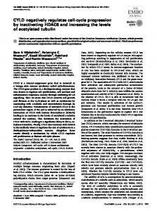
RESULTS Genetic analysis between htz1⌬ and replication genes. We have previously demonstrated that H2A.Z and the N-terminal tail of histone H4 have an overlapping role required for viability in yeast (17). Given that the N-terminal tail of H4 is important for both transcription and cell cycle progression (32, 53), we investigated whether H2A.Z, in addition to its role in transcription, also affected cell cycle progression. As a first step in determining whether it had a role outside of transcription, we investigated genetic interactions between HTZ1 and mutations in genes involved in chromatin assembly, chromosome structure, replication, repair, cell cycle progression, and checkpoint control. We systematically crossed htz1⌬ strains with either deletions or conditional mutations in nearly 90 genes (see Table S1 in the supplemental material). Visual examination of the double mutants after tetrad dissection for significant growth defects identified several genes involved in replication and checkpoint control to be synthetically lethal or to cause extreme sickness in combination with htz1⌬. Genetic interactions were observed between htz1⌬ and conditional mutants in orc5-1, orc2-1, rnr1-1 (Fig. 1A), sld5-1, psf1-1, or mcm3-1 (data not shown). Interestingly, mutations in the DNA helicases srs2⌬, rrm3⌬, and sgs1⌬ (data not shown); DNA polymerases; the cyclins clb2⌬, cln3⌬, clb5⌬, or clb6⌬; or the kinase cdc7-1 had no synthetic phenotypes when combined with htz1⌬. Since Clb5p and Clb6p are redundant for replication initiation (70), we tested the phenotype of a clb5⌬ clb6⌬ htz1⌬ triple mutant and observed a severe growth defect in this strain (Fig. 1A).
VOL. 26, 2006
H2A.Z AFFECTS CELL CYCLE PROGRESSION
491
FIG. 1. Genetic interactions between mutations in cell cycle genes and htz1⌬. A. H2A.Z genetically interacted with replication proteins. Analysis of growth of various single, double, and triple mutants on rich medium. Appropriate strains were grown to mid-log phase, and 10-fold serial dilutions were spotted on YPD plates. Plates with orc2-1, orc5-1, and rnr1-1 were incubated at 25°C (permissive temperature), while the remaining were grown at 30°C for 2 to 3 days prior to documentation. B. A simple schematic representation of the DNA damage and S-phase checkpoint pathways (54). C. Single mutants rad53-1 (ROY2083) and htz1⌬ (ROY3436) as well as rad53-1 htz1⌬ (ROY2532 and ROY2535) double mutants (top row) or single pol2-12 (ROY2251) or htz1⌬ (ROY3436) mutants as well as double pol2-12 htz1⌬ (ROY2248 and ROY2250) mutants (bottom row) carrying a URA3 plasmid-borne copy of H2A.Z were streaked on YMD-Ura (70a) and YMD plus fluoroorotic acid (FOA) plates to induce plasmid loss. Plates were incubated for 2 to 3 days prior to documentation. D. Growth of various single and htz1⌬ double mutants. Strains grown to mid-log phase were serially diluted and spotted on YPD plates. Plates were incubated at 30°C for 2 to 3 days prior to documentation.
Our genetic interaction data reveal synthetic phenotypes between proteins required for replication and mutations in Htz1p. These genetic interactions can be understood based on the functions of the interacting genes. The origin recognition
complex (ORC) (including Orc2p and Orc5p) binds to replication origins and is required for initiation of replication and the S-phase checkpoint (7, 71, 86). The GINS (Go, Ichi, Ni, and San) complex (77) (including Sld5p and Psf1p) is required
492
DHILLON ET AL.
for the establishment of replication forks and progression of the forks from the origins, while the MCM (minichromosome maintenance) complex (92) (including Mcm3p) is believed to be a DNA helicase that binds to the ORC at origins prior to initiation while replication initiation is regulated by the Cdc28/ cyclin-dependent modification of various complexes (18). Thus, proteins that function in replication had genetic interactions with this histone variant. The S-phase checkpoint pathway was essential in the absence of H2A.Z. We showed that HTZ1 genetically interacted with genes involved in replication, and mutants in replication proteins have previously been shown to be synthetically lethal with checkpoint mutants (9, 31, 74). We therefore asked whether cells lacking H2A.Z also had genetic interactions with checkpoint proteins. We generated and analyzed double mutants between htz1⌬ and mutations in genes involved in checkpoint control (reviewed in reference 54). We systematically crossed htz1⌬ strains with either deletions or conditional mutations in the genes known to be involved in DNA damage, S-phase, and spindle checkpoint control (Fig. 1B). We identified several genes involved in replication checkpoint control to be synthetically lethal or to cause extreme sickness in combination with htz1⌬. We identified rad53-1 and pol2-12 to be lethal with htz1⌬, while mrc1-1 (Fig. 1D) and asf1⌬ (data not shown) cells were synthetically sick. The systematic genetic analysis with H2A.Z has also identified Mrc1p to be synthetically sick with H2A.Z (81). The synthetic lethality between htz1⌬ and rad53-1 or pol2-12 was confirmed by plasmid-shuffle experiments (Fig. 1C). The interaction between htz1⌬ and pol2-12 was allele specific, as pol2-18 was not lethal with htz1⌬ (data not shown). The pol2-12 allele of DNA polymerase epsilon is defective in the S-phase checkpoint (59), while pol2-18 is a replication-defective allele of this gene (6). The synthetic interactions with Rad53p, Pol2p, Mrc1p, and Asf1p reflect a requirement for the checkpoint in cell survival when replication is defective. Compared with the replication checkpoint, mutations in the DNA damage checkpoint genes did not have any synthetic growth defects in the absence of Htz1p (Fig. 1D), suggesting that the defect was being generated during replication in these mutant cells. The S-phase checkpoint is functional in htz1⌬ cells. Since H2A.Z mutants had genetic interactions with the S-phase checkpoint genes, we inquired whether these cells had a fully functional S-phase checkpoint pathway. Yeast cells grown in the presence of a sublethal dose of hydroxyurea (HU; 0.2 M) initiate replication but block cell cycle progression in S phase via the S-phase checkpoint (60), and we investigated whether htz1⌬ cells were able to arrest in S phase, since this checkpoint was necessary for the viability of htz1⌬ cells. Wild-type and mutant cells were first synchronized in the G1 phase of the cell cycle with ␣ factor and then released into medium containing HU. Wild-type cells shifted to HU-containing media arrest in the S phase of the cell cycle with short mitotic spindles, a single nucleus, and large buds (Fig. 2A and B). Identically treated htz1⌬ cells showed that these were also proficient in activating the checkpoint because they also arrested in the S phase of the cell cycle (as monitored by flow cytometry) with short spindles, a single nucleus, and large buds (Fig. 2A and B). Activation of the S-phase checkpoint also results in the
MOL. CELL. BIOL.
phosphorylation of checkpoint proteins such as Rad53p. We also investigated the Rad53p phosphorylation response of htz1⌬ cells in the presence of HU. Wild-type and mutant cells arrested with ␣ factor were released into various concentrations of HU (0 to 25 mM) for 30 or 60 min, and the phosphorylation of Rad53p was monitored as an index of checkpoint activation (Fig. 2C). We found that Rad53p was phosphorylated in the mutant within 30 min of exposure to low concentrations of HU, while this was seen only after 60 min in wildtype cells, suggesting that cells lacking H2A.Z were hypersensitive to activation of the checkpoint. The hypersensitivity may be related to the observation that cells exposed to small doses of DNA damage more rapidly activate the checkpoint pathway to additional damage (61). When cells are exposed to HU, early origins of replication fire but the firing of late origins is delayed. To determine if the firing of late origins in HU was also delayed in htz1⌬ mutants, we used an assay that detects replication intermediates generated at origins (68). Normally, ARS305 is an early firing origin whose activation is not affected by HU, whereas ARS501 is a late origin that does not fire in the presence of HU within 120 min (2). Cells synchronized in ␣ factor were released into YPD with 0.2 M HU and harvested at 30-min intervals, and the appearance of replication intermediates was analyzed (Fig. 2D). The ARS305 replication intermediates were clearly visible in wild-type and htz1⌬ cells, while ARS501, a late origin, failed to fire in wild-type and htz1⌬ cells in the presence of HU within the first 120 min. All of these results suggested that htz1⌬ cells, exposed to HU, triggered the appropriate checkpoint to block cell cycle progression. Removal of HU and restoration of nucleotide pools results in the resumption of DNA synthesis from stalled forks and new forks from late firing origins (80). If htz1⌬ cells were defective in recovery from DNA damage during S-phase arrest, their viability should be reduced after exposure to HU. The wild type and H2A.Z mutants were maintained for various lengths of time (up to 8 h) in 0.2 M HU and then plated onto YPD plates lacking HU to determine the viability of those cells (Fig. 2E). These analyses revealed that htz1⌬ cells retained their viability to the same extent as wild-type cells in HU, indicating that the S-phase checkpoint that monitored completion of replication functioned properly to arrest these cells until the HU-induced replication stress had subsided. Timing of replication was delayed in htz1⌬ cells. The genetic interactions observed with htz1⌬ mutants suggested that this histone variant might play a role in replication. As a first step in determining if this was indeed the case, we measured the replication timing of several different loci in wild-type and mutant cells at 30°C (27). Consistent with previously published data (23), in wild-type cells a region adjacent to the ARS1 locus replicated early in S phase (35 min after release from ␣ factor), and a region adjacent to the HMR locus replicated later (45 min after release from ␣ factor) (Fig. 3A), and the difference in replication timing between these two loci was approximately 10 min. In cells lacking H2A.Z, ARS1 and HMR both replicated 15 min later than in the wild type. These results suggested that H2A.Z might be influencing the timing of replication from origins. Notably, H2A.Z loss led to a delay in the entire replication program, and unlike other determinants that affect the differ-
VOL. 26, 2006
H2A.Z AFFECTS CELL CYCLE PROGRESSION
493
FIG. 2. Replication checkpoint was functional in htz1⌬ cells. A. FACS profiles of asynchronous and HU-arrested wild-type (ROY1944) and mutant (ROY3443) htz1⌬ strains. B. Microtubule staining with a monoclonal antibody against ␣-tubulin and nuclear staining with 4⬘,6⬘-diamidino-2-phenylindole of wild-type (ROY1944) and htz1⌬ (ROY3443) strains arrested with 0.2 M HU for 2 h. C. Rad53 phosphorylation as a function of time of exposure and dose of HU. Mutant (ROY3443) and wild-type (ROY1945) strains were synchronized in G1 phase and released into various concentrations of HU in YPD. Cells were collected at 30 min and 60 min and processed for immunoblotting. Antibodies specific to Rad53p were used for detection. D. Early but not late origins fired in htz1⌬ cells in HU. G1-phase-synchronized wild-type (ROY1945) and htz1⌬ (ROY3442) strains were released into 0.2 M HU at 25°C. Cells were collected at different times, and replication intermediates were isolated and subjected to DNA blot analysis with probes against an early (ARS305) and a late (ARS501) origin. E. Survival curves of wild-type (ROY1944) and mutant htz1⌬ (ROY3433) cells exposed to 0.2 M HU. Cells were initially arrested with ␣ factor and released into YPD containing 0.2 M HU and held in HU for various lengths of time. Samples removed at the indicated times were serially diluted and plated on YPD. Plates were incubated at 30°C for 2 days before CFU were counted.
ential firing of origins during replication (4, 20, 26), this mutation did not cause the early and late replication program to be disrupted. The profiles also suggested that in the mutant, the regions examined took longer to replicate since the peaks were significantly broader at all loci examined. This could be due to slower fork progression or a consequence of a loss of synchrony in replication initiation. To distinguish between the two, we measured fork progression rates of a replication fork
that initiated from ARS607 and replicated a ⬃75-kbp stretch of DNA (79). The time of replication of three EcoRI restriction fragments (2.8 kb, 2.1 kb, and 1.7 kb long) located ⬃4.5 kbp, ⬃39 kbp, and ⬃63 kbp from ARS607 was determined and plotted as a function of distance, and the data demonstrated that the rate of fork progression was not significantly different in the mutant compared to the wild-type cells (Fig. 3B). These results indicated that fork progression per se was not affected
494
VOL. 26, 2006
in the mutant and that the defect was likely in replication initiation. Cdc45p release from origins was affected in htz1⌬ cells. One of the steps in replication initiation is the association of Cdc45p with origins (4, 5, 94), which then migrates with the replication fork as replication commences. Thus, the association of Cdc45 with, and its release from, origins are good indicators of replication initiation. We therefore investigated the loading and release of Cdc45p at early and late origins in wild-type cells and htz1⌬ mutants by ChIP. In wild-type cells grown at 15°C, we found that Cdc45p was loaded at ARS305 (an early origin) at 75 min while it was present at ARS501 (a late origin) at 135 min (Fig. 3C). The difference in time of replication in Fig. 3C compared to that in Fig. 3A is due to the fact that cells were grown at 15°C. Since Cdc45p is a component of the replication fork, upon replication initiation it moves with the fork and occupancy at the origin sequence is lost. Compared to wild-type cells, loading of Cdc45p at both origins was not dramatically delayed in htz1⌬ cells. While there was no major delay in the binding of Cdc45p to the origin, in the mutant the residence time of Cdc45p at the origin increased by an additional 30 min before release from the origin (Fig. 3C). Our results would argue either that replication was initiating asynchronously in the mutant population or replication initiation or events soon thereafter were delayed. These two scenarios are consistent with the data showing that regions of the genome took longer to replicate in the mutant. Origin firing was delayed in cells lacking Htz1p. We further examined the replication delay in htz1⌬ cells by analyzing replication origin firing and fork progression using two-dimensional agarose gel electrophoresis (2D gels) (8). We released G1-synchronized cells into S phase at 15°C and analyzed replication structures at the early-origin ARS305. In wild-type cells, bubble structures indicative of origin firing were first observed at 60 min after release; the abundance of initiation bubbles declined significantly by 75 min and were almost gone by 90 min (Fig. 3D). In htz1⌬ cells, initiation bubbles also were first observed at 60 min. However, in contrast to wild-type cells, the signal was significantly weaker in the htz1⌬ cells at 60 min, and initiation events at ARS305 occurred over a much broader period, lasting until about 120 min. Thus, replication initiation events appear to occur asynchronously throughout the popu-
H2A.Z AFFECTS CELL CYCLE PROGRESSION
495
lation of htz1⌬ cells, with most cells being delayed in ARS305 initiation. Nevertheless, replication initiation remained highly efficient in htz1⌬ cells because virtually no fork structures were detected. The 2D gels also suggest that fork movement within the vicinity of the origin is normal, as there is no accumulation of smaller bubble structures or any evidence of fork pausing. Analysis of a natural pause site (the tRNA gene, SUP6) also suggests there is no defect in replication fork pausing or progression through natural pause sites in htz1⌬ cells. These data are consistent with the analyses of replication timing (Fig. 3A) and Cdc45 chromatin association (Fig. 3C) and suggest that cells lacking H2A.Z have a lengthened S phase. H2A.Z localization at early and late origins. We therefore determined if there was any correlation between the presence of the variant at origins and origin function. If H2A.Z was present in the vicinity of origins, then it might influence origin firing and S-phase progression by affecting the local chromatin structure. We mapped H2A.Z at all known origins of replication on chromosome VI using quantitative ChIP (Fig. 3E). We determined that H2A.Z was present in various amounts at different origins. We next compared the levels of H2A.Z at various origins to their time of replication and the usage of these origins (25, 91). Surprisingly, we did not find any correlation between H2A.Z occupancy at origins and replication timing or usage of those origins. H2A.Z was required for normal cell cycle progression. Cells lacking H2A.Z showed a delay or asynchrony in replication initiation that could be the culmination of a delay in the entire program that is normally initiated at Start. We therefore investigated the kinetics of cell cycle progression in cells lacking H2A.Z by FACS analysis. Wild-type and mutant cells were initially synchronized in the G1 phase of the cell cycle with ␣ factor and then released into YPD medium. Cells were harvested at 10-min intervals, and their DNA content was analyzed by flow cytometry (Fig. 4). The analysis revealed that cells lacking this histone variant progressed more slowly through the cell cycle, with an approximate delay of 10 min compared to wild-type cells (compare the wild type to the mutant at 40 min and 50 min after release from ␣ factor). These results suggested that in the absence of H2A.Z cell cycle progression was delayed, presumably due to the longer S
FIG. 3. Timing of replication. A. Loci were delayed in the timing of their replication in htz1⌬ cells. Alpha factor-arrested wild-type and mutant cells stably and constitutively expressing DNA adenine methylase (ROY2245 and ROY3444) were released into YPD at 30°C, and time points were collected every 5 min. Genomic DNA isolated from these samples was digested with DpnI and EcoRI and analyzed by DNA blots using probes that hybridized adjacent to ARS1 and HMR. The y axis represents the amounts of hemimethylated (newly replicated) DNA that hybridized to the probes. B. Fork progression rate on chromosome VI. Wild-type and mutant cells (ROY3759 and ROY3760) were treated and analyzed as described in the legend to Fig. 6A, except that the genomic DNA was digested with DpnI and EcoRI and the blots were probed with probes that hybridized to EcoR1 fragments located ⬃4.5 kb (probe 2.8), ⬃39 kb (probe 2.1), and ⬃63 kb (probe 1.7) from ARS607. The time of replication for each probe was calculated by determining the peak from the replication profiles, and these values were plotted against distance from ARS607. C. The kinetics of loading of HA-Cdc45p at ARS305 and ARS501. Alpha factor-arrested wild-type (ROY2980) and htz1⌬ (ROY3236) strains carrying HA-Cdc45p were released in YPD at 15°C, and time points collected every 15 min were subjected to quantitative ChIP analysis. ChIP experiments were performed using the HA.11 monoclonal antibody. The y axis represents the ratio of immunoprecipitate (IP)/input DNA for each sample. The wild-type and mutant profiles are depicted as dashed and solid lines, respectively. D. Initiation of replication occurred over a longer duration in htz1⌬ cells. Alpha factor-arrested wild-type (ROY1945) and htz1⌬ (ROY3442) strains were released in YPD at 15°C, and time points were collected every 15 min. Replication intermediates were resolved by 2D gel electrophoresis, and the gels were blotted and probed with a probe specific to ARS305 and SUP6. E. H2A.Z occupancy near origins. Quantitative ChIP analysis of HA-H2A.Z localization at all known origins on chromosome VI (ChrVI) in ROY2987 cells. The time of replication (Trep ) and the usage of each replication are shown below the x axis and are derived from references 25 and 91.
496
DHILLON ET AL.
MOL. CELL. BIOL.
phase, which is consistent with our data showing that S phase may be lengthened in this mutant. Start was delayed in htz1⌬ cells. The commitment to cell division versus growth occurs at the end of G1 at an execution point called Start. Cells initiate budding and spindle pole body duplication and commit to initiating DNA replication (66), and these events depend upon the activation of Cdk activity (58). Progression through Start can therefore be measured by bud emergence and the degradation of the Cdk inhibitor Sic1p (57, 84). We initially measured the kinetics of bud emergence in ␣ factor-arrested cells that were released into fresh YPD medium at 15°C. Samples were collected every 15 min and were monitored for bud emergence by microscopy. There was a striking lag in bud emergence in the mutant (Fig. 5A). Since bud emergence is a subjective assay, we also examined the degradation of Sic1p-3xHA in ␣ factor-arrested cells released into fresh medium by immunoblotting (Fig. 5B). The kinetics of Sic1p degradation was delayed by 15 min in cells lacking H2A.Z (Fig. 5B). Our loading controls (histone H3) indicate that the differences in Sic1p observed on our protein blots were not due to differences in loading but reflect differences in the kinetics of degradation of Sic1p. Our data suggest that at a minimum, H2A.Z was directly or indirectly regulating events at Start rather than solely affecting replication. Transcriptional induction of specific cyclin genes was delayed in htz1⌬ mutants. One of the key events at Start is the Cdk kinase-dependent activation of transcription of genes required for G1/S progression (34), including the G1- and Sphase cyclin genes CLN1 CLN2 and CLB5 CLB6, respectively (3). Previous studies on genome-wide steady-state transcript levels have suggested that H2A.Z is predominantly involved in the activation of subtelomeric genes but not cell cycle-regulated genes (55). However, our observations that events in Start might be delayed in htz1⌬ cells and, given that bud emergence and replication initiation are temporally controlled via periodic fluctuations in the transcription of the cyclin genes (88, 89) and since H2A.Z has previously been shown to regulate the transcription of some inducible genes (1), led us to determine the effects of H2A.Z loss on the transcription of a few cell cycle-regulated genes. We chose CLN2 and CLB5, and we isolated total RNA from asynchronously grown cultures and quantitated the levels of CLN2 and CLB5 mRNA by RT-PCR using radionuclides. These were normalized to TUB2 mRNA, which has previously been shown to be unaffected in htz1⌬ cells (67). Similar to previous observations (55), we observed a less than twofold decrease in the transcript levels of these genes in asynchronous cultures (Fig. 6A). H2A.Z has been shown to affect the kinetics of induction of inducible genes (1). We therefore analyzed the induction of the CLN2 and CLB5 transcripts as a function of the cell cycle in wild-type and htz1⌬ cells. Cells were arrested with ␣ factor and released into fresh YPD medium at 16°C, and samples
FIG. 4. S phase was lengthened in htz1⌬ mutants. FACS profiles of wild-type (ROY1945) and htz1⌬ (ROY3442) cells through the cell cycle. Cells were arrested with ␣ factor and released into YPD at room temperature, and samples collected every 10 min were processed for FACS analysis.
VOL. 26, 2006
FIG. 5. Cells lacking Htz1p were delayed in Start. A. Bud index of htz1⌬ cells was delayed. Alpha factor-arrested wild-type (ROY1945) and htz1⌬ (ROY3442) strains were released into YPD medium at 16°C. Cells were collected at regular intervals and visualized microscopically. At least 100 cells were counted per time point, and the percentages of cells containing buds were quantitated and plotted as a function of time. The solid lines represent wild-type cells, and the dashed lines represent mutant cells. B. Sic1p degradation was delayed in htz1⌬ mutants. Wild-type (ROY3757) and htz1⌬ (ROY3758) strains containing tagged Sic1p were synchronized in G1 phase and released into YPD. Cells were collected every 15 min at 16°C and were processed for immunoblot analysis. Antibodies against the HA epitope were used to monitor Sic1p-3xHA. Commercial antibodies against histone H3 were used to monitor H3.
were collected every 30 min followed by quantitation of the transcripts. We analyzed the appearance of CLN2 and CLB5 transcripts after release from ␣ factor and normalized these to the TUB2 mRNA (Fig. 6B). In wild-type cells, the levels of CLN2 and CLB5 transcripts peaked around 80 min after release from ␣ factor, and, interestingly, the induction of both transcripts was delayed in the htz1⌬ mutant. While additional experiments will be necessary to determine all of the G1/S-phase genes that are affected in htz1⌬ cells, these results suggest that a delay in the transcriptional induction of genes at Start may feed into the S-phase delay seen in cells lacking H2A.Z. H2A.Z localized to the promoters of the CLN2 and CLB5 genes. H2A.Z could be either directly regulating the induction of CLN2 and CLB5 transcript levels, or it may be exerting this effect indirectly. To distinguish between the two possibilities, we determined whether promoters of these genes contained H2A.Z using ChIP experiments. We mapped 3xHA-Htz1p adjacent to the CLN2, CLB5, and GIT1 promoters using an anti-HA monoclonal antibody by quantitative ChIP using quantitative PCR with SyBr Green. The data were normalized to the Tel VIR 0.5-kb locus that is depleted of this histone variant (55). We found that H2A.Z was present at both the CLN2 and the CLB5 promoters (Fig. 6C), and the amount of H2A.Z at the CLN2 and CLB5 promoters was equivalent to that seen with the GIT1 promoter, where H2A.Z has previ-
H2A.Z AFFECTS CELL CYCLE PROGRESSION
497
ously been shown to localize (55). These data are consistent with results demonstrating that H2A.Z influenced the rapid induction of the cyclin genes. Overexpression of Cln2p suppresses the delay in cell cycle progression. Expression of Cln2p is a key driver of events at Start, and overexpression of CLN2 accelerates Start (82). It has even been suggested that the G1 cyclins may be the only proteins limiting for Start (28). If the cell cycle delay in Htz1p mutants is due to a delay in the induction of G1 cyclins, then overexpression of Cln2p might alleviate the delay. We transformed wild-type and htz1⌬ cells with vector alone or vector carrying Cln2p under the control of the GAL1 promoter. Cells were arrested with alpha factor, CLN2 was induced by a shift of cells to medium containing galactose, and 30 min after the induction cells were released into medium lacking ␣ factor. We monitored progression through the cell cycle by measuring bud emergence. The results of this analysis are shown in Fig. 7 and indicate that overexpression of CLN2 is able to significantly alleviate the delay in budding that is observed in htz1⌬ cells, demonstrating that the overexpression can bypass the htz1⌬ defect. However, it is unlikely that misregulation of CLN2 is the sole cause for the defects observed in htz1⌬ cells, since CLN2 overexpression suppresses an htz1 null mutant and is most likely a bypass suppressor. This would be consistent with the observation that overexpression of CLN2 also advances Start in wild-type cells (82). DISCUSSION S-phase delay in H2A.Z mutants. H2A.Z is known to affect transcription of genes and chromosome segregation (37). Our observations indicate that DNA replication was also affected in cells that did not contain H2A.Z, since htz1⌬ cells were slow in their progression through S phase, and the timing of replication of both an early- and a late-replicating region was delayed in these cells. Furthermore, the induction of transcription of cyclin genes was affected in htz1⌬ cells. Chromatin plays an important role in replication, since nucleosomes positioned over an origin affect origin function (72) and nucleosomes located adjacent to origins facilitate replication initiation (48). Furthermore, origins localized near silenced chromatin domains are usually late replicating (52, 73), while acetylation of histones around a late origin induces it to fire early (85). Similarly, mutant phenotypes of Orc2p are suppressed by mutations in the histone acetyltransferase Sas2p (24, 36, 86). We have now shown that the histone variant H2A.Z is one additional factor that directly or indirectly influenced replication and cell cycle progression. There are several models to explain how H2A.Z may affect S phase. Genome-wide expression analyses have suggested that H2A.Z acts as an activator and repressor of genes (38, 55), and while it does not significantly affect steady-state transcript levels of cell cycle-regulatory proteins (55 and our unpublished data), we have clearly shown that the kinetics of induction of two cell cycle-regulatory genes, CLN2 and CLB5, are affected in cells lacking Htz1p. It is therefore possible that cells lacking this variant have reduced levels of proteins involved in cell cycle progression during a specific point in the cell cycle Start. The down-regulation of these proteins, in htz1⌬ cells, could therefore result in the delay in replication that we observe. The
498
DHILLON ET AL.
FIG. 6. CLN2 and CLB5 transcription induction was delayed in htz1⌬ mutants. A. Measurement of steady-state transcript levels in asynchronous wild-type and htz1⌬ strains. Wild-type (ROY1945) and htz1⌬ (ROY3442) strains were grown in YPD medium at 16°C. Cells were collected, and total RNA was extracted from these cells. cDNA prepared from the RNA was analyzed by PCR using radionuclides and primers specific for CLN2, CLB5, and TUB2. The levels of transcript for CLN2 and CLB5 were quantitated by PhosphorImager analysis of the gels and are plotted normalized to TUB2. B. Alpha factor-arrested wild-type (ROY1945) and htz1⌬ (ROY3442) strains were released into YPD medium at 16°C. Cells were collected at 30-min intervals, and total RNA was extracted from these cells. cDNA prepared for each time point was analyzed by PCR using radionuclides and primers specific for CLN2, CLB5, and TUB2. Panels on the left are images of gels with the CLN2 and CLB5 PCR products (the time points are marked above the gel image, and “A” represents asynchronous cultures). The levels of transcript for CLN2 and CLB5 were quantitated by PhosphorImager analysis of the gels and are plotted as a function of time, normalized to TUB2. The solid lines represent wild-type cells,
MOL. CELL. BIOL.
synthetic sickness that we observe between H2A.Z and replication proteins can also be explained by the possibility that reduced levels of replication proteins coupled with mutations that weaken these proteins would be expected to lead to slower growth rates. An alternative model to explain our current observations is that the localization of H2A.Z at intergenic regions throughout the genome could conceivably create a chromatin environment that facilitates all nuclear processes, including transcription, replication, repair, and segregation. Loss of this variant might create more condensed chromatin, thereby slowing the kinetics of most nuclear processes in the cell. A third possibility is that H2A.Z has multiple and distinct functions in the cell: its enrichment at pericentric chromatin could affect chromosome segregation (41) via interactions with kinetochore proteins; its localization in the intergenic regions at the promoters of genes could aid in the recruitment of specific transcription factors affecting activation and repression of genes (38, 55, 67); and it could also directly affect the assembly and function of replication complexes at origins, especially since origins are located in intergenic regions (90). While it is likely that H2A.Z alters the chromatin structure in intergenic regions of the genome and it is possible that it helps in recruiting the replication machinery to origins, we have analyzed the distribution of H2A.Z at all of the origins on chromosome VI and do not find any correlation between H2A.Z occupancy and replication timing or usage (25, 91). Furthermore, the binding of ORC at origins is not significantly affected in cells lacking H2A.Z (see Fig. S1 in the supplemental material). The final possibility is that cells lacking H2A.Z have increased damage of a specific nature that is recognized by the S-phase checkpoint. We observe genetic interactions between htz1⌬ mutants and mutants in genes involved in the S-phase checkpoint but not with genes involved in the DNA damage checkpoint. This result suggested that even in the absence of any genotoxic stress, a specific kind of lesion was being induced in the absence of this histone variant, which necessitated a functional S-phase checkpoint pathway. The necessity to repair the damage in S phase could result in the delay that we observe. The S-phase checkpoint is necessary in the presence of stalled replication forks (49) or when replication initiation is impaired (21, 31, 71, 86). The misregulation of the cyclins has also been shown to activate the checkpoint, presumably due to defects in replication initiation (21, 31, 47, 78). It is therefore possible that the genetic interactions observed between htz1⌬ and the S-phase checkpoint proteins are a consequence of increased damage that needs to be repaired before cells can finish replication and progress through the cell cycle. H2A.Z and the regulation of cyclin genes. In budding yeast, H2A.Z has been shown to be necessary for transcriptional
and the dashed lines represent mutant cells. C. Localization of H2A.Z at various loci. ChIP experiments using the HA.11 monoclonal antibody were performed in asynchronous wild-type (ROY2987) strains carrying HA-Htz1p. Localization was measured by real-time PCR using probes adjacent to the promoters of CLN2, CLB5, and GIT1 as well as primers specific to telomere 6R. The y axis represents the ratio of immunoprecipitate (IP)/input DNA for each sample.
VOL. 26, 2006
H2A.Z AFFECTS CELL CYCLE PROGRESSION
499
level equivalent to that observed at the GIT1 gene. The presence of this variant at the promoter of the cyclin genes and the delayed induction of these genes in the absence of H2A.Z suggest that this histone variant was required for the correct temporal activation of these genes during the cell cycle. The mechanism of regulation of the cyclin promoters by H2A.Z was likely to be similar to that observed at the GAL1 promoter (1, 67), though this needs to be further investigated. While the histone variant is directly or indirectly required for the proper temporal regulation of the cyclin genes, at this point we are unable to determine the precise reason for the delay in progression through the cell cycle that is observed in htz1⌬ cells. We find that double mutants between htz1⌬ and various cyclins are not sick, though a triple mutant between clb5⌬, clb6⌬, and htz1⌬ is very sick. The clb5⌬ clb6⌬ double mutants are also inviable in the absence of Cln1p and Cln2p (19) or Clb3p and Clb4p (16). It is therefore possible that H2A.Z regulates multiple cyclin genes, but our genetic results suggest an even more complicated picture. Spt16 regulates transcription of the G1 cyclins, and spt16-197 mutants grown at the nonpermissive temperature exhibit a decrease in the levels of CLN1 and CLN2 transcripts (51, 65). However, we do not observe any growth defect in a spt16-197 htz1⌬ double mutant (at the permissive temperature). It is therefore possible that the cell cycle phenotypes described in this work are due to the delay in the induction of cyclin genes as well as defects in the induction of other G1/S-phase genes or even other processes not related to the regulation of these genes and will need to be investigated further. Numerous models can explain the phenotypes associated with htz1⌬ cells: H2A.Z affects the kinetics of induction of cell cycle-regulated genes, H2A.Z regulates global chromatin accessibility, and H2A.Z is required to repair damage at the G1/S transition. Additional experiments will be necessary to determine the role for this enigmatic variant. FIG. 7. Start was accelerated in cells overexpressing CLN2. A. The bud index of htz1⌬ (ROY3442) cells overexpressing CLN2 or with the vector alone. GAL1p::CLN2 expression was induced by adding galactose to G1-arrested cells prior to their release from alpha factor. Cells were released into fresh medium at 20°C, and samples were collected into 0.1% sodium azide every 15 min to monitor bud emergence. B. The bud index of wild-type (ROY1945) cells overexpressing CLN2 or with the vector alone. GAL1p::CLN2 expression was induced by adding galactose to G1-arrested cells prior to their release from alpha factor. Cells were released into fresh medium at 20°C, and samples were collected into 0.1% sodium azide every 15 min to monitor bud emergence.
activation of genes (55, 67). It is depleted from silenced regions and is elevated in the intergenic regions of inducible genes (1, 38, 42, 46, 56, 67). The absence of this variant leads to compromised recruitment of TBP and RNA polymerase II to the promoters of genes and a delay in the induction of these genes. Interestingly, measurements of the expression of the cyclin genes in asynchronous cultures suggest that this variant played a minor role in the expression of these genes (consistent with the microarray data [55]). However, in synchronized cells traversing Start, the loss of this variant significantly affected the induction of CLN2 and CLB5 transcription. We also find that this variant was present at the promoters of these genes at a
ACKNOWLEDGMENTS We thank Steve Bell, Doug Koshland, John Diffley, Paul Kaufman, Tom Petes, Judy Berman, Rodney Rothstein, Jasper Rine, Hiroyuki Araki, Judy Campbell, Mary Ann Osley, Rolf Sternglanz, Serge Gangloff, David Shore, Stefan Hohman, Akio Sugino, Peter Burger, Sue Biggins, Molly Fitzgerald-Hayes, Lorraine Pillus, Mary Bryk, Kim Nasmyth, David Stillman, Michael Grunstein, Carl Wu, Orna Cohen-Fix, Steve Elledge, and Munira Basrai for various plasmids, strains, and antibodies. We also thank Steve Bell, Doug Koshland, and John Diffley for protocols and technical help. We acknowledge help from the NCI FACS facility. We also thank Jasper Rine, Alan Hinnebusch, Mitch Smith, Munira Basrai, Lourdes Valenzuela, and Alex Strunnikov for helpful comments and suggestions. REFERENCES 1. Adam, M., F. Robert, M. Larochelle, and L. Gaudreau. 2001. H2A.Z is required for global chromatin integrity and for recruitment of RNA polymerase II under specific conditions. Mol. Cell. Biol. 21:6270–6279. 2. Alcasabas, A. A., A. J. Osborn, J. Bachant, F. Hu, P. J. Werler, K. Bousset, K. Furuya, J. F. Diffley, A. M. Carr, and S. J. Elledge. 2001. Mrc1 transduces signals of DNA replication stress to activate Rad53. Nat. Cell Biol. 3:958– 965. 3. Andrews, B., and V. Measday. 1998. The cyclin family of budding yeast: abundant use of a good idea. Trends Genet. 14:66–72. 4. Aparicio, O. M., A. M. Stout, and S. P. Bell. 1999. Differential assembly of Cdc45p and DNA polymerases at early and late origins of DNA replication. Proc. Natl. Acad. Sci. USA 96:9130–9135. 5. Aparicio, O. M., D. M. Weinstein, and S. P. Bell. 1997. Components and dynamics of DNA replication complexes in S. cerevisiae: redistribution of MCM proteins and Cdc45p during S phase. Cell 91:59–69.
500
DHILLON ET AL.
6. Araki, H., P. A. Ropp, A. L. Johnson, L. H. Johnston, A. Morrison, and A. Sugino. 1992. DNA polymerase II, the probable homolog of mammalian DNA polymerase epsilon, replicates chromosomal DNA in the yeast Saccharomyces cerevisiae. EMBO J. 11:733–740. 7. Bell, S. P., and B. Stillman. 1992. ATP-dependent recognition of eukaryotic origins of DNA replication by a multiprotein complex. Nature 357:128–134. 8. Brewer, B. J., and W. L. Fangman. 1991. Mapping replication origins in yeast chromosomes. Bioessays 13:317–322. 9. Chanet, R., and M. Heude. 2003. Characterization of mutations that are synthetic lethal with pol3-13, a mutated allele of DNA polymerase delta in Saccharomyces cerevisiae. Curr. Genet. 43:337–350. 10. Choy, J. S., and S. J. Kron. 2002. NuA4 subunit Yng2 function in intra-Sphase DNA damage response. Mol. Cell. Biol. 22:8215–8225. 11. Clarkson, M. J., J. R. Wells, F. Gibson, R. Saint, and D. J. Tremethick. 1999. Regions of variant histone His2AvD required for Drosophila development. Nature 399:694–697. 12. Cosgrove, A. J., C. A. Nieduszynski, and A. D. Donaldson. 2002. Ku complex controls the replication time of DNA in telomere regions. Genes Dev. 16:2485–2490. 13. Cosma, M. P. 2002. Ordered recruitment: gene-specific mechanism of transcription activation. Mol. Cell 10:227–236. 14. Cosma, M. P., S. Panizza, and K. Nasmyth. 2001. Cdk1 triggers association of RNA polymerase to cell cycle promoters only after recruitment of the mediator by SBF. Mol. Cell 7:1213–1220. 15. Cosma, M. P., T. Tanaka, and K. Nasmyth. 1999. Ordered recruitment of transcription and chromatin remodeling factors to a cell cycle- and developmentally regulated promoter. Cell 97:299–311. 16. Cross, F. R., and M. D. Jacobson. 2000. Conservation and function of a potential substrate-binding domain in the yeast Clb5 B-type cyclin. Mol. Cell. Biol. 20:4782–4790. 17. Dhillon, N., and R. T. Kamakaka. 2000. A histone variant, Htz1p, and a Sir1p-like protein, Esc2p, mediate silencing at HMR. Mol. Cell 6:769–780. 18. Diffley, J. F. 2004. Regulation of early events in chromosome replication. Curr. Biol. 14:R778—R786. 19. Dirick, L., T. Bohm, and K. Nasmyth. 1995. Roles and regulation of ClnCdc28 kinases at the start of the cell cycle of Saccharomyces cerevisiae. EMBO J. 14:4803–4813. 20. Donaldson, A. D., M. K. Raghuraman, K. L. Friedman, F. R. Cross, B. J. Brewer, and W. L. Fangman. 1998. CLB5-dependent activation of late replication origins in S. cerevisiae. Mol. Cell 2:173–182. 21. Early, A., L. S. Drury, and J. F. Diffley. 2004. Mechanisms involved in regulating DNA replication origins during the cell cycle and in response to DNA damage. Philos. Trans. R Soc. Lond. B. Biol. Sci. 359:31–38. 22. Ehrenhofer-Murray, A. E. 2004. Chromatin dynamics at DNA replication, transcription and repair. Eur. J. Biochem. 271:2335–2349. 23. Ehrenhofer-Murray, A. E., R. T. Kamakaka, and J. Rine. 1999. A role for the replication proteins PCNA, RF-C, polymerase epsilon and Cdc45 in transcriptional silencing in Saccharomyces cerevisiae. Genetics 153:1171–1182. 24. Ehrenhofer-Murray, A. E., D. H. Rivier, and J. Rine. 1997. The role of Sas2, an acetyltransferase homologue of Saccharomyces cerevisiae, in silencing and ORC function. Genetics 145:923–934. 25. Friedman, K. L., B. J. Brewer, and W. L. Fangman. 1997. Replication profile of Saccharomyces cerevisiae chromosome VI. Genes Cells 2:667–678. 26. Friedman, K. L., J. D. Diller, B. M. Ferguson, S. V. Nyland, B. J. Brewer, and W. L. Fangman. 1996. Multiple determinants controlling activation of yeast replication origins late in S phase. Genes Dev. 10:1595–1607. 27. Friedman, K. L., M. K. Raghuraman, W. L. Fangman, and B. J. Brewer. 1995. Analysis of the temporal program of replication initiation in yeast chromosomes. J. Cell Sci. Suppl. 19:51–58. 28. Futcher, B. 1996. Cyclins and the wiring of the yeast cell cycle. Yeast 12: 1635–1646. 29. Garber, P. M., and J. Rine. 2002. Overlapping roles of the spindle assembly and DNA damage checkpoints in the cell-cycle response to altered chromosomes in Saccharomyces cerevisiae. Genetics 161:521–534. 30. Ghidelli, S., D. Donze, N. Dhillon, and R. T. Kamakaka. 2001. Sir2p exists in two nucleosome-binding complexes with distinct deacetylase activities. EMBO J. 20:4522–4535. 31. Gibson, D. G., J. G. Aparicio, F. Hu, and O. M. Aparicio. 2004. Diminished S-phase cyclin-dependent kinase function elicits vital Rad53-dependent checkpoint responses in Saccharomyces cerevisiae. Mol. Cell. Biol. 24:10208– 10222. 32. Grunstein, M. 1990. Histone function in transcription. Annu. Rev. Cell Biol. 6:643–678. 33. Hanawalt, P. C. 1994. Transcription-coupled repair and human disease. Science 266:1957–1958. 34. Horak, C. E., N. M. Luscombe, J. Qian, P. Bertone, S. Piccirrillo, M. Gerstein, and M. Snyder. 2002. Complex transcriptional circuitry at the G1/S transition in Saccharomyces cerevisiae. Genes Dev. 16:3017–3033. 35. Howe, L., D. Auston, P. Grant, S. John, R. G. Cook, J. L. Workman, and L. Pillus. 2001. Histone H3 specific acetyltransferases are essential for cell cycle progression. Genes Dev. 15:3144–3154. 36. Iizuka, M., and B. Stillman. 1999. Histone acetyltransferase HBO1 interacts
MOL. CELL. BIOL.
37. 38.
39. 40. 41.
42.
43.
44. 45.
46.
47. 48. 49. 50. 51. 52. 53. 54. 55. 56. 57.
58. 59. 60. 61. 62.
with the ORC1 subunit of the human initiator protein. J. Biol. Chem. 274: 23027–23034. Kamakaka, R. T., and S. Biggins. 2005. Histone variants: deviants? Genes Dev. 19:295–310. Kobor, M. S., S. Venkatasubrahmanyam, M. D. Meneghini, J. W. Gin, J. L. Jennings, A. J. Link, H. D. Madhani, and J. Rine. 2004. A protein complex Containing the conserved Swi2/Snf2-Related ATPase Swr1p deposits histone variant H2A.Z into euchromatin. PLoS Biol. 2:E131. Krebs, J. E., C. J. Fry, M. L. Samuels, and C. L. Peterson. 2000. Global role for chromatin remodeling enzymes in mitotic gene expression. Cell 102:587– 598. Krebs, J. E., M. H. Kuo, C. D. Allis, and C. L. Peterson. 1999. Cell cycleregulated histone acetylation required for expression of the yeast HO gene. Genes Dev. 13:1412–1421. Krogan, N. J., K. Baetz, M. C. Keogh, N. Datta, C. Sawa, T. C. Kwok, N. J. Thompson, M. G. Davey, J. Pootoolal, T. R. Hughes, A. Emili, S. Buratowski, P. Hieter, and J. F. Greenblatt. 2004. Regulation of chromosome stability by the histone H2A variant Htz1, the Swr1 chromatin remodeling complex, and the histone acetyltransferase NuA4. Proc. Natl. Acad. Sci. USA 101:13513– 13518. Krogan, N. J., M. C. Keogh, N. Datta, C. Sawa, O. W. Ryan, H. Ding, R. A. Haw, J. Pootoolal, A. Tong, V. Canadien, D. P. Richards, X. Wu, A. Emili, T. R. Hughes, S. Buratowski, and J. F. Greenblatt. 2003. A Snf2 family ATPase complex required for recruitment of the histone H2A variant Htz1. Mol. Cell 12:1565–1576. Kusch, T., L. Florens, W. H. Macdonald, S. K. Swanson, R. L. Glaser, J. R. Yates III, S. M. Abmayr, M. P. Washburn, and J. L. Workman. 2004. Acetylation by Tip60 is required for selective histone variant exchange at DNA lesions. Science 306:2084–2087. Larochelle, M., and L. Gaudreau. 2003. H2A.Z has a function reminiscent of an activator required for preferential binding to intergenic DNA. EMBO J. 22:4512–4522. Le Masson, I., D. Y. Yu, K. Jensen, A. Chevalier, R. Courbeyrette, Y. Boulard, M. M. Smith, and C. Mann. 2003. Yaf9, a novel NuA4 histone acetyltransferase subunit, is required for the cellular response to spindle stress in yeast. Mol. Cell. Biol. 23:6086–6102. Leach, T. J., M. Mazzeo, H. L. Chotkowski, J. P. Madigan, M. G. Wotring, and R. L. Glaser. 2000. Histone H2A.Z is widely but nonrandomly distributed in chromosomes of Drosophila melanogaster. J. Biol. Chem. 275:23267– 23272. Lengronne, A., and E. Schwob. 2002. The yeast CDK inhibitor Sic1 prevents genomic instability by promoting replication origin licensing in late G(1). Mol. Cell 9:1067–1078. Lipford, J. R., and S. P. Bell. 2001. Nucleosomes positioned by ORC facilitate the initiation of DNA replication. Mol. Cell 7:21–30. Lopes, M., C. Cotta-Ramusino, A. Pellicioli, G. Liberi, P. Plevani, M. MuziFalconi, C. S. Newlon, and M. Foiani. 2001. The DNA replication checkpoint response stabilizes stalled replication forks. Nature 412:557–561. Madigan, J. P., H. L. Chotkowski, and R. L. Glaser. 2002. DNA doublestrand break-induced phosphorylation of Drosophila histone variant H2Av helps prevent radiation-induced apoptosis. Nucleic Acids Res. 30:3698–3705. Malone, E. A., C. D. Clark, A. Chiang, and F. Winston. 1991. Mutations in SPT16/CDC68 suppress cis- and trans-acting mutations that affect promoter function in Saccharomyces cerevisiae. Mol. Cell. Biol. 11:5710–5717. McCarroll, R. M., and W. L. Fangman. 1988. Time of replication of yeast centromeres and telomeres. Cell 54:505–513. Megee, P. C., B. A. Morgan, and M. M. Smith. 1995. Histone H4 and the maintenance of genome integrity. Genes Dev. 9:1716–1727. Melo, J., and D. Toczyski. 2002. A unified view of the DNA-damage checkpoint. Curr. Opin. Cell Biol. 14:237–245. Meneghini, M. D., M. Wu, and H. D. Madhani. 2003. Conserved histone variant H2A.Z protects euchromatin from the ectopic spread of silent heterochromatin. Cell 112:725–736. Mizuguchi, G., X. Shen, J. Landry, W. H. Wu, S. Sen, and C. Wu. 2004. ATP-driven exchange of histone H2A.Z variant catalyzed by SWR1 chromatin remodeling complex. Science 303:343–348. Nash, P., X. Tang, S. Orlicky, Q. Chen, F. B. Gertler, M. D. Mendenhall, F. Sicheri, T. Pawson, and M. Tyers. 2001. Multisite phosphorylation of a CDK inhibitor sets a threshold for the onset of DNA replication. Nature 414:514– 521. Nasmyth, K. 1993. Control of the yeast cell cycle by the Cdc28 protein kinase. Curr. Opin. Cell Biol. 5:166–179. Navas, T. A., Z. Zhou, and S. J. Elledge. 1995. DNA polymerase epsilon links the DNA replication machinery to the S phase checkpoint. Cell 80:29–39. Pasero, P., K. Shimada, and B. P. Duncker. 2003. Multiple roles of replication forks in S phase checkpoints: sensors, effectors and targets. Cell Cycle 2:568–572. Paulovich, A. G., and L. H. Hartwell. 1995. A checkpoint regulates the rate of progression through S phase in S. cerevisiae in response to DNA damage. Cell 82:841–847. Peterson, C. L., and J. Cote. 2004. Cellular machineries for chromosomal DNA repair. Genes Dev. 18:602–616.
VOL. 26, 2006 63. Raghuraman, M. K., E. A. Winzeler, D. Collingwood, S. Hunt, L. Wodicka, A. Conway, D. J. Lockhart, R. W. Davis, B. J. Brewer, and W. L. Fangman. 2001. Replication dynamics of the yeast genome. Science 294: 115–121. 64. Rangasamy, D., L. Berven, P. Ridgway, and D. J. Tremethick. 2003. Pericentric heterochromatin becomes enriched with H2A.Z during early mammalian development. EMBO J. 22:1599–1607. 65. Rowley, A., R. A. Singer, and G. C. Johnston. 1991. CDC68, a yeast gene that affects regulation of cell proliferation and transcription, encodes a protein with a highly acidic carboxyl terminus. Mol. Cell. Biol. 11:5718– 5726. 66. Rupes, I. 2002. Checking cell size in yeast. Trends Genet. 18:479–485. 66a.Sambrook J., and D. W. Russell. 2001. Molecular cloning: a laboratory manual, 3rd ed. Cold Spring Harbor Laboratory Press, Cold Spring Harbor, N.Y. 67. Santisteban, M. S., T. Kalashnikova, and M. M. Smith. 2000. Histone H2A.Z regulates transcription and is partially redundant with nucleosome remodeling complexes. Cell 103:411–422. 68. Santocanale, C., and J. F. Diffley. 1998. A Mec1- and Rad53-dependent checkpoint controls late-firing origins of DNA replication. Nature 395:615–618. 69. Schmitt, M. E., T. A. Brown, and B. L. Trumpower. 1990. A rapid and simple method for preparation of RNA from Saccharomyces cerevisiae. Nucleic Acids Res. 18:3091–3092. 70. Schwob, E., and K. Nasmyth. 1993. CLB5 and CLB6, a new pair of B cyclins involved in DNA replication in Saccharomyces cerevisiae. Genes Dev. 7:1160–1175. 70a.Sherman, F. 1991. Getting started with yeast. Methods Enzymol. 194:3–21. 71. Shimada, K., P. Pasero, and S. M. Gasser. 2002. ORC and the intra-S-phase checkpoint: a threshold regulates Rad53p activation in S phase. Genes Dev. 16:3236–3252. 72. Simpson, R. T. 1990. Nucleosome positioning can affect the function of a cis-acting DNA element in vivo. Nature 343:387–389. 73. Stevenson, J. B., and D. E. Gottschling. 1999. Telomeric chromatin modulates replication timing near chromosome ends. Genes Dev. 13:146–151. 74. Suter, B., A. Tong, M. Chang, L. Yu, G. W. Brown, C. Boone, and J. Rine. 2004. The origin recognition complex links replication, sister chromatid cohesion and transcriptional silencing in Saccharomyces cerevisiae. Genetics 167:579–591. 75. Svejstrup, J. Q. 2002. Mechanisms of transcription-coupled DNA repair. Nat. Rev. Mol. Cell. Biol. 3:21–29. 76. Swaminathan, J., E. M. Baxter, and V. G. Corces. 2005. The role of H2Av variant replacement and histone H4 acetylation in the establishment of Drosophila heterochromatin. Genes Dev., in press.19:65–76. 77. Takayama, Y., Y. Kamimura, M. Okawa, S. Muramatsu, A. Sugino, and H. Araki. 2003. GINS, a novel multiprotein complex required for chromosomal DNA replication in budding yeast. Genes Dev. 17:1153–1165. 78. Tanaka, S., and J. F. Diffley. 2002. Deregulated G1-cyclin expression induces genomic instability by preventing efficient pre-RC formation. Genes Dev. 16:2639–2649. 79. Tercero, J. A., and J. F. Diffley. 2001. Regulation of DNA replication fork progression through damaged DNA by the Mec1/Rad53 checkpoint. Nature 412:553–557. 80. Tercero, J. A., M. P. Longhese, and J. F. Diffley. 2003. A central role for
H2A.Z AFFECTS CELL CYCLE PROGRESSION
81.
82. 83. 84. 85. 86.
87. 88. 89. 90.
91.
92. 93.
94.
501
DNA replication forks in checkpoint activation and response. Mol. Cell 11:1323–1336. Tong, A. H., G. Lesage, G. D. Bader, H. Ding, H. Xu, X. Xin, J. Young, G. F. Berriz, R. L. Brost, M. Chang, Y. Chen, X. Cheng, G. Chua, H. Friesen, D. S. Goldberg, J. Haynes, C. Humphries, G. He, S. Hussein, L. Ke, N. Krogan, Z. Li, J. N. Levinson, H. Lu, P. Menard, C. Munyana, A. B. Parsons, O. Ryan,R. Tonikian, T. Roberts, A. M. Sdicu, J. Shapiro, B. Sheikh, B. Suter, S. L. Wong, L. V. Zhang, H. Zhu, C. G. Burd, S. Munro, C. Sander, J. Rine, J. Greenblatt, M. Peter, A. Bretscher, G. Bell, F. P. Roth, G. W. Brown, B. Andrews, H. Bussey, and C. Boone. 2004. Global mapping of the yeast genetic interaction network. Science 303: 808–813. Tyers, M., G. Tokiwa, and B. Futcher. 1993. Comparison of the Saccharomyces cerevisiae G1 cyclins: Cln3 may be an upstream activator of Cln1, Cln2 and other cyclins. EMBO J. 12:1955–1968. Verger, A., and M. Crossley. 2004. Chromatin modifiers in transcription and DNA repair. Cell Mol. Life Sci. 61:2154–2162. Verma, R., H. McDonald, J. R. Yates III, and R. J. Deshaies. 2001. Selective degradation of ubiquitinated Sic1 by purified 26S proteasome yields active S phase cyclin-Cdk. Mol. Cell 8:439–448. Vogelauer, M., L. Rubbi, I. Lucas, B. J. Brewer, and M. Grunstein. 2002. Histone acetylation regulates the time of replication origin firing. Mol. Cell 10:1223–1233. Weinberger, M., P. A. Trabold, M. Lu, K. Sharma, J. A. Huberman, and W. C. Burhans. 1999. Induction by adozelesin and hydroxyurea of origin recognition complex-dependent DNA damage and DNA replication checkpoints in Saccharomyces cerevisiae. J. Biol. Chem. 274:35975–35984. Weinreich, M., M. A. Palacios DeBeer, and C. A. Fox. 2004. The activities of eukaryotic replication origins in chromatin. Biochim. Biophys. Acta 1677: 142–157. Wittenberg, C., and S. I. Reed. 2005. Cell cycle-dependent transcription in yeast: promoters, transcription factors, and transcriptomes. Oncogene 24: 2746–2755. Wittenberg, C., K. Sugimoto, and S. I. Reed. 1990. G1-specific cyclins of S. cerevisiae: cell cycle periodicity, regulation by mating pheromone, and association with the p34CDC28 protein kinase. Cell 62:225–237. Wyrick, J. J., J. G. Aparicio, T. Chen, J. D. Barnett, E. G. Jennings, R. A. Young, S. P. Bell, and O. M. Aparicio. 2001. Genome-wide distribution of ORC and MCM proteins in S. cerevisiae: high-resolution mapping of replication origins. Science 294:2357–2360. Yamashita, M., Y. Hori, T. Shinomiya, C. Obuse, T. Tsurimoto, H. Yoshikawa, and K. Shirahige. 1997. The efficiency and timing of initiation of replication of multiple replicons of Saccharomyces cerevisiae chromosome VI. Genes Cells 2:655–665. Yan, H., S. Gibson, and B. K. Tye. 1991. Mcm2 and Mcm3, two proteins important for ARS activity, are related in structure and function. Genes Dev. 5:944–957. Zhang, W., J. R. Bone, D. G. Edmondson, B. M. Turner, and S. Y. Roth. 1998. Essential and redundant functions of histone acetylation revealed by mutation of target lysines and loss of the Gcn5p acetyltransferase. EMBO J. 17:3155–3167. Zou, L., and B. Stillman. 1998. Formation of a preinitiation complex by S-phase cyclin CDK-dependent loading of Cdc45p onto chromatin. Science 280:593–596.