coding for E. coli ribonucleotide reductase are encoded in an operon of known ... additions: 1 mM PMSF; 1 mM benzamidine; 6 wg of trypsin inhibitor;. 0.05 pg of ...
THEJOURNALOF
VOl. 262, No 20, Issue of July
BIOLOGICAL CHEMISTRY
C) 1987 by The American Society of Biological Chemists, Inc
pp. 9736-9743,1987 Printed in U.S A.
Half-site Reactivity of the Tyrosyl Radical of Ribonucleotide Reductase fromEscherichia coli* (Received for publication, October 24, 1986)
Britt-Marie SjobergSll, Margareta KarlssonSll, and Hans Jornvallg From the $Department of MoIecuhr Bwhgy, Swedish University ojAgricultura1 Sciences, Uppsalu BiomedicalCenter, S-75124 Uppsala, Sweden and the §Department of Chemistry I, Karolinska Institute, S-104 01 Stockholm,Sweden
A C-terminally truncated form of protein B2, the homodimeric small subunit of ribonucleotide reductase from Escherichia coli, was found as the result of an apparently specific proteolysis. Truncated homodimers contain an intact binuclear iron center and a normal tyrosyl radical but have no binding capacity for the other ribonucleotide reductase subunit, protein B 1, and are consequently enzymatically inactive. Heterodimers, consisting of one full-length and one truncated polypeptide, formed spontaneously during a chelation-reconstitution cycle and were easily separated from the two homodimeric variants. The heterodimeric formof B2shows a weak interaction with the B1 subunit resulting in low enzyme activity. Using heterodimers containing deuterated tyrosine on the fulllength side and protonated tyrosine on the truncated side, we could demonstrate that the tyrosyl radical was randomly generated in one or the other of the two polypeptide chains of the heterodimeric B2 subunit. The small subunit of ribonucleotide reductase thus conforms to a half-site reactivity.
and compared to the primary structure of protein B2 (Sjoberg et al., 1985). The homologous subunit from clam (Standart et al., 1985), mouse (Thelander andBerg, 1986), herpessimplex virus(Dutia, 1983; McLauchlanandClements,1983),Epstein-Barr virus (Gibson et al., 1984), and bacteriophage T4 (Sjoberg et al., 1986b) all display a conservedtyrosine residue in a position equivalent to Tyr-122 in E . coli B2. Protein engineering techniques were used t o identify the tyrosyl radical to this position inB2 (Larsson andSjoberg, 1986). Recently we separately cloned the structuralgene of E. coli coding for the B1 and B2 subunits in runaway replication vectors andachieved highly efficientexpressions of the cloned products (Larsson, 1984; Sjoberg et al., 1986a). The basis for the present investigation was an accidental discovery of a limited site-specific proteolysis of protein B2 in the extracts of our overproducing bacteria. Theavailability of a specifically truncated B2 polypeptide has allowed a detailed study of the location of the tyrosyl radical within protein B2. In addition, our studies with the truncated B2 polypeptide have led to an increased knowledge of the folding of protein B2 and its interaction with protein B1. MATERIALS ANDMETHODS
Ribonucleoside-diphosphate reductase (EC 1.17.4.1) is an E. coli protein B1 and thioredoxin reductase were prepared as oligomeric enzyme responsible for a balanced supply of nucleotide precursors for DNA synthesis inliving organisms. It described by Thelander et al. (1978) and Pigiet and Conley (1977), respectively. E. coli thioredoxin was a kind gift of A. Holmgren, consists of two nonidentical homodimeric subunits, in Esch- Department of Physiological Chemistry, Karolinska Institute, Stockerichia coli denoted proteins B1 and B2, and the holoenzyme holm, Sweden. Trypsin (tosylphenylalanyl chloromethane-treated) hasanstructure(ThelanderandReichard, 1979). The was obtained from Worthington, and carboxypeptidases A and Y active site is at the interphase of the two subunits. The B1 were from Boehringer Mannheim GmbH. CzDP’ was a kind gift of subunit ( a 2 )contains one redox-active dithiol group/polypep- F. Eckstein, Department of Chemistry, Max-Planck-Institut, Gottinobtained from tide chain. The B2 subunit ( p2), on the other hand, contributesgen, Federal Republic of Germany. /3,/3-D2-tyrosine was Uppsala, Sweden. PMSF was from Sigma. only one tyrosyl radical for the two polypeptide chains. This Reagenta, Nomenclature-The native B2 polypeptide is denoted 0 and the radical is necessary for enzyme activity and is stabilized by truncated polypeptide 0’. Three dimeric combinations are possible: a n adjacent F-oxo bridged binuclearironcenter(recently the native B2(/3/3)as well as B2(/3/3‘)and B2(/3’/3’). reviewed in Graslund et al., 1985). The reduction of ribonuBacterial Strain-E. coli C600 thr-1, leu-6, thi-1, supE44, lacYI, cleotides most likely proceeds via a radical cation substrate gal, pro, rpsL, hsrK-, hsmK+was a kind gift of G. Magnusson, Departintermediate, initiated by abstraction of the 3“hydrogen of ment of Virology, University of Uppsala, Sweden. General Methods-Iron was determined by the method of Massey the ribose moiety by the tyrosyl radicalof protein B2 (Stubbe (1957) as modified by Atkin et al. (1973). Absorption spectra were and Ackles, 1980; Stubbe et al., 1983). The structural genes recorded on a Cary 219 spectrophotometer. EPR spectroscopy and coding for E. coli ribonucleotide reductase are encoded in an spin quantitation were made as described by Peterson et al. (1980). Protease Inhibitors-Aliquots of 21 pg ofprotein B2 wereincubated operon of known nucleotide sequence (Carlson et al., 1984). In recentyears the aminoacid sequences of the small subunit in 50 ~1 of 50 mM Tris-HC1, pH 7.6, containing one of the following of ribonucleotide reductase fromseveral other organismswere additions: 1 mM PMSF; 1 mM benzamidine; 6 wg of trypsin inhibitor; pg of leupeptin; 1 mM sodium bisulfite; or 1 mM 2-mercaptoethdeduced from the DNAsequences of the correspondinggenes 0.05 anol. An aliquot of B2 without additions served as a control. Samples * This work was supported by grants from the Swedish Medical Research Councils (Projects 03X-6801 and -3532) and the Magn. Bergvall Foundation. The costs of publication of this article were defrayed in part by the payment of page charges. This article must therefore be hereby marked “aduertisement” in accordance with 18 U.S.C. Section 1734 solely to indicate this fact. TI Present address: Dept. of Molecular Biology, University of Stockholm, s-106 91 Stockholm, Sweden.
were incubated at 20 “C for 16 h. At both the beginning and the end of an experiment, samples of 6 pgofB2 from each aliquot were precipitated in 5% trichloroacetic acid and stored at -20 “C for -
-
’ The
abbreviations used are: CzDP, 2’-azido-2’-deoxy-CDP; PMSF, phenylmethylsulfonyl fluoride; SDS, sodium dodecyl sulfate; PAGE, polyacrylamide gel electrophoresis; HPLC, high-performance liquid chromatography.
9736
Radical Tyrosyl
of Ribonucleotide Reductase
further analysis by SDS/PAGE by the method of Laemmli (19701, as modified by O’Farrell (1975). Zon Exchange HPLC-Proteins B2(PP), B2(PP’), and B2(P’P’) were separated on a MonoQ HR5/5 (Pharmacia P-L Biochemicals) anion exchange column. The gradient was 0-0.7 M NaCl in 50 mM Tris-HC1, pH 7.6. Elution of protein was followed bythe absorbance at 280 nm. Amino AcidSequence Analysis-Homogeneous protein BZ(PP) and B2(P’P’) were reduced with dithiothreitol and carboxymethylated with 14C-labelediodoacetate (Thelander, 1973) prior to proteolytic digestions and sequence analyses. Carboxypeptidase digestions were performed in a volume of 0.45 ml with 5 nmol of B2(@P)or B2(P’@’)and a mixture of carboxypeptidases Aand Y (0.01and 0.25 mg, respectively) in 50 mM ammonium bicarbonate, 0.8 M urea, at 25 “C. Aliquots containing 0.8 nmol of BZ(@P)or B2(@’P’)were sampled at 0, 1,2,3,5, and8 h of incubation into an equal volume of acetic acid and freeze-dried prior to analysis on a Beckman 121M amino acid analyzer. A sample without protein B2 was incubated for 8 h and served as a blank. Tryptic digestion was performed with 10 nmol of BZ(8’P’) and 4.3 pg of trypsin in 0.1 M ammonium bicarbonate for 5 ha t 37 “C. Tryptic peptides were separated with a gradient of0-100% acetonitrile in 0.1% trifluoroacetic acid on a PepRPC 5/5 (Pharmacia P-L Biochemicals) HPLC column. This chromatogram differed at four positions from a similarly treated sample of B2(8P), and the peptides from these peaks in the BZ(P’P’) chromatogram were further analyzed. N-terminal amino acid sequences were determined by liquid-phase Sequencer degradation (Beckman 890D) using a 0.1 M Quadrol peptide program and application into glycine-precycledPolybrene (Jornvall and Philipson, 1980).Phenylthiohydantoin derivatives were identified by HPLC (Zimmerman et al., 1977). Chelation and Reconstitution of Heterodimers-Aliquots of B2(@@) and B2(P‘P’) were mixed, precipitated with ammonium sulfate, dissolved in 50 mM Tris-HC1, pH 7.6, and chelated in the presence of imidazole and 8-hydroxyquinoline 5-sulfonate. Metal-free B2 preparations (apo-B2) were reconstituted in the presence of Fe(I1) ascorbate. The procedures for chelation and reconstitution of B2were according to Atkin et al. (1973). Enzyme Assays-The enzymatic activity of protein B2 was determined spectrophotometrically or with [3H]CDP (Thelander et al., 1978) with an excess of protein B l (1-2 p M ) . ATP (1.5 mM) or dTTP (40 p ~ were ) used as effectors andCDP or GDP (0.5 mM) as substrates. One enzyme unit is the amount of B2 preparation that catalyzes the reduction of 1 nmol of CDP or GDP/min at 25 “C and in the presence of an excess of protein B1. When enzymatic measurements were coupled with EPR measurements equimolar amounts of ) used. Further details are given protein B1 and B2 (10-20 p ~ were under CzDP inactivation. Glycerol Gradient Sedimentation-Gradients were 4.8 ml of2540% glycerol in 50 mM Tris-HC1, pH 7.6, 0.1 M KCl, 5 mM dithiothreitol, 10 mM MgCIZ,and, where indicated, 60 p~ dTTP. Each tube contained a 0.2-ml cushion of 60% glycerol in 50 mM Tris-HCI, pH 7.6, 50 mM KCl, 10 mM MgCl,. Samples were loaded in 0.1 ml of 50 mM Tris-HC1, pH 7.6, 5 mM dithiothreitol, 10 mM MgCl,, and, where indicated, 60 p~ dTTP. Amounts loaded were 0.6 mg of protein B1 and 0.24-0.25 mg of protein B2 preparations. Catalase (sz0,+ = 11.4 S) was added as a reference. Samples were centrifuged at +2 “C in a VTi65 rotor a t 50,000 rpm for 4.5 h. Fractions of approximately 0.13 ml were collected manually. Protein was determined according to Lowry et al. (1951). and the amount of protein B l and B2, respectively, was determined by SDS/PAGE and tracing in a Joyce-Loebl microdensitometer. Preparation of DeuteratedProtein B2”The B2 overproducing strain C6OO/pBS1 (Sjoberg et al., 1986a) wasgrown a t 30 “C in minimal medium (Davis and Mingioli, 1950) supplemented with 80 mg/liter leucine, 50 mg/liter threonine, 30 mg/liter proline, 40 mg/ liter @@-D,-tyrosine,and 1 mg/liter thiamine. At a cell density of approximately 10’ cells/ml, the temperature was raised to 42 “C. After 7 hof cultivation a t 42 “C, 1.6 g of cells were harvested by centrifugation at +4 “C and thenstored at -70 “C. Cells were disintegrated by grinding with alumina (Eriksson et al., 1977), and protein B2 was purified as described (Sjoberg et al., 1986a) except that in the last step anion exchange HPLC was used as described above. CzDP Znactiuation-Mixtures of 0.72 mg of protein B1 and 0.27 mg of protein B2(PP) or 0.23 mg of protein B2(@@’)/0.175ml of 50 mM Tris-HC1, pH 7.6, 15 mM MgCl,, 5 mM dithiothreitol, 80 p~ dTTP were frozen in liquid N, in EPR tubes. After a zero time recording of the EPR spectra, samples were thawed and CDP or
9737
CzDP was added to 5 or 1.5 mM, respectively. Each sample was used for a complete time curve by repeated freeze-thaw cycles. Thus, samples were incubated at 25 “C afterwhich they were quickly frozen in liquid N, at the times indicated, EPR spectra were recorded, and the samples were then thawed at 25 “C for continued incubation. RESULTS
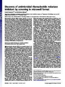
Proteolytic Degradation of Protein B2 i n Overproducing Cells The basis for the present investigationwas the finding that apparently homogeneous preparations of the small subunit of E. coli ribonucleotide reductase, obtained from an overproducingstrain (Sjoberg et al., 1986a),acquireddetectable amounts of a 40-kDa species in addition to the original protein band at 43.5 kDa, as seen by SDS/PAGE. The new band appeared during the ultradialysis concentration, which lasted for approximately 24 h at +4 “C. The relative amount of fulllength protein B2 was further reduced in concentrated preparations that had been thawed andrefrozen several times. It was found that a site-specific degradation of protein B2 occurred during the entire purification procedure even though all these steps were performed at +4 “C. A series of protease inhibitors was tested forpossible inhibitory action on the conversion of full-length B2 polypeptides to the 40-kDa molecular mass species. Onlythe serine protease inhibitor PMSF not shown). The degradation was found to be inhibitory (data of protein B2 in crude extractsis thus inhibited by routinely including PMSFinthe very firstpurificationstepand throughout (Sjoberg et al., 1986a). Separation of Protein B 2 and Its Proteolytic Products by Anion Exchange HPLC A partly degraded preparation of B2 was chromatographed on the anion exchange column MonoQ and three different protein fractions eluted at 0.24, 0.27, and 0.29 M NaC1, respectively (cf. Fig. 2B). Analysis by SDS/PAGE showed that the first protein fraction contained exclusively P’-polypeptides,the second containedequalamounts of P- and p’polypeptides and the last fraction containedonly P-polypeptides (cf. Fig. 2B, inset). Thus, it is possible to isolate the three different dimeric combinations of P- and P’-polypeptides. 60
1
mAYTTFS~TKNDOLKEP~FFG~PVNVARYDQGKKDIFEKLIEKQLSFFYRPEEVDVSRDR1
I + (Tl)120 61 DYaALPEHEKHIFISNLKYaTLLDSIoCRSPNVALLPLISIPELETWVETWAFSETIHSR (T4)121 180
SYTHIIRNIVNDPSVVFDDIVTNEQI~KRAEGISSYYDELIEMTSYVHLLGEGTHTVN~K 181
240
TVTVSLRELKKKLYLCLMSVNALEAIRFYVSFACSFAFAERELMECN~IIRLI~DEAL 241
300
HLTGTOHMLNLLRSGADDPEMAEECKOEC~GECYDLFVa~aaEKDWADYLFRDGSMIGLN
-
( T 3 a ) ~ 301
‘
.
1
‘
360
KDILCQYVEYITNIRHaAVGLDLPFQTRSNPIPWINTVLVSD~aVAPQEVEVSSYLVGa (T3b)361 . 375 IDSEVDTDDLSNFQL tl
( T 2 )-
U
FIG. 1. Results of amino acid sequence determination of protein BZ(@’@’)in relation to the deduced complete amino acid sequence (Carlson et d.,1984) of protein BZ(j30). Rightward arrows denote residues proven by Sequencer degradations of the protein or tryptic (T) fragments. Leftward arrows denote sequences established by carboxypeptidase degradations. The vertical arrow marks the assumed C-terminal end of the @‘-polypeptide.The lower case m denotes the initiator methionine, which is absent in the mature protein.
Tyrosyl Radical of Ribonucleotide Reductase
9738
FIG. 2. Anion exchange HPLC of protein B2 and its proteolytic products. A , chromatography of equimolar amounts of BZ(PP) and BZ(P’P’). B , chromatography of equimolar amounts of B2(&3) and BZ(P’P’) after iron chelation as described under “Materials and Methods.” Inset, SDS/PAGE of peak fractions from panel B: 1, BZ(P’P’); 2, BZ(PP’1; 3, BZ(PP).
10
20 30 Elution volume (ml)
deduced amino acid sequence of protein B2. Peptide peak T 3 contained two peptides (T3a and T3b) of different stoichiometry. They could be interpreted to match positions 255N-terminal Analysis-Carboxymethylated @‘-polypeptides 263 (T3a)and 303-314 (T3b), respectively. These results were subjected to N-terminal Sequencer degradations. Phen- indicate that the apparent differences in the chromatograms ylthiohydantoin derivatives were identified into a single se- of trypsin-digested @- and @‘-polypeptides originated from quence for 40 cycles (Fig. 1).This confirms that B2(@’@’) nonstoichiometric or pseudo-specific tryptic degradation of consists of polypeptide chains with identical N-terminal se- the @’-preparationas compared to the@-preparationand thus quences. Comparison with the amino acid sequence of native emphasize the extensive correspondence of the two polypepprotein B2 (Sjoberg et al., 1986a) shows that @- and @’-tides. Taken together our sequence analyses thus farindicate polypeptides have identical N-terminal sequences. Moreover, that @’-polypeptidesare derived from a C-terminalprocessing residues 1-40of @’-polypeptidesagree with the predicted event beyond position 333. amino acid sequence of protein B2 as deduced from the C-terminal Analyses-Carboxymethylated @- and @’-polyreported nucleotide sequence of the nrdB gene (Carlson et al., peptides were digested separately with carboxypeptidases. 1984) but also show that the N-terminal processing has re- Digestion of @-polypeptidesresulted in a time-dependent removed the initiator methionine. lease of amino acid residues leucine, asparagine/glutamine/ Analysis of Tryptic Peptides-To further verify that the@’-serine,’ and phenylalanine, whereas asparagine/glutamine/ polypeptides originate from @-polypeptides, carboxymeth- serine’ and valine were released from @’-polypeptides(Fig. 1). ylated p- and @’-polypeptideswere digested separately with The result obtained with the @-polypeptidefits very well with trypsin, and the resulting peptides were separated by HPLC. the predicted C-terminal sequence -Phe-Gln-Leu of the nrdB We expected to find chromatograms differing only in one product as deduced from the nucleotide sequence. The result position, e.g. one peptide peak in the @-digestbeing replaced obtained with the @‘-polypeptidefits with amino acid residues by another peptide peak in the @’-digest.Instead, the chro- Asn-343, Val-344, and Gln-345 from the same predicted sematograms differed in four positions, in which additional quence and implies that @’-polypeptides have arisen by a peptide peaks were present in the @’-digestbut absent in the specific endoproteolytic cleavage between residue Gln-345 and @-digest.These four @‘-derivedpeptide peaks were analyzed Val-346 of a @-polypeptideto give a @‘-polypeptidewhich is by N-terminal Sequencer degradations. As shown in Fig. 1 The conditions used will not separate asparagine, glutamine, and peptide T1 corresponded to positions 47-57, peptide T2 to positions 329-333, and peptide T4 topositions 90-105 of the serine. Amino Acid Sequence Comparison of the @- and @‘-Polypeptides
Tyrosyl Radical of Ribonucleotide Reductase
9739
dimers are formed spontaneously in a random fashion, and this implies that the@‘-polypeptides are as competent as the @-polypeptidesin dimer formation. It can furtherconcluded be that iron- and radical-freeB2, the apo-B2 form, folds into a dimer of very similar nature to native protein B2.
61
Characterization of DifferentB2 Dimers A preparation of B2( pp’) was chelated, desalted, and then reconstituted in the presence of ferrous iron and dioxygen. The resulting dimers were isolated by HPLC and characterized as follows: ( a ) molecular mass and polypeptide composition (cf. Fig. 2B, inset); ( b ) tyrosyl radical structure (cf.Fig. 5 ) and content; ( c ) iron content and structure of iron center, ( d )B2 specific activity (i.e.the enzyme activity in the presence of an excess of protein Bl); and ( e ) complex formation with protein B1 (Fig. 3). The results summarizedin Table I show that B2( p@’)and B2( @’@’)are identical toB2( p@)regarding the tyrosyl radical content and the structure of the iron center. However, a major difference is obvious in the specific activities of the different dimers. Compared to nativeB2( p@),B2( p@’)obtained in the sameexperimenthad onlya few percentactivity,and B2( @’p’)had no detectableenzyme activity at all. The defective catalytic activity of BZ(p’p’) correlated to an impaired binding of protein B1 (Fig. 3). In the absence of allosteric effectors the complex formation between B2( @p)and protein B1 gave a species sedimenting at 9.1 S in a glycerol gradient, corresponding approximately to a 1:l complex of B1 and B2 (Brown and Reichard, 1969a). Inclusion of the allosteric effector d T T P gave rise to some oligomerization observed as Om4 species sedimenting between 9.4 and 12.2 S. When B2( @’@’) was incubated with proteinB l in the presenceof dTTP, both proteins sedimented as single components. The results with B2( PO’) under the same conditionsclearly show that binding 10 20 30 10 20 30 does occur, but the absence of oligomerization in this case Fraction number could indicate that bindingis less efficient than withB2( @&). It is thus obvious that the unique prostethic group of protein FIG. 3. Glycerol gradient sedimentation of protein B2(&3), B2(&3’), and B2(@‘@’) in the absence and presence of equi- B2, the tyrosyl radical stabilized by the binuclear iron center, molar amounts of protein B1. 0, total protein; X, protein B1; A, is identical in B2(po, @p’,and @’@’)dimers. In contrast, the B2 preparation. In the right panels, B2 areas have been specifically binding capacity to protein B1 is lost as a consequence of the indicated. The arrows denote the 11.4 S position of catalase. specific proteolytic degradation of the @’-polypeptides and results in a defective catalytic activity of BZ(p’@’)d’ lmers. 30 residues shorter than the@-polypeptide at the C-terminal The reduced catalytic activity of B2(@@’) dimers can be exend. plained if the C-terminal part of both @-polypeptides in a B1. dimer is needed for proper binding to protein Iron Chelation of Protein B2 Involves Dissociation of Polypeptides Further Characterization ofBP(@@’) 00.2 - 4 L ! ! L
t
Equimolar amounts of B2(@@) and BZ(p’p’) were mixed and carried through a cycle of ironchelation followed by removal of the resulting iron-hydroxyquinolinecomplex on a Sephadex G-50 column. This procedure generated a mixture of the three different dimers in the relative proportions 0.27, 0.45, and 0.28, as seen by HPLC analysis (Fig. 2). It is thus clear that the iron chelation procedure, which e.g. involves dialysis against 1 M imidazolebuffer, pH 7.0, transiently produces the monomeric form of protein B2. After chelation,
B2( p@’)heterodimers do not rearrange spontaneously 50 in mM Tris-HC1, pH 7.6. Rechromatography of the sa(@@’) preparationafterrepeated cycles of thawingand freezing showed a major peak corresponding to the B2(@p’)dimer and only trace amounts of pp and @‘@’ dimers. Fig. 4 shows that a low enzyme activity is an intrinsic property of the B2( pp’) heterodimer,sincethe B2 specific activityisconstant throughout the B2( PO’) peak. The activity obtained with B2( p@’)responded differently
TABLEI Properties of protein B2 and its proteolytic products Iron-dependent Type Ironof content composition protein
B2(PP) B2(PP’) B2(P‘P‘)
RadicalPolypeptide
visible absorption
kDA
rnolfrnol
rnol/rnol
2 x 43.5 43.5 40.0 2 X 40.0
1.0
2.4 3.0 2.6
+
0.8 0.8
Specific activity
Complex formation with protein B1
unitslrng
Yes Yes Yes
3600 70 t 5-
Normal Weak No
Tyrosyl Radical of Ribonucleotide Reductase
9740
sponded differently than that measured in the presence of B2(@@)toward changes in allostericeffectors and substrates.
Selective Deuteration of B2(@@’) Heterodimers The fully random choice of polypeptides for dimer formation during the chelation and reconstitution reaction offers a possibility of selectivelabeling of each half of a B2(@@’) heterodimer. B2 overproducing cells were grown in the presence of deuterated tyrosine, and full-lengthB2 was purified. The EPR spectrum of this preparation had collapsed from a doublet to a single splitting due to the lost exchange coupling of the hydrogens at the C-2 position of tyrosine. The deuter10 20 30 ated B2(@@)was mixed with a n equimolar amount of protonElution volume(ml) a chelation-reconstiated B2(@’@’)and then carried through FIG. 4. Rechromatography of B2(&3’)with anion exchange @@’, HPLC. Enzyme activity was assayed as described under “Materials tution cycle (Fig. 5). The three different B2 dimers (@@, in which @-polypeptides contained deuterated tyand Methods” with the following modifications: protein B1, 6 p ~ ; and thioredoxin, 0.2 mM; thioredoxin reductase, 8 p ~ . rosine residues and @’-polypeptidescontained protonated tyrosine residues were separated by HPLC. As expected, the TABLE I1 dimer exhibiteda singlet EPR spectrum charisolated B2(@@) Effect of assay conditions on the enzyme activity of BS(P0) a n d acteristic of a deuterated tyrosyl radical and the B2(@’@’) BS(l3B’) dimer a doublet E P R signal characteristic of a protonated Specific tyrosylradical. Theheterodimer sa(@@’)showed amixed Additions Experiactivity EPR spectrum composed of contributions from 58% doublet ment no. splitting and 42%single splitting. All three dimer species Effector Substrate showed close to one radicalfiinuclear iron center. @’@I)
rT2&
1
2 3’
ATP ATP dTTP dTTP” dTTP dTTP
CDP CDP CDP CDP GDP CDP
+ + + + -
W
0.2 0.2 0.2 0.2 0.2 10
unitslmg
3900 800 2390 2350 630 30
50 50 180 80 70 7
Activity was measured in the presence of 0.8 M sodium acetate, pH 8.1. Activity measurements were coupled with EPR measurements, and the following changes were made: dTTP was 100 p~ instead of 40 pM; CDP was 5 mM instead of 0.5 mM; B1 was 10 p M instead of 0.2 pM; B2(PP) was 10 p M instead of 0.9 nM; B2(PP’) was 10 p M instead of 17 nM.
Inactivation of B2(@@’) Heterodimers with the SuicidalK,, Inhibitor CzDP The Kcatsubstrate analog CzDP waspreviouslyused to probe aradical cation intermediate in the reaction mechanism of ribonucleotide reductase (Thelanderet al., 1976; Sjoberg et al., 1983). The suicidal reaction with CzDP leads toa preferential inactivation of the B2 subunit in theholoenzyme com-
than that of B2( @@)toward changes in theassay conditions. First,theB2(@@)activityisdependentuponan efficient reduction of protein B1, which is oxidized during catalysis. A 5-fold lower activity was observedwhen thioredoxin was omitted from the assay mixture and dithiothreitol was used as thesole reducing agent. The activityof B2( @@’)was unaffected by this change (Table 11, experiment 1).Second, the B2( @@)activity is generally lower when the allostericeffector mix. chelate iron and reconstitute d T T P is used in place of ATP. This was most pronounced when GDP was used as substrate place in of CDP. The activity of B2( ($3’) was generally higher with dTTP aseffector (Table 11, experiments 1 and 2). Third, enzyme activities were also measuredinincubationsmonitored by EPR spectroscopy, whichrequireconsiderablyhigher proteinconcentrations. Suchexperiments involve equimolar concentrations of B1 and B2 in the absence of thioredoxin. As compared to the normal conditions the B2(@@)activity was decreased 80-fold in these experiments, and the activity of B2( p@’)was lowered approximately 25-fold (Table 11, experiments 2 and 3). In summary, B2(@@’) is a stable dimer, which in the presence of B1 gives rise to a low enzyme activity. In comparison with B2(@@), the activity obtained with B2(@@’) is less af42% D-tyr+58%H-tYr fected by extrinsic factors such as concentration and reducFIG. 5. Reconstitution of a mixture of deuterated B2(&3) tion of protein B1. Allosteric effectors bind exclusively to the and protonated B2(@’B’).Below each schematic representation of B1subunit(BrownandReichard,1969b),andbinding of dimeric B2 preparation the corresponding EPR signal recorded at 77 substrates can be demonstrated with the isolated B1 subunit K is shown. The EPR signal of the heterodimer B2(/3/3’)was resolved (von Dobeln and Reichard, 1976). Despite this we observed into a mixture of singlet and doublet signals (as shown by the values that the activity measured in the presence of B2(@@’) re- of the relative proportions indicated in the figure).
Tyrosyl Radical of Ribonucleotide Reductase
9741
plex. As expected, we presently find that the tyrosyl radical amounting to 58% of the original radical content. We intercan occur of B2(P@)is almost completely inhibited after 5-10 min of pret this to mean that substrate-like reactions only at the full-length side of a B2( pp’) dimer, most likely because reaction with CzDP in the presence of equimolar amountsof protein B1 (Fig. 6).In B2(@6’),ontheotherhand, only the proper composite active site cannot be formed between approximately 50% of the radical signal disappears after 5- the p’-polypeptide of B2(PP’) and proteinB1. Ourresults alsoshow that there is noredistribution of 10 min of incubation, and virtually no further decrease occurs upon prolonged incubation. These experiments were moni- tyrosyl radical from its original location within the B2(p@’) dimer during the CzDP inactivation. This point was further tored with EPR spectroscopy, during which conditions the studied in an experiment with the normal substrate CDP. overallenzyme activityis considerably lower thanduring s a ( @ ’ ) preparation optimal conditions (Table 11, experiments 1 and 3). In addi- The half-deuterated and half-inactivated separated fromexcess CzDP by passage tion, these conditions result ainCDP reductionwhich is only fromabovewas four times faster in the presence of B2(pp) as compared to through a column of Sephadex G-50 and then incubatedwith B2(&3’) (Table 11, experiment 3). The CzDP-dependent rate the normal substrate CDP. There was no change or decrease of decay, on the other hand, is approximately the same for of the doublet EPR signal for 60 min of incubation nor was measured by the B2(@p’)as for B2(@P).This indicates that the kinetics of there any formationof the product dCDP as CzDP inactivation is different from that obtained with turn- isotope assay. Asample of B2( pp’) incubated with CDP both over conditions. before and after the desalting step served as a control. In this The CzDP-dependent inactivationof B2( pp’) was also fol- casethe mixed EPR spectrumsustained,andthedCDP B2(D-p, H-8’) heterodimer (Figs. product was formed.It is thus clear also lowed in the half-deuterated thatduring conditions mixed E P R signal of product formation from the CDP substrate, there is no 6 and 7). Duringtheinactivationthe gradually lost the singlet contribution until, after 20 min of preferential distribution of tyrosyl radical from one side to reactionwithCzDP,theEPRsignal was a puredoublet B2( (I@’). the other within the heterodimer DISCUSSION
*
C
I
I 10 Tim.
20
01 incubation (min)
FIG. 6. Time-dependent inactivation of B2(@/3)and B2(88’) in the presence of the substrate analog CzDP. A, B2(pp) +
CzDP; 0 , BP(pp’) + CzDP; H, B2((3@)+ CDP; 0, half-deuterated B2(D-p, H-p’) + CzDP; 0, half-deuterated BZ(D-p, H-0’) + CDP. For filled symbols the amplitude of the B2-specific EPR signal was used as a measure of radical concentration, and for open symbols the double integral of each spectrum was calculated.
V
FIG. 7. Time-dependent inactivation of half-deuterated B2(D+, H-8’) in the presence of the substrate analog CzDP. EPR spectra were recorded a t 77 K.
The proteolytic degradation of the small subunit of ribonucleotide reductase reported hereoccurred in extractsof the E. coli K12 strain C600. It probably also occursto some extent i n uiuo, since crude extracts prepared in the presence of PMSF showed traces of a 40-kDa band upon SDS/PAGE and gave a B2(@’/3’)peakupon ionexchange chromatography.Our data suggest that the proteolysis occurs at a site 30 residues from the C-terminal end of the B2 polypeptide. Several soluble serine endoproteases are known in E. coli (Swamy and Goldberg, 1981). In addition, it has been reported thatE. coli K12 strains, a t least in some cases, appear to show more potent proteolytic breakdown thanE. coli B strains (Nath and Koch, 1971; Boss et al., 1984). Proteolytic breakdown of enzymes is a common regulatory pathway in prokaryotes, as well as in eukaryotes, and plays an important and essential role in the disposal of abnormal polypeptide products (Hershko andCiechanover, 1982). The proteolysis inthis caseworks on a natural E. coli product, albeit produced in 200 times larger quantities than under normal bacterial growth. It is tempting to speculate that this specific degradation and concomitant inactivation of one subunitof ribonucleotide reductase is part of a general regulatorymechanism of ribonucleotide reductase i n uiuo. The ribonucleotide reductase activity in eukaryotes varies with growth phase, and the peak of activity closely 1979). precedes the DNA synthesis (Thelander and Reichard, Two widely diverse eukaryotes, mouse and sea urchin, appear to regulate theirenzyme activity by variations in the amount of the small subunitof ribonucleotide reductase whereas that of the large subunit remains constant (Engstromet al., 1985; Standart et al., 1985). As in eukaryotes synchronized E. coli cells showed a variation in ribonucleotide reductase activity with growth phase. In this case, most of the variation was explained by a concomitant variation of the level of specific mRNA (Hanke and Fuchs,1983). The specific cleavage of B2 suggests that the B2 polypeptides may fold into domains connectedvia an exposed stretch of residuescomprising the susceptible bond. We have not been able to detect fragments corresponding toa possible CB2 preparations. Such a fragment terminal domain in our may in fact be rapidly degraded once the initial proteolysis has taken place. The @’-polypeptide, as shown here, has a stable structure with an overall similarity to the native con-
9742
Tyrosyl Radical of Ribonucleotide Reductase
formation. The soledifference we could establish was the ically competent B2 dimer, by site-directed protein engineergreatly impaired binding to protein B1. It is obvious from the ing of the cloned nrdB gene coding for protein B2, or by results with CzDP inactivationof half-deuterated BZ(D-@,H- noncovalentrestoration of thenativeB2dimer froma 8’) that only the native full-lengthside of the heterodimer is B2(@@‘)dimer by addition of a peptide fragment correspondcatalytically competent. Formation of the active site at the ing to the C-terminal part. The efficient chromatographicseparation of homo- and subunit interphaseof ribonucleotide reductase shouldinvolve a specific interaction between its two nonidentical subunits. heterodimers reported here was probably achieved because Our present datasuggest that someof the C-terminalresidues the C-terminal partof B2 has a net charge of -7 (cf. Fig. 1). of full-length @-polypeptidesare responsible for this interac- For the dimeric enzyme tyrosyl tRNA synthetase, heterodifrom tion of protein B2 with protein B1. A further support for the mers were formed after urea denaturation and separated possibility that the C-terminal part of B2 is mainly respon- homodimeric forms by native PAGE (Carter et al., 1986). In enzyme, activeheterodimers sible for the binding to the B1 subunit comes from experi- another study with the same homodimeric forms (Bedouelle ments with herpes simplex virus ribonucleotide reductase. were constructed from inactive Two different research groups independently showed that a and Winter, 1986). In analogy with the results of this report, synthetic nonapeptide, equivalent to positions 356-364 in the the different heterodimers of tyrosyl tRNA synthetase also proved useful in elucidating interactionsat the macromolecE. coli residue numbering system and corresponding to the Cterminal partof herpes simplex virus B2, inhibited the stable ular level. There are numerous examplesof interallelic cominteraction between the two nonidentical subunitsof the virus plementation in uiuo; for the complex oligomeric enzyme enzyme (Dutia et al., 1986; Cohen et al., 1986). These findings aspartate transcarbamoylase a few such reconstitutions have form an important starting point for a protein engineering been successfully interpreted in structural and dynamic terms study specifically addressing the question of which residues (Schachman et al., 1984). With the possibility of designing altered enzymes by oligonucleotide-directed mutagenesis, hetin B2 are engaged in the subunit interaction. and the fact that ero-oligomeric complementation in vitro will undoubtedly The stabilityof the proteolytic product@‘ the of complex the iron chelation treatment of B2 involves transient disso- prove to be an important method in elucidation ciation of B2 dimers into monomers have permitted a detailed catalytic pathways and allosteric transitions across subunit interphases. study of structure-function relationships for ribonucleotide reductase. Itis obvious that p’-polypeptides are as competent wish to thank Gunnel Strom for help with as @-polypeptides indimer formation anddevelopment of the theAcknowledgments-We spin quantitation and the visible spectra, and Ella Cederlund and iron center and the tyrosyl radical. Regarding the explanation Jane Barros-Soderling for help with the protein analyses. for the stoichiometry of one tyrosyl radical/iron center, we REFERENCES prefer the assumption that each preparation a homogeneous is mixture of dimers containing only onetyrosyl radical/dimer Atkin, C. L., Thelander, L., Reichard, P., and Lang, G. (1973) J. Biol. instead of the possibility of a mixed population containing0, Chem. 2 4 8 , 7464-7472 1, or 2 tyrosyl radicals. This assumption isbased on the fact Bedouelle, H.,and Winter, G. (1986) Nature 3 2 0 , 371-373 that the same radical content is found repeatedly in both Boss, M. A., Kenten, J. H., Wood, C. R., and Emtage, J. S. (1984) Nucleic Acids Res. 1 2 , 3791-3806 purified andreconstitutedB2preparations from different Brown, N. C., and Reichard, P. (1969a) J.Mol. Biol. 4 6 , 25-38 types of derepressed (Ehrenberg and Reichard, 1972) or ov- Brown, N. C., and Reichard, P. (1969b) J. Mol. Biol. 46,39-55 erproducing (Eriksson et al., 1977; Platz and Sjoberg, 1984; Carbon, J., Fuchs, J. A., and Messing, J. (1984) Proc. Natl. Acad. Sci. Sjoberg et al., 1986a; Larsson and Sjoberg,1986) bacterial U.S. A . 8 1,4294-4297 @@’) Carter, P., Bedouelle, H., and Winter, G. (1986) Proc. Natl. Acad. Sci. cells. Based on this assumption, the half-deuterated B2( U.S. A. 83,1189-1192 dimers have a random distribution of the tyrosyl radical in Cohen, E. A., Gaudreau, P., Brazeau, P., and Langelier, Y . (1986) eitherthe @- or the @’-polypeptide chain.Thissituation Nature 3 2 1 , 441-443 closely resemblesa half-site reactivity.A symmetrically Davis, B. D.,and Mingioli, E. S. (1950) J . Bacteriol. 60, 17-28 formed iron center at the interphaseof two B2 polypeptides Dutia, B. (1983) J. Gen. Virol. 6 4 , 513-521 would form the basis for a random generation of the tyrosyl Dutia, B. M., Frame, M. C., Subak-Sharp, J. H., Clark, W. N., and radical. Prior to activationof apo-B2, two equivalent tyrosine Marsden, H. S. (1986) Nature 321,439-441 A,, and Reichard, P. (1972) J. Biol. Chem. 2 4 7 , 3485residues should be available for radical generation. Once the Ehrenberg, 3488 radical is introduced in one of the tyrosine residues the iron Engstrom, Y., Eriksson, S., Jildevik, I., Skog, S., Thelander, L., and center can no longer promote the abstraction of a n electron Tribukait, B. (1985) J. Biol. Chem. 260,9114-9116 from the other tyrosine residue. This provides a structural Eriksson, S., Sjoberg, B-M., Hahne, S., and Karlstrom, 0. (1977) J. Biol. Chem. 252,6132-6138 explanation for theapparenthalf-site reactivity. Half-site Gibson, T., Stockwell, P., Ginsburg, M., and Barrell, B. (1984) Nucleic reactivity is known tobe found predominantly in oligomeric Acids Res. 1 2 , 5087-5099 allostericallyregulatedenzymes(Levitzki andKoshland, Griislund, A,, Sahlin, M., and Sjoberg, B-M. (1985) Enuiron. Health 1976; Herzfeld et al., 1981). Ribonucleotide reductase deviates Perspect. 64, 139-149 marginally from this by having the allostericregulation exe- Hanke, P. D., and Fuchs, J. A. (1983) J. Bacteriol. 154,1040-1045 Hershko, A,, and Ciechanover, A. (1982) Annu. Reu. Biochem. 51, cuted a t one dimeric subunitandthehalf-sitereactivity 335-364 apparently executed by the other dimeric subunit. Herzfeld, J., Ichiye, T., and Jung, D. (1981) Biochemistry 2 0 , 4936We looked carefullyfora redistribution of the tyrosyl 4941 radical from its original position to the other potential tyro- Jornvall, H., and Philipson, L. (1980) Eur. J . Biochem. 104, 237sine candidate but found no evidence for this in the normal 247 reduction mechanism or in the suicidal model reaction. This Laemmli, U. K. (1970) Nature 227, 680-685 Larsson, A. (1984) Acta Chem. Scand. B38,905-907 may be a specific characteristic of thepartially defective Larsson, A., and Sjoberg, B-M. (1986) EMBO J. 5,2037-2040 B2(p@’)heterodimer. If this apparent inertness of the ran- Levitzki, A,, and Koshland, D. E., Jr. (1976) Curr. Top. Cell. Regul. domly generated tyrosyl radical is true also for a normal 10.1-40 B2(p@) dimeris still unknown. However, this question may Lowry, 0. H., Rosebrough, N. J., Farr, A. L., and Randall, R. J. (1951) J . Biol. Chem. 1 9 3 , 265-275 be addressed by preferential labeling of one side of a catalyt-
Radical Tyrosyl
of Ribonucleotide Reductase
Massey, V. (1957) J. Bwl. Chem. 229, 763-770 McLauchlan, J., and Clements, J . B. (1983) J. Gen. Virol. 64, 9971006 Nath, K., and Koch, A. L. (1971) J. Bwl. Chem. 2 4 6 , 6956-6967 O’Farrell, P. H. (1975) J. Biol. Chem. 250,4007-4021 Peterson, L., Graslund, A., Ehrenberg, A., Sjoberg, B-M., and Reichard, P. (1980) J. Biol. Chem. 255,6706-6712 Pigiet, V. P., and Conley, R.R. (1977) J. Biol. Chem. 252, 63676372 Platz, A., and Sjoberg, B-M. (1984) J. Bacteriol. 160, 1010-1016 Schachman, H. K., Panza, C. D., Navre, M., Karels, M. J., Wu, L., andYang, Y.R. (1984) Proc. Natl. Acad. Sci. U. S. A. 81,115-119 Sjoberg, B-M., Graslund, A., and Eckstein, F. (1983) J. Biol. Chem. 258,8060-8067 Sjoberg, B-M., Eklund, H., Fuchs, J. A., Carlson, J., Standart, N. M., Ruderman, J. V., Bray, S. J., and Hunt, T.(1985) FEBS Lett. 183, 99-102 Sjoberg, B-M., Hahne, S., Karlsson, M., Jornvall, H., Goransson, M., and Uhlin, B-E. (1986a) J. Biol. Chem. 261, 5658-5662
9743
Sjoberg, B-M., Hahne, S., Mathews, C . Z., Mathews, C. K., Rand, K. N., and Gait, M. J. (1986b) EMBO J. 5,2031-2036 Standart, N. M., Bray, S. J., George, E. L., Hunt, T., and Ruderman, J. V. (1985) J. Cell Biol. 100, 1968-1976 Stubbe, J., and Ackles, D.(1980) J. Biol. Chem. 255,8027-8030 Stubbe, J., Ator, M., and Krenitsky, T. (1983) J. Biol. Chem. 258, 1625-1631 Swamy, K. H. S., and Goldberg, A. L. (1981) Nature 2 9 2 , 652-654 Thelander, L. (1973) J. Bwl. C k m . 248,4591-4601 Thelander, L., and Berg, P. (1986) Mol. Cell. Biol. 6, 3433-3442 Thelander, L., and Reichard, P. (1979)Annu. Reu. Biochen. 48,133158 Thelander, L., Larsson, B., Hobbs, J., andEckstein, F. (1976)J.Biol. Chem. 2 5 1 , 1398-1405 Thelander, L., Sjoberg, B-M., and Eriksson, S. (1978) Methods Enzymol. 51, 227-237 von Dobeln, U., and Reichard, P. (1976) J. Biol. Chem. 2 5 1 , 36163622 Zimmerman, C. L., Appella, E., and Pisano, J. J. (1977) Anal. Biochem. 77,569-573