The adenosine-5-bromouridine complex was crystallized in clusters of thin .... Comparison of a complex of 9-ethyl adenine and 1-methyl-5-bromouracil currently.
872
BIOCHEMISTRY: HASCHEMEYER AND SOBELL
PROC. N. A. S.
2 3
Freyd, P. J., thesis: Functor Theory, Princeton University, 1960. Kan, D. M., "Adjoint functors," Trans. Amer. Math. Soc., 87, 294-329 (1958). 4 Lawvere, F. W., "The convolution ring of a small category," Notices Amer. Math. Soc., 10, 280 (1963); Errata, Notices Amer. Math. Soc., 10, 516 (1963).
THE CRYSTAL STRUCTURE OF AN INTERMOLECULAR NUCLEOSIDE COMPLEX: ADENOSINE AND ,5-BROMOURIDINE* BY A. E. V. HASCHEMEYERt AND HENRY M. SOBELLT DEPARTMENT OF BIOLOGY, MASSACHUSETTS INSTITUTE OF TECHNOLOGY
Communicated by Linus Pauling, September 9, 1963
The concept of hydrogen-bonding specificity between the purine and pyrimidine bases, adenine and thymine, guanine and cytosine, is fundamental in the present theory of nucleic acid structure and replication. The base-pairing scheme proposed by Watson and Crick' in their structural hypothesis for the DNA molecule has gained wide acceptance among biologists and recently has received strong support from three-dimensional Fourier analysis of DNA fiber X-ray diffraction data,2 and from structure analysis of single crystals containing guanine and cytosine derivatives in an intermolecular complex.A 4 On the other hand, Hoogsteen5 has found a different base-pairing configuration in a crystalline complex of 9-methyl adenine and 1-methyl thymine. Here, the ring nitrogen N3 of thymine hydrogenbonds to the imidazole nitrogen N7 of adenine instead of bonding to Ni as in the Watson-Crick model. A similar pairing configuration has recently been found in a crystalline complex containing 9-ethyl adenine and 1-methyl uracil.6 This pairing is of considerable interest since it is thought to occur in the triple-stranded 2:1 complex of polyuridylic acid and polyadenylic acid.7 The information derived from these structure investigations has prompted us to investigate other possibilities for cocrystallization of important compounds known to interact in biological systems. The present work describes a single crystal analysis of a nucleoside intermolecular complex between adenosine and 5-bromouridine. The presence of the sugars on the purine and pyrimidine bases brings this model system close to the biological systems of interest. Furthermore, the brominesubstituted derivative is of particular interest since the closely related molecule bromodeoxyuridine is a well-known mutagenic agent. The results show the existence of a third type of base-pair configuration. Methods.-The adenosine-5-bromouridine complex was crystallized in clusters of thin needles by slow evaporation from an aqueous solution containing equimolar quantities of these compounds. Ultraviolet absorption measurements on aqueous solutions made from single crystals confirmed the presence of the two nucleosides in approximately equal proportions. The crystals were found to be orthorhombic, space group P22,21 with a = 4.80 4 0.01, b = 15.19 i 0.01, and c = 31.76 i 0.03 A; the density determined in benzene-methyliodide solutions was 1.706 i 0.010 gm/cc. The unit cell contains four asymmetric units, each consisting of an adenosine-bromouridine pair and a water molecule. Equi-inclination Weissenberg photographs were taken about the a axis with filtered CuKa radiation, using the multiple film technique. The intensities were estimated visually and corrected with the appropriate Lorentz-polarization factors. A total of 2,511 reflections were indexed, of which 2,015 were nonzero, representing about 90 per cent of the data accessible in the copper sphere. No correction was made for absorption effects.
VOL. 50, 1963
BIOCHEMISTRY: HASCHEMEYER AND SOBELL
873
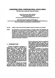
Determination of the structure: The bromine atom position was determined from the threedimensional Patterson function. The heavy atom method was first applied to the centrosymmetric (Okl) projection yielding a trial structure for the bromouridine molecule. Successive Fourier syntheses revealed the complete two-dimensional structure, as shown in Figure 1. A three-dimensional Fourier based on the bromine phases was then easily interpretable, and most of the atomic positions could be determined unambiguously. A second three-dimensional Fourier based on this structure established the complete structure of the nucleoside pair and revealed an extra peak of low electron density. This was tentatively identified as a water molecule. The structure was refined by the method of least squares using a modified version of the full matrix least squares program for the IBM 7090 computer devised by Busing, Martin, and Levy.8 The presence of a water molecule associated with the asymmetric unit was confirmed during the course of refinement; variation of the atom multiplier led to a final value of 0.6 water molecules per nucleoside pair, in good agreement with the value of 0.4 obtained from the crystal density. Successive cycles of individual atom isotropic refinement has reduced the over-all residual factor to 12 per cent. The standard deviation for light atom bond lengths is 410.03 A based on the standard errors of atomic coordinates calculated from the full normal matrix.8
Description of the Structure.-Figure 2 shows the crystal structure viewed down the a axis. Presumed hydrogen-bonding contacts are indicated by dashed lines, and the distances are given. Dotted lines indicate other distances of interest. The existence of a strong hydrogen bond between the bases is clearly indicated between N3 of bromouridine and N7 of adenosine with a length of 2.80 A. A second weak hydrogen bond appears to occur between 02 of bromouridine and N6 of adenosine. The C6-N6-02 angle of 1260 is favorable for hydrogen bonding; however, the distance of 3.10 A is too long. It is possible that the bond is strained by the interactions of the bromouridine 04 and adenosine 05' with an adjacent sugar or that the proximity of the water, 2.90A from 02 of uracil and 2.99 A from N6 of adenine, perturbs the base pairing.
0
0~
c/2
b/2 0
2
3
4A
FIG. 1.-(Okl) Fourier projection of the adenosine-5-bromouridine crystal structure. Contours are drawn at equal but arbitrary levels of electron density. The bromine atom contours are drawn for every fourth level of electron density. The extra peak shown in the lower left-hand corner is a water molecule; its low level of electron density is attributed to incomplete occupancy in the crystal lattice.
BIOCHEMISTRY: HASCHEMEYER AND SOBELL
874
0J«0
2~
1