Curr Infect Dis Rep (2014) 16:399 DOI 10.1007/s11908-014-0399-8
INTRA-ABDOMINAL INFECTIONS, HEPATITIS, AND GASTROENTERITIS (D BOBAK, SECTION EDITOR)
Hepatitis E Virus: Current Concepts and Future Perspectives Harry R. Dalton & Suzan D. Pas & Richie G. Madden & Annemiek A. van der Eijk
# Springer Science+Business Media New York 2014
Abstract For many years, hepatitis E was considered a disease found only in certain developing countries. In these geographical settings, hepatitis E virus (HEV) causes a selflimiting hepatitis in young adults, except in pregnant females, in whom the mortality is 25 %. Our understanding of HEV has changed radically in the past decade. It is now evident that HEV is a threat to global health. This review article considers the current concepts and future perspectives of HEV and its effects on human health, with particular reference to developed countries.
Hepatitis E virus is currently the only species classified within the genus Hepevirus, family Hepeviridae. The hepatitis E virion is nonenveloped and 27–35 nm in size. The viral genome is single-stranded positive sense RNA of ~7.2 Kb. The genome consists of a 5’ m7G-cap, short 5’ untranslated regions (UTRs), three partially overlapping open-reading frames (ORFs) of
positive polarity, a short 3’UTR, and a 3’ polyA tail [1]. ORF1 encodes a nonstructural polyprotein of 1,693 aminoacids, with methyltransferase, papain-like cysteine protease, RNA helicase, and RNA-dependent RNA polymerase activity [2, 3]. A bicistronic 2.2-Kb subgenomic RNA fragment was found to encode both pORF3 and pORF2 in separate reading frames [4]. ORF2 is translated into the viral capsid protein of 660 aminoacids/88 kDa [5]. This multifunctional protein is involved in virion assembly [6•] (encapsidating the viral genome) and host–virus interaction by heparin sulfate proteoglycans (syndecans) [7] and harbors virus-neutralizing epitopes within the aa458-607 region of the P domain [5, 8–10]. ORF3 encodes a 114-aminoacids/13-kDa phosphoprotein, which can interact with Src homology (SH3) domains of host intracellular signaling proteins and bind and inhibit mitogen-activating phosphokinase (MAPK) via proline-rich motifs (PXXP) [4, 11]. These observations and the association of pORF3 to epidermal growth factor receptor (EGFR) [12] suggest that pORF3 regulates transcription by interacting in MAPK/ERK and JAK/STAT pathways [13], thereby hampering immune activation. In addition, pORF3 is thought to play an important role in virion release (budding) by associating with lipids of the vacuolar protein sorting pathway [14].
This article is part of the Topical Collection on Intra-abdominal Infections, Hepatitis, and Gastroenteritis
Laboratory Testing
Keywords Hepatitis E . Zoonosis . Hepatitis . Chronic liver disease . Epidemiology
Basic Virology
H. R. Dalton (*) : R. G. Madden Cornwall Gastrointestinal Unit, Royal Cornwall Hospital Trust, TR1 3LJ Truro, UK e-mail:
[email protected] H. R. Dalton European Centre for the Environment and Human Health, University of Exeter Medical School, Truro, UK S. D. Pas : A. A. van der Eijk Department of Viroscience, Erasmus MC, University Medical Center Rotterdam, Rotterdam, The Netherlands
Confirmation of HEV infection relies on the detection of HEV-specific serum IgM and IgG antibodies and/or HEV RNA testing in serum or feces [15]. In addition, an HEV antigen detection assay may be of use if HEV RNA testing is not available and has a sensitivity of 60 % and a specificity of 86 % [16]. HEV has proven difficult to culture and is not used for diagnostic purposes, and real-time PCR techniques are more sensitive than virus culture. In immunocompromised patients, antibody production may be delayed or absent [17,
399, Page 2 of 10
Curr Infect Dis Rep (2014) 16:399
18], and so detection of HEV RNA is essential to diagnosing HEV infection in this patient group [19]. In immunocompetent individuals, there is a narrow window (mean 28, range 17–48 days) [20] in which HEV RNA can be detected in serum or feces. Therefore, a combination of serology and PCR is required to diagnose HEV infection in such patients. The diagnosis of reinfection can be problematic and may be more common than currently appreciated. Such patients are typically anti-HEV IgM negative, and so the diagnosis depends on the demonstration of HEV RNA by PCR [21••]. When patients with reinfection present after the viraemic period, they will be PCR and anti-HEV IgM negative but anti-HEV IgG positive. These results are indistinguishable from those found in previous/distant infection, and so the only way of making a diagnosis of reinfection in this situation is by demonstrating IgG antibodies of high avidity and/or a rising IgG-titer in a convalescent sample. These additional serological assays are rarely performed in routine clinical practice. HEV Serology Performance of anti-HEV IgM and IgG assays have been compared [22–29], and there is discordance between the results of some assays. Some commercially available antiHEV IgG assays have low sensitivities, particularly to distant infection of over 1 year [22]. This almost certainly has resulted in an underestimation of seroprevalence in many of the earlier studies (Table 1). Differences in assay design are considerable and include (1) the format used, either (in)direct or one μ Table 1 HEV viraemia and seroprevalence in blood donors
The above data are from blood donors, with the exception of those marked *, which are from “healthly adults.”
chain capture assay; (2) the genotype of the recombinant antigens used (most ELISAs use genotype 1 and 2 antigens; only one assay currently uses genotype 1 and 3 antigens); (3) the corresponding structural region of the recombinant antigens, which are all ORF2+ORF3); and (4) the use of dimers in antigen presentation. An international effort to validate existing serology is urgently needed. HEV Molecular Testing Until recently, molecular testing for HEV had also been problematic, since a range of in-house assays have been used. A comparison study [30] among 20 European laboratories showed a wide variability (up to 3log) in quantitative assays. To overcome this, a WHO international standard (genotype 3) for nucleic acid testing has been available since 2011 [31••]. The recently marketed commercial HEV viral load assays appear to have a good sensitivity (20–100 IU/ml) and specificity [32, 33]. HEV genotyping is of most importance for research and epidemiologic purposes [34]. Conventional RT-PCR products are used for genotyping HEV on different genome segments. Partial ORF2 sequences are historically widely used, although Zhai et al. [35] showed that a 306-bp region of RdRp (ORF1) was statically representative for complete HEV genome flanked by conserved primer sites. The widely accepted classification of HEV genotypes is currently defined by 20 % nucleotide divergence of the ORF2, but the utility of HEV subtypes is a subject of debate. Inconsistencies of the previously suggested 24 subtypes [36] have been observed [37,
Country
Blood Donors HEV RNA Positive
HEV IgG Seroprevalence
Assay
Reference
SW France
No data
52.5 % 16 %*
Wantai Adaltis
Germany
1:1200 1:4525 29.5 %* 18.0 %* 4.5 %* 27.0 %* 1.1 %*
Wantai Mikrogen MP diagnostics Wantai Abbott
12.0 %* 5.3 %*
Wantai Abbott
Mansuy et al., 2011 [45••] Mansuy et al., 2008 [97] Vollmer et al., 2012 [98] Baylis et al., 2012 [99•] Wenzel et al., 2013 [23] Wenzel et al., 2013 [23] Wenzel et al., 2013 [23] Slot et al., 2013 [50•] Zaaijer et al., 1993 [100] Ijaz et al., 2012 [101] Beale et al., 2011 [51] Bernal et al., 1996 [102] Baylis et al., 2012 [99] Olsen et al., 2006 [103]
Netherlands
1:2671
England
1:7000
Sweden
1:7986
Scotland USA
1:14520 Nil
9.2 %*
Abbott
4.7 %*
Wantai
16.0 %*
Wantai
Cleland et al., 2013 [104] Baylis et al., 2012 [99•] Xu et al., 2012 [105] Xu et al., 2012 [105]
Curr Infect Dis Rep (2014) 16:399
38], probably due to the small part of ORF2 not being representative for the complete genome sequence variation. A recent phylogenetic study [37], on the basis of complete genomes, suggests that the current taxonomy should be refined. The suggested new taxonomy would include the four established genotypes (1–4), with only genotype 3 separated into three subgroups (3.1–3.3), a novel genotype 5 (Japanese wild boars), and 5 new genera for ferret, rat, chicken/avian, and bat HEV. The authors also suggested not classifying cutthroat trout virus [39] within the Hepeviridae.
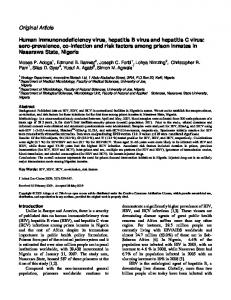
Epidemiology of HEV in Developed Countries HEV genotype 3 is found in pigs worldwide, and genotype 4 is found in pigs in China and Japan. Infected swine herds are asymptomatic; however, they excrete extremely large quantities of HEV in their feces. The pig is considered to be a primary host, and there is a close similarity between HEV strains obtained from pigs and humans. HEV is highly infectious in pig herds, since the basic reproductive ratio of the virus (R0) is up to 8; so once HEV has been introduced into a pig herd, infection is almost universal [40]. Of fattening pigs studied in the Netherlands, 73 % were found to be excreting HEV in their stool [40]. HEV has also been found in many other animals, including deer and rabbits [41•], which can also be a source of human infection. In comparison with pigs, they are a less important reservoir for human infection. HEV has also been documented in a range of other mammals, including rats, bats, and ferrets. It is uncertain whether these animal reservoirs hold a negative consequence for human health. For example, rat HEV is not transmissible to rhesus monkeys, indicating that it is unlikely to be pathogenic to humans [42]. There are a number of possible routes of infection (see Fig. 1), but in most cases of hepatitis E contracted in humans, it is not possible to determine the source or route of infection. However, consumption of infected pork products has been well documented. HEV has been found in retail pork in grocery stores in several European countries, Japan, and the U.S. In 2012, HEV genotype 3 was identified throughout the human food chain in the U.K. and was found to contaminate 10 % of retail sausages that were tested [43••]. HEV requires cooking temperatures of 71 °C for 20 min to inactivate it, which is much longer than sausages usually get cooked [44]. HEV has also been found in foods such as a traditional French air-dried pig liver sausage, which is not cooked [45••]. A recent case–control study from the U.K. suggests that processed pork products, such as ham and pork pies, might also be a source of infection [46]. A route of infection that may be important is via infected water (see Fig. 1), since HEV has been found in watercourses and seawater [47]. In southwest England, 50 % of the cases of hepatitis E were found to live within 2 km of the coast [48]. It
Page 3 of 10, 399
is thought that this could relate to recreational use with water contaminated with HEV. In addition, a recent study has found HEV in shellfish [49]. It was previously documented that the prevalence of antiHEV IgG in many developed countries was low, ranging from 1 % to 2 %. These studies have a major flaw, since they used IgG assays of poor sensitivity. More recent studies, using more sensitive assays, have produced higher estimates of seroprevalence, with rates of 12 % in England, 27 % in the Netherlands [50•], 29 % in Germany [23], and 52 % in southern France [45••]. These data suggest that HEV is hyperendemic in southwest France, and these very high seroprevalence rates are congruent with worryingly large numbers of blood donors who are viraemic at the time of donation (Table 1). The incidence of hepatitis E has been poorly documented but almost certainly varies between and within countries and over time. The incidence in the U.K. has been estimated at 0.2 % [51]. In the U.S., the Incidence is 0.7 % [52]. Since the population of the U.S. is 316 million, this implies that there are over 2.2 million infections per year, nearly all of which are asymptomatic or unrecognized, since there are currently no FDA-approved HEV diagnostics in the U.S.
Acute HEV in Developed Countries: Clinical Aspects Zoonotic autochthonous hepatitis E has been found in [21••] Japan (genotypes 3 and 4) Europe, and New Zealand, and most recently, a small number of cases have been found in the U.S. [53]. The majority of cases of hepatitis E in Europe are caused by HEV genotype 3. A recent study has documented HEV genotype 4 in European pigs, and there have been two clusters of human infection caused by genotype 4 in France and Italy [54, 55]. Genotypes 3 and 4 produce a similar clinical picture, and excess mortality in pregnancy is not seen with either genotype [56]. In developed countries, hepatitis E appears to cause symptomatic hepatitis more commonly in middle-aged/ elderly males (median age 60 years, M:F ratio 3:1). The reason for this observation is uncertain but might be explained by subclinical hepatic fibrosis in such individuals [57]. With the exception of neurological syndromes (see below), the symptoms of hepatitis E are nonspecific and indistinguishable from other causes of viral hepatitis (Table 2). Typical laboratory parameters and the differential diagnosis [58, 59•] are shown in Table 3. Most cases recover within 4–6 weeks; however, in patients with underlying chronic liver disease, the prognosis is poor, with excess mortality from hepatic failure [21••].
Chronic HEV: Clinical Aspects Chronic HEV infection caused by HEV genotype 3 is defined by HEV RNA in plasma and/or stool detected by RT-PCR for
399, Page 4 of 10
Curr Infect Dis Rep (2014) 16:399
Fig. 1 Possible, probable, and definite routes of transmission of hepatitis E virus
6 months or more. To date, no chronic hepatitis has been reported with genotypes 1, 2, or 4. Chronic infection has been reported in immunocompromised patients, with most cases in solid organ transplant recipients, HIV-infected patients, and recipients of allogeneic stem cell transplantation (alloHSCT) [19, 60, 61•, Table 2 Acute autochthonous hepatitis E: symptoms Common
Less Common
No symptoms* Jaundice Anorexia Lethargy Abdominal pain Vomiting Fever
Myalgia Pruritis Weight loss Headaches Arthralgia Neurological**
Note. The data in this table are derived from a large number of sporadic cases of autochthonous hepatitis E documented in southwest England (1999–2013) [72]. *Asymptomatic infection is very common, and accounts for the large numbers of blood donors who are viraemic at the time of donation (Table 1). In an outbreak of hepatitis E genotype 3 on a cruise ship, 67 % of patients had no symptoms [106]. **5 % of patients present with a range of neurological symptoms, including Guillain-Barre syndrome and brachial neuritis [78••].
62, 63]. A prevalence of 1 %–3 % of hepatitis E viremia in recipients of solid organ transplants has been reported, with 47 %–83 % of the patients developing chronic hepatitis [19, 63, 64]. Most immunocompromised patients have no clinical symptoms at presentation but usually show modest transaminitis [61, 65•, 66]. Riezebos-Brilman et al. recently studied the clinical course of HEV infection in 34 immunosuppressed patients. The median ALT levels in patients with a chronic infection were significantly lower (106 U/l), as compared with the levels found in patients who had spontaneous viral clearance within 6 months (1,087 U/l) (p=.002) [67]. Similar findings were reported from a larger cohort of mainly European transplant recipients [65•]. Several case reports have described chronic HEV infections in HIV-infected patients with low CD4 counts (2,000 and may be higher • HAV is now rare, and the age of infected individuals is increasing. It can occur in the elderly.
HEV
Autoimmune hepatitis EBV
Acute HBV HAV
Note. The data in this table are derived from >2,000 consecutive patients attending the Jaundice Hotline clinic, Truro, England (1998–2012) [59•] and are listed in order of incidence. ALT: alanine aminotransferase, normal range: 3–35 IU/L. EBV: Epstein Barr virus; HBV: hepatitis B virus; HAV: hepatitis A virus. *In studies from the U.K. [58] and U.S. [107] of cohorts of patients with ‘criterion-referenced’ drug-induced liver injury, 13 % and 3 % (respectively) were found to have hepatitis E caused by HEV genotype 3.
infection of 2.4 % was reported [61•]. This study had a median follow-up time of 41 months, and chronic hepatitis occurred in five out of eight acute HEV cases (63 %). In the HEV-infected patients, liver enzyme abnormalities were thought to be related to hepatic graft-versus-host disease in 5 (63 %) patients, and druginduced liver injury in 3 (38 %) patients. One patient was diagnosed with HEV reactivation after a preceding infection prior to alloHSCT. This is the second case of HEV reactivation after alloHSCT described so far in the literature [61•, 71]. Diagnosis of HEV in these patients is hampered by relatively low peak aminotransferase levels, as compared with nonimmunocompromised patients [72], which may be explained by intensive immunosuppressive therapy suppressing inflammation. In recipients of transplanted solid organs, the use of tacrolimus, rather than cyclosporine A, as a main immunosuppressant and a low platelet count at diagnosis of HEV infection are two independent variables that predict the development of
Page 5 of 10, 399
chronic HEV infection [65•]. Progression to chronic HEV in alloHSCT patients is less well characterized but might be explained by impaired immune reconstitution, including insufficient lymphocyte recovery, which are well-known risk factors for posttransplantation infections [73–75]. In particular, impaired reconstitution of CD4+ and CD8+ T-cells predisposes for infectious morbidity [76]. Rapid progression of fibrosis and cirrhosis in chronically infected HEV transplant recipients has been described [77]. Liver histology in chronic HEV-infected heart transplant recipients showed advanced fibrosis within 2 years after infection [66]. Biopsies of patients with chronic HEV showed typical signs of acute viral hepatitis with inflammatory activity, councilman bodies, and acidophilic degeneration [61•, 66]. However, no distinct pathognomic features differentiating HEV infections from hepatitis B or C have been identified.
Extrahepatic Manifestations of HEV There have been a number of case reports of HEV being associated with extra-hepatic pathology. Hepatitis E causes a number of extra-hepatic manifestations, including pancreatitis, acute thrombocytopenia, aplastic anaemia, and renal disease [21••]. Of cases of hepatitis E, 5 % present with a neurological illness [78••]. HEV-associated neurological injury has been documented in both acute and chronic HEV infection, and in some cases, HEV has been found in the cerebrospinal fluid [79]. There is considerable current research interest in the role of HEV in Guillain-Barre syndrome and brachial neuritis. A recent case–control study showed that 10 (5 %) of 201 GuillainBarre syndrome patients from The Netherlands had hepatitis E infection at the start of their illness [80••]. The clinical features and outcome in these patients were similar to those found in Guillain-Barre syndrome cases not associated with HEV. A further study of U.K. and Dutch patients with brachial neuritis showed that 5 of 47 (10 %) of cases had an associated HEV genotype 3 infection at the start of their illness [81••]. Patients affected were in the 30- to 40-year range and had bilateral involvement of the brachial plexus. All of the above patients were anicteric with only mildly raised ALTs (in some cases, the ALT was normal), and some were viraemic at presentation. The latter observation raises the possibility of whether early antiviral therapy might improve the natural history of either condition. The pathophysiological mechanisms of HEV-associated neurological injury are uncertain. Neurological features dominate the clinical picture, and so the diagnosis is easily overlooked.
HEVand the Blood Supply HEV RNA has been found in donated blood in a number of countries (Table 1). Several reports have shown that hepatitis
399, Page 6 of 10
E virus can be transmitted via blood transfusion [82–86], although risk factors for transmission—for example, viral load or recipient’s immune status—are not yet known. The clinical consequences of using blood and blood products contaminated with HEV are also uncertain. Currently, blood donors are not routinely screened for HEV. Whether they should be is the subject of lively debate and on-going research.
Treatment and Prevention Acute HEV Most cases of acute hepatitis E infection have a self-limiting illness requiring no treatment. Patients with severe hepatitis and underlying chronic liver disease have a poor prognosis; several of these patients have been treated successfully with ribavirin [87]. Chronic HEV Treatment of HEV infection after transplantation includes reduction of immunosuppressive therapy and treatment with antiviral agents. Pegylated interferon-alpha-2a (Peg-IFN-α-2a) and oral ribavirin have been successfully used for treating HEV infection in immunocompromised patients [61•, 66, 88–92]. Reducing the dose of immunosuppressive drugs targeting T cells can lead to HEV clearance in up to one third of patients [93], Other studies have shown more limited utility of reducing immunosuppressive therapy, since one study showed that only 2 out of 18 solid organ transplant recipients were able to achieve viral clearance using this approach [65•, 67, 93]. A possible explanation for this may be that the evolution of the HEV infection could be related to the type of solid organ transplant and level of immunosuppression employed [67]. Peg-IFN-α-2a has been successfully used for treating HEV infection in liver and kidney transplant recipients, as well as an HIV-infected patient [17, 89, 90, 92].Tapering immunosuppressive drugs or treatment with Peg-IFN-α-2a is not always possible or desirable, due to the high risk for rejection, which may lead to chronic allograft dysfunction and death. For these patients, treatment with ribavirin should be considered. Rapid clearance of HEV RNA in plasma with normalizing ALT levels are observed after the start of therapy, and a sustained virological response can be induced in patients with chronic HEV infection [61•, 66, 88, 91, 94]. The optimal daily dose and treatment duration of ribavirin are unknown. In case reports and small case series, sustained viral response has been described with daily dosages between 200 and 1,200 mg [66, 95•]. Treatment durations shorter than 3 months and dose reduction of ribavirin have been associated with viral relapses or breakthrough [88]. No viral relapse was observed following 5 months of ribavirin therapy for treatment of chronic HEV in 9 transplant recipients [88, 94].
Curr Infect Dis Rep (2014) 16:399
Conclusions and Future Directions A key characteristic of zoonotic HEV is its ability to hide from human scrutiny. Biological time-clock studies show that HEV diverged into its four genotypes hundreds of years ago, suggesting that HEV genotypes 3 and 4 have remained hidden in its primary host, the pig, for centuries. It wasn’t until as recently as 1997 that HEV was first found in pigs, in the U.S. [96]. More recently, HEV has been found in asymptomatic blood donors and chronic infection in the immunosuppressed. It is almost certain that HEV has been circulating unnoticed in these populations for decades. One key reason for the previous underestimation of the effect of zoonotic HEV on human health is the poor quality of first-generation diagnostics. Initial studies in developed countries grossly underestimated the anti-HEV IgG seroprevalence, which led to the mistaken notion that it was of little relevance. With the standardization of molecular studies, our ability to detect HEV has improved considerably, but there remains an urgent need to standardize serological assays and develop reference materials for global use. In the U.S.; there are no FDA-approved diagnostic assays for use in humans. This issue needs to be addressed as a priority. Now that HEV is on our “radar,” its effects on human health are beginning to emerge. Hepatitis E has always been considered to be a primarily hepatotropic virus and can cause acute hepatitis in the immunocompetent and acute and chronic infection in the immunosuppressed. However, it is now clear that HEV can cause a range of extrahepatic manifestations, the most interesting of which is its association with a number of neurological syndromes. In such patients the degree of hepatological injury, as judged by the serum ALT, is usually minor or even absent altogether. HEV has been documented in a minority of patients with Guillan-Barre syndrome and brachial neuritis at the onset of neurological symptoms. The role of HEV in other neurological illnesses requires urgent investigation. Perhaps, in due course, zoonotic HEV might even come to be recognized as primarily a neurotropic, rather than hepatotropic. pathogen.
Compliance with Ethics Guidelines Conflict of Interest Harry R. Dalton consults for GSK, Wantai, and Aptalis. Dalton has received honoraria from Merck and GFE Blut MBh, as well as travel accommodations/reimbursements from GSK, Merck, GFE Blut MBh, and Wantai. Richie G. Madden has no conflicts. Suzan D. Pas has received grants from the Dutch government, the Netherlands Genomics Initiative and the European Community Seventh Framework Programme. Pas received travel reimbursements from Mikrogen. Annemiek van der Eijk received grants from the Dutch government and the Netherlands Genomics Initiative. Human and Animal Rights and Informed Consent This article does not contain any studies with human or animal subjects performed by the authors.
Curr Infect Dis Rep (2014) 16:399
Page 7 of 10, 399
References
16.
Papers of particular interest, published recently have been highlighted as: • Of importance •• Of major importance
17.
1.
2.
3.
4.
5.
6.•
7.
8.
9.
10.
11.
12.
13.
14.
15.
Tam AW, Smith MM, Guerra ME, Huang CC, Bradley DW, Fry KE, et al. Hepatitis E virus (HEV): molecular cloning and sequencing of the full-length viral genome. Virology. 1991;185:120–31. Koonin EV, Gorbalenya AE, Purdy MA, Rozanov MN, Reyes GR, Bradley DW. Computer-assisted assignment of functional domains in the nonstructural polyprotein of hepatitis E virus: delineation of an additional group of positive-strand RNA plant and animal viruses. Proc Natl Acad Sci U S A. 1992;89:8259–63. Reyes GR, Purdy MA, Kim JP, Luk KC, Young LM, Fry KE, et al. Isolation of a cDNA from the virus responsible for enterically transmitted non-A, non-B hepatitis. Science. 1990;247:1335–9. Graff J, Torian U, Nguyen H, Emerson SU. A bicistronic subgenomic mRNA encodes both the ORF2 and ORF3 proteins of hepatitis E virus. J Virol. 2006;80:5919–26. Yamashita T, Mori Y, Miyazaki N, Cheng RH, Yoshimura M, Unno H, et al. Biological and immunological characteristics of hepatitis E virus-like particles based on the crystal structure. Proc Natl Acad Sci U S A. 2009;106:12986–91. Li SW, Zhang J, He ZQ, Gu Y, Liu RS, Lin J, et al. Mutational analysis of essential interactions involved in the assembly of hepatitis E virus capsid. J Biol Chem. 2005;280:3400–6. This is the first structural and funtional study on hepatitis E virus using crystallography, giving insight on virus–host interactions. Kalia M, Chandra V, Rahman SA, Sehgal D, Jameel S. Heparan sulfate proteoglycans are required for cellular binding of the hepatitis E virus ORF2 capsid protein and for viral infection. J Virol. 2009;83:12714–24. Tang X, Yang C, Gu Y, Song C, Zhang X, Wang Y, et al. Structural basis for the neutralization and genotype specificity of hepatitis E virus. Proc Natl Acad Sci U S A. 2011;108:10266–71. Zhang J, Li SW, Wu T, Zhao Q, Ng MH, Xia NS. Hepatitis E virus: neutralizing sites, diagnosis, and protective immunity. Rev Med Virol. 2012;22:339–49. Meng J, Dai X, Chang JC, Lopareva E, Pillot J, Fields HA, et al. Identification and characterization of the neutralization epitope(s) of the hepatitis E virus. Virology. 2001;288:203–11. Korkaya H, Jameel S, Gupta D, Tyagi S, Kumar R, Zafrullah M, et al. The ORF3 protein of hepatitis E virus binds to Src homology 3 domains and activates MAPK. J Biol Chem. 2001;276:42389–400. Chandra V, Kar-Roy A, Kumari S, Mayor S, Jameel S. The hepatitis E virus ORF3 protein modulates epidermal growth factor receptor trafficking, STAT3 translocation, and the acute-phase response. J Virol. 2008;82:7100–10. Kar-Roy A, Korkaya H, Oberoi R, Lal SK, Jameel S. The hepatitis E virus open reading frame 3 protein activates ERK through binding and inhibition of the MAPK phosphatase. J Biol Chem. 2004;279:28345–57. Nagashima S, Takahashi M, Jirintai, Tanaka T, Yamada K, Nishizawa T, et al. A PSAP motif in the ORF3 protein of hepatitis E virus is necessary for virion release from infected cells. J Gen Virol. 2011;92:269–78. Huang S, Zhang X, Jiang H, Yan Q, Ai X, Wang Y, et al. Profile of acute infectious markers in sporadic hepatitis E. PLoS One. 2010;5:e13560.
18.
19.
20.
21.••
22.
23.
24.
25.
26.
27.
28.
29.
30.
31.••
Gupta E, Pandey P, Pandey S, Sharma MK, Sarin SK. Role of hepatitis E virus antigen in confirming active viral replication in patients with acute viral hepatitis E infection. J Clin Virol 2013. Legrand-Abravanel F, Kamar N, Sandres-Saune K, Garrouste C, Dubois M, Mansuy JM, et al. Characteristics of autochthonous hepatitis E virus infection in solid-organ transplant recipients in France. J Infect Dis. 2010;202:835–44. Pas SD, Streefkerk HRA, Pronk M, de Man RA, Beersma MF, Osterhaus ADME, et al. Diagnostic performance of selected commercial HEV IgM and IgG ELISAs for immunocompromised and immunocompetent patients. Journal of Clinical Virology Accepted. Pas SD, de Man RA, Mulders C, Balk AH, van Hal PT, Weimar W, et al. Hepatitis E Virus Infection among Solid Organ Transplant Recipients, the Netherlands. Emerg Infect Dis. 2012;18:869–72. Dalton HR, Bendall R, Ijaz S, Banks M. Hepatitis E: an emerging infection in developed countries. Lancet Infect Dis. 2008;8:698– 709. Kamar N, Bendall R, Legrand-Abravanel F, Xia NS, Ijaz S, Izopet J, et al. Hepatitis E. Lancet. 2012;379:2477–88. Comprehensive review of hepatitis E from a multinational group of opinion leaders. This is a “must read” for those interested in the field. Bendall R, Ellis V, Ijaz S, Ali R, Dalton H. A comparison of two commercially available anti-HEV IgG kits and a re-evaluation of anti-HEV IgG seroprevalence data in developed countries. J Med Virol. 2010;82:799–805. Wenzel JJ, Preiss J, Schemmerer M, Huber B, Jilg W. Test performance characteristics of Anti-HEV IgG assays strongly influence hepatitis E seroprevalence estimates. J Infect Dis. 2013;207: 497–500. Herremans M, Bakker J, Duizer E, Vennema H, Koopmans MP. Use of serological assays for diagnosis of hepatitis E virus genotype 1 and 3 infections in a setting of low endemicity. Clin Vaccine Immunol. 2007;14:562–8. Elkady A, Tanaka Y, Kurbanov F, Hirashima N, Sugiyama M, Khan A, et al. Evaluation of anti-hepatitis E virus (HEV) immunoglobulin A in a serological screening for HEV infection. J Gastroenterol. 2007;42:911–7. Legrand-Abravanel F, Thevenet I, Mansuy JM, Saune K, Vischi F, Peron JM, et al. Good performance of immunoglobulin M assays in diagnosing genotype 3 hepatitis E virus infections. Clin Vaccine Immunol. 2009;16:772–4. Drobeniuc J, Meng J, Reuter G, Greene-Montfort T, Khudyakova N, Dimitrova Z, et al. Serologic assays specific to immunoglobulin M antibodies against hepatitis E virus: pangenotypic evaluation of performances. Clin Infect Dis. 2010;51:e24–7. Schnegg A, Burgisser P, Andre C, Kenfak-Foguena A, Canellini G, Moradpour D, et al. An analysis of the benefit of using HEV genotype 3 antigens in detecting anti-HEV IgG in a European population. PLoS One. 2013;8:e62980. Pas SD, Streefkerk HRA, Pronk M, de Man RA, Beersma MF, Osterhaus ADME, et al. Diagnostic performance of selected commercial HEV IgM and IgG ELISAs for immunocompromised and immunocompetent patients. submitted. Baylis SA, Hanschmann KM, Blumel J, Nubling CM. Standardization of hepatitis E virus (HEV) nucleic acid amplification technique-based assays: an initial study to evaluate a panel of HEV strains and investigate laboratory performance. J Clin Microbiol. 2011;49:1234–9. Baylis SA, Blumel J, Mizusawa S, Matsubayashi K, Sakata H, Okada Y, et al. World health organization international standard to harmonize assays for detection of hepatitis e virus RNA. Emerg Infect Dis. 2013;19:729–35. Previous studies have shown very little interlaboratory concordance for HEV quantitative PCR. This paper defines WHO standards to harmonise HEV PCR assays and is a key paper in terms of laboratory diagnosis.
399, Page 8 of 10 Abravanel F, Chapuy-Regaud S, Lhomme S, Dubois M, Peron JM, Alric L, et al. Performance of two commercial assays for detecting hepatitis E virus RNA in acute or chronic infections. J Clin Microbiol. 2013;51:1913–6. 33. Mokhtari C, Marchadier E, Haim-Boukobza S, Jeblaoui A, Tesse S, Savary J, et al. Comparison of real-time RT-PCR assays for hepatitis E virus RNA detection. J Clin Virol. 2013;58:36–40. 34. Aggarwal R. Hepatitis E: clinical presentation in disease-endemic areas and diagnosis. Semin Liver Dis. 2013;33:30–40. 35. Zhai L, Dai X, Meng J. Hepatitis E virus genotyping based on full-length genome and partial genomic regions. Virus Res. 2006;120:57–69. 36. Lu L, Li C, Hagedorn CH. Phylogenetic analysis of global hepatitis E virus sequences: genetic diversity, subtypes and zoonosis. Rev Med Virol. 2006;16:5–36. 37. Oliveira-Filho EF, Konig M, Thiel HJ. Genetic variability of HEV isolates: inconsistencies of current classification. Vet Microbiol. 2013;165:148–54. 38. Fu H, Wang L, Zhu Y, Geng J, Li L, Wang X, et al. Analysing complete genome sequence of swine hepatitis E virus (HEV), strain CHN-XJ-SW13 isolated from Xinjiang, China: putative host range, and disease severity determinants in HEV. Infect Gen Evol J Mol Epidemiol Evol Genetics Infect Dis. 2011;11:618–23. 39. Batts W, Yun S, Hedrick R, Winton J. A novel member of the family Hepeviridae from cutthroat trout (Oncorhynchus clarkii). Virus Res. 2011;158:116–23. 40. Backer JA, Berto A, McCreary C, Martelli F, van der Poel WH. Transmission dynamics of hepatitis E virus in pigs: estimation from field data and effect of vaccination. Epidemics. 2012;4:86–92. 41.• Izopet J, Dubois M, Bertagnoli S, Lhomme S, Marchandeau S, Boucher S, et al. Hepatitis E virus strains in rabbits and evidence of a closely related strain in humans, France. Emerg Infect Dis. 2012;18:1274–81. HEV is demonstrated in farmed and wild rabbits in France and may have the potential to cause human disease. 42. Purcell RH, Engle RE, Rood MP, Kabrane-Lazizi Y, Nguyen HT, Govindarajan S, et al. Hepatitis E virus in rats, Los Angeles, California, USA. Emerg Infect Dis. 2011;17:2216–22. 43.•• Berto A, Martelli F, Grierson S, Banks M. Hepatitis E virus in pork food chain, United Kingdom, 2009-2010. Emerg Infect Dis. 2012;18:1358–60. A comprehensive study of HEV in pig products in the human food chain from slaughter to retail stores used by the public. HEV is demonstrated in every step of this chain and was found in 10% of pork sausages that were tested. 44. Barnaud E, Rogee S, Garry P, Rose N, Pavio N. Thermal inactivation of infectious hepatitis E virus in experimentally contaminated food. Appl Environ Microbiol. 2012;78:5153–9. 45.•• Mansuy JM, Bendall R, Legrand-Abravanel F, Saune K, Miedouge M, Ellis V, et al. Hepatitis E virus antibodies in blood donors, France. Emerg Infect Dis. 2011;17:2309–12. Using a highly sensitive assay, this paper shows that the HEV IgG seroprevalence in blood donors from the Southwest of France was 52%. This is much higher than previous estimates and suggests that HEV is hyperendemic in this region. These findings are congruent with the extrememy high incidence of hepatitis E that has been found in the Toulouse transplant population. 46. Said B, Ijaz S, Chand MA, Kafatos G, Tedder R, Morgan D. Hepatitis E virus in England and Wales: indigenous infection is associated with the consumption of processed pork products. Epidemiol Infect 2013:1-9. 47. Ishida S, Yoshizumi S, Ikeda T, Miyoshi M, Goto A, Matsubayashi K, et al. Detection and molecular characterization of hepatitis E virus in clinical, environmental and putative animal sources. Arch Virol. 2012;157:2363–8. 48. Madden RG, Hunter JG, Stone AM, Thornton C, Barlow M, Lewis J, et al. Locally acquired hepatitis E. Geographical
Curr Infect Dis Rep (2014) 16:399
32.
49.
50.•
51.
52.
53.
54.
55.
56.
57.
58.
59.•
60.
61.•
62.
63.
64.
65.•
clustering and environmental factors: a nested case control study. J Hepatol. 2012;56:S64–5. Crossan C, Baker PJ, Craft J, Takeuchi Y, Dalton HR, Scobie L. Hepatitis E virus genotype 3 in shellfish, United Kingdom. Emerg Infect Dis. 2012;18:2085–7. Slot E, Hogema B, Riezebos-Brilman A, Kok T, Molier M, Zaaijer H. Silent hepatitis E virus infection in Dutch blood donors, 2011 to 2012. Euro Surveill 2013; 18. HEV was denonstrated in 1 of 2,671 blood donors in the Netherlands by PCR. Beale MA, Tettmar K, Szypulska R, Tedder RS, Ijaz S. Is there evidence of recent hepatitis E virus infection in English and North Welsh blood donors? Vox Sang. 2011;100:340–2. Faramawi MF, Johnson E, Chen S, Pannala PR. The incidence of hepatitis E virus infection in the general population of the USA. Epidemiol Infect. 2011;139:1145–50. Drobeniuc J, Greene-Montfort T, Le NT, Mixson-Hayden TR, Ganova-Raeva L, Dong C, et al. Laboratory-based surveillance for hepatitis E virus infection, United States, 2005-2012. Emerg Infect Dis. 2013;19:218–22. This is the first structural and funtional study on hepatitis E virus using crystallography, giving insight on virus–host interactions.. Colson P, Romanet P, Moal V, Borentain P, Purgus R, Benezech A, et al. Autochthonous infections with hepatitis E virus genotype 4, France. Emerg Infect Dis. 2012;18:1361–4. Garbuglia AR, Scognamiglio P, Petrosillo N, Mastroianni CM, Sordillo P, Gentile D, et al. Hepatitis E virus genotype 4 outbreak, Italy, 2011. Emerg Infect Dis. 2013;19:110–4. Anty R, Ollier L, Peron JM, Nicand E, Cannavo I, Bongain A, et al. First case report of an acute genotype 3 hepatitis E infected pregnant woman living in South-Eastern France. J Clin Virol. 2012;54:76–8. Dalton HR, Bendall RP, Rashid M, Ellis V, Ali R, Ramnarace R, et al. Host risk factors and autochthonous hepatitis E infection. Eur J Gastroenterol Hepatol. 2011;23:1200–5. Dalton HR, Fellows HJ, Stableforth W, Joseph M, Thurairajah PH, Warshow U, et al. The role of hepatitis E virus testing in druginduced liver injury. Aliment Pharmacol Ther. 2007;26:1429–35. Vine LJ, Shepherd K, Hunter JG, Madden R, Thornton C, Ellis V, et al. Characteristics of Epstein-Barr virus hepatitis among patients with jaundice or acute hepatitis. Aliment Pharmacol Ther. 2012;36: 16–21. HEV is the commonest cause of acute viral hepatitis in Cornwall, U.K. Dalton HR, Bendall RP, Keane FE, Tedder RS, Ijaz S. Persistent carriage of hepatitis E virus in patients with HIV infection. N Engl J Med. 2009;361:1025–7. Versluis J, Pas SD, Agteresch HJ, de Man RA, Maaskant J, Schipper ME, et al. Hepatitis E virus: an underestimated opportunistic pathogen in recipients of allogeneic hematopoietic stem cell transplantation. Blood. 2013;122:1079–86. This is the first cohort study that describes chronic hepatitis E infections and reactivation of hematopoietic stem cell transplant recipients and the clinical presentation. Haagsma EB, Niesters HG, van den Berg AP, Riezebos-Brilman A, Porte RJ, Vennema H, et al. Prevalence of hepatitis E virus infection in liver transplant recipients. Liver Transpl. 2009;15: 1225–8. Kamar N, Selves J, Mansuy JM, Ouezzani L, Peron JM, Guitard J, et al. Hepatitis E virus and chronic hepatitis in organ-transplant recipients. N Engl J Med. 2008;358:811–7. Haagsma EB, van den Berg AP, Porte RJ, Benne CA, Vennema H, Reimerink JH, et al. Chronic hepatitis E virus infection in liver transplant recipients. Liver Transpl. 2008;14:547–53. Kamar N, Garrouste C, Haagsma EB, Garrigue V, Pischke S, Chauvet C, et al. Factors associated with chronic hepatitis in patients with hepatitis E virus infection who have received solid organ transplants. Gastroenterology. 2011;140:1481–9. This study provides the progression, outcomes, and factors associated with
Curr Infect Dis Rep (2014) 16:399 development of chronic HEV infection in recipients of transplanted solid organs. 66. Koning L, Pas SD, de Man RA, Balk AH, de Knegt RJ, ten Kate FJ, et al. Clinical implications of chronic hepatitis E virus infection in heart transplant recipients. J heart Lung Transplant Off Pub Int Soc Heart Transplant. 2013;32:78–85. 67. Riezebos-Brilman A, Verschuuren EA, van Son WJ, van Imhoff GW, Brugemann J, Blokzijl H, et al. The clinical course of hepatitis E virus infection in patients of a tertiary Dutch hospital over a 5-year period. J Clin Virol 2013. 68. Colson P, Kaba M, Moreau J, Brouqui P. Hepatitis E in an HIVinfected patient. J Clin Virol. 2009;45:269–71. 69. Abravanel F, Mansuy JM, Huynh A, Kamar N, Alric L, Peron JM, et al. Low risk of hepatitis E virus reactivation after haematopoietic stem cell transplantation. J Clin Virol. 2012;54:152–5. 70. Koenecke C, Pischke S, Heim A, Raggub L, Bremer B, Raupach R, et al. Chronic hepatitis E in hematopoietic stem cell transplant patients in a low-endemic country? Transpl Infect Dis. 2012;14:103–6. 71. le Coutre P, Meisel H, Hofmann J, Rocken C, Vuong GL, Neuburger S, et al. Reactivation of hepatitis E infection in a patient with acute lymphoblastic leukaemia after allogeneic stem cell transplantation. Gut. 2009;58:699–702. 72. Dalton HR, Stableforth W, Thurairajah P, Hazeldine S, Remnarace R, Usama W, et al. Autochthonous hepatitis E in Southwest England: natural history, complications and seasonal variation, and hepatitis E virus IgG seroprevalence in blood donors, the elderly and patients with chronic liver disease. Eur J Gastroenterol Hepatol. 2008;20:784–90. 73. Storek J, Espino G, Dawson MA, Storer B, Flowers ME, Maloney DG. Low B-cell and monocyte counts on day 80 are associated with high infection rates between days 100 and 365 after allogeneic marrow transplantation. Blood. 2000;96:3290–3. 74. Wils EJ, van der Holt B, Broers AE, Posthumus-van Sluijs SJ, Gratama JW, Braakman E, et al. Insufficient recovery of thymopoiesis predicts for opportunistic infections in allogeneic hematopoietic stem cell transplant recipients. Haematologica. 2011;96:1846–54. 75. Wils EJ, Cornelissen JJ. Thymopoiesis following allogeneic stem cell transplantation: new possibilities for improvement. Blood Rev. 2005;19:89–98. 76. Storek J, Gooley T, Witherspoon RP, Sullivan KM, Storb R. Infectious morbidity in long-term survivors of allogeneic marrow transplantation is associated with low CD4 T cell counts. Am J Hematol. 1997;54:131–8. 77. Gerolami R, Moal V, Colson P. Chronic hepatitis E with cirrhosis in a kidney-transplant recipient. N Engl J Med. 2008;358:859–60. 78.•• Kamar N, Bendall RP, Peron JM, Cintas P, Prudhomme L, Mansuy JM, et al. Hepatitis E virus and neurologic disorders. Emerg Infect Dis. 2011;17:173–9. In a large Anglo-French cohort of acute and chronic hepatitis E patients, neurological complications were seen in 5 %. In these cases, the LFTs were only mildly abnormal, and the neurological symptoms and signs completely dominated the clinical picture. 79. Comont T, Bonnet D, Sigur N, Gerdelat A, Legrand-Abravanel F, Kamar N, et al. [Acute hepatitis E infection associated with Guillain-Barre syndrome in an immunocompetent patient.]. La Revue de medecine interne / fondee par la Societe nationale francaise de medecine interne 2013. 80.•• van den Berg B, van der Eijk AA, Pas SD, Hunter JG, Madden RG, Thio-Gillen AP, et al. Guillain-Barré syndrome associated with preceding hepatitis E virus infection. In press, Neurology. A case control study of 201 patients with Guillain-Barré syndrome from the Netherlands. Of patients with Guillain-Barré syndrome, 5 % were shown to have hepatitis E at the onset of their neurological illness. The LFTs were mildly abnormal, neurological symptoms and signs dominated the clinical picture, and HEV-
Page 9 of 10, 399 associated Guillain-Barré syndrome was clinically indistinguishable from cases not associated with HEV. 81.•• van Eijk JJ, Madden RG, van der Eijk AA, Hunter JG, Reimerink JHJ, Bendall RP, et al. Neuralgic amyotrophy and hepatitis E virus infection. submitted. An Anglo-Dutch cohort study of 47 patients with neuralgic amyotrophy. Of patients with neuralgic amyotrophy, 10 % were shown to have hepatitis E at the onset of their neurological illness. The LFTs were mildly abnormal; neurological symptoms and signs dominated the clinical picture. 82. Tamura A, Shimizu YK, Tanaka T, Kuroda K, Arakawa Y, Takahashi K, et al. Persistent infection of hepatitis E virus transmitted by blood transfusion in a patient with T-cell lymphoma. Hepatol Res. 2007;37:113–20. 83. Boxall E, Herborn A, Kochethu G, Pratt G, Adams D, Ijaz S, et al. Transfusion-transmitted hepatitis E in a 'nonhyperendemic' country. Transf Med (Oxf Engl). 2006;16:79–83. 84. Matsubayashi K, Kang JH, Sakata H, Takahashi K, Shindo M, Kato M, et al. A case of transfusion-transmitted hepatitis E caused by blood from a donor infected with hepatitis E virus via zoonotic food-borne route. Transfusion. 2008;48:1368–75. 85. Matsubayashi K, Nagaoka Y, Sakata H, Sato S, Fukai K, Kato T, et al. Transfusion-transmitted hepatitis E caused by apparently indigenous hepatitis E virus strain in Hokkaido, Japan. Transfusion. 2004;44:934–40. 86. Colson P, Coze C, Gallian P, Henry M, De Micco P, Tamalet C. Transfusion-associated hepatitis E, France. Emerg Infect Dis. 2007;13:648–9. 87. Peron JM, Dalton H, Izopet J, Kamar N. Acute autochthonous hepatitis E in western patients with underlying chronic liver disease: a role for ribavirin? J hepatol. 2011;54:1323–4. This is the first structural and funtional study on hepatitis E virus using crystallography, giving insight on virus–host interactions.. 88. Pischke S, Hardtke S, Bode U, Birkner S, Chatzikyrkou C, Kauffmann W, et al. Ribavirin treatment of acute and chronic hepatitis E: a single-centre experience. Liver Int. 2013;33:722–6. 89. Haagsma EB, Riezebos-Brilman A, van den Berg AP, Porte RJ, Niesters HG. Treatment of chronic hepatitis E in liver transplant recipients with pegylated interferon alpha-2b. Liver Transpl. 2010;16:474–7. 90. Kamar N, Rostaing L, Abravanel F, Garrouste C, Esposito L, Cardeau-Desangles I, et al. Pegylated interferon-alpha for treating chronic hepatitis E virus infection after liver transplantation. Clin Infect Dis. 2010;50:e30–3. 91. Kamar N, Rostaing L, Izopet J. Hepatitis E virus infection in immunosuppressed patients: natural history and therapy. Semin Liver Dis. 2013;33:62–70. 92. Dalton HR, Keane FE, Bendall R, Mathew J, Ijaz S. Treatment of chronic hepatitis E in a patient with HIV infection. Ann Intern Med. 2011;155:479–80. 93. Kamar N, Abravanel F, Selves J, Garrouste C, Esposito L, Lavayssiere L, et al. Influence of immunosuppressive therapy on the natural history of genotype 3 hepatitis-E virus infection after organ transplantation. Transplantation. 2010;89:353–60. 94. Selves J, Kamar N, Mansuy JM, Peron JM. Hepatitis E virus: a new entity. Ann Pathol. 2010;30:432–8. 95.• Kamar N, Rostaing L, Abravanel F, Garrouste C, Lhomme S, Esposito L, et al. Ribavirin therapy inhibits viral replication on patients with chronic hepatitis e virus infection. Gastroenterology. 2010;139:1612–8. This is the first study to describe ribavirine as an option for hepatitis E virus infection treatment. 96. Meng XJ, Purcell RH, Halbur PG, Lehman JR, Webb DM, Tsareva TS, et al. A novel virus in swine is closely related to the human hepatitis E virus. Proc Natl Acad Sci U S A. 1997;94:9860–5. 97. Mansuy JM, Legrand-Abravanel F, Calot JP, Peron JM, Alric L, Agudo S, et al. High prevalence of anti-hepatitis E virus antibodies in blood donors from South West France. J Med Virol. 2008;80:289–93.
399, Page 10 of 10 98.
99.•
100.
101.
102.
Vollmer T, Diekmann J, Johne R, Eberhardt M, Knabbe C, Dreier J. Novel approach for detection of hepatitis E virus infection in German blood donors. J Clin Microbiol. 2012;50:2708–13. Baylis SA, Gartner T, Nick S, Ovemyr J, Blumel J. Occurrence of hepatitis E virus RNA in plasma donations from Sweden, Germany and the United States. Vox Sang. 2012;103:89–90. This is a very nice overview of the incidence of hepatitis E virus in blood donors in industrialized countries. Zaaijer HL, Kok M, Lelie PN, Timmerman RJ, Chau K, van der Pal HJ. Hepatitis E in The Netherlands: imported and endemic. Lancet. 1993;341:826. Ijaz S, Szypulska R, Tettmar KI, Kitchen A, Tedder RS. Detection of hepatitis E virus RNA in plasma mini-pools from blood donors in England. Vox Sang. 2012;102:272. Bernal W, Smith HM, Williams R. A community prevalence study of antibodies to hepatitis A and E in inner-city London. J Med Virol. 1996;49:230–4.
Curr Infect Dis Rep (2014) 16:399 103.
104.
105.
106.
107.
Olsen B, Axelsson-Olsson D, Thelin A, Weiland O. Unexpected high prevalence of IgG-antibodies to hepatitis E virus in Swedish pig farmers and controls. Scand J Infect Dis. 2006;38:55–8. Cleland A, Smith L, Crossan C, Blatchford O, Dalton HR, Scobie L, et al. Hepatitis E virus in Scottish blood donors. Vox Sang 2013. Xu C, Wang RY, Schechterly CA, Ge S, Shih JW, Xia NS, et al. An assessment of hepatitis E virus (HEV) in US blood donors and recipients: no detectable HEV RNA in 1939 donors tested and no evidence for HEV transmission to 362 prospectively followed recipients. Transfusion 2013. Said B, Ijaz S, Kafatos G, Booth L, Thomas HL, Walsh A, et al. Hepatitis E outbreak on cruise ship. Emerg Infect Dis. 2009;15: 1738–44. Davern TJ, Chalasani N, Fontana RJ, Hayashi PH, Protiva P, Kleiner DE, et al. Acute hepatitis E infection accounts for some cases of suspected drug-induced liver injury. Gastroenterology. 2011;141:1665–72.e9.


![[Download]PDF Curing Hepatitis C: Current and Future ... - Google Sites](https://m.moam.info/img/260x300/downloadpdf-curing-hepatitis-c-current-and-future-_64771797097c474d228ba6e9.jpg)