Hereditary Genetics: Current Research
Moura, et al., Hereditary Genet 2014, 3:3 http://dx.doi.org/10.4172/2161-1041.1000132
Mini Review
Open Access
Genetic and Pathoanatomical Features of the Bovine Prenatal Lethal Chondrodysplasia Moura E1*, Prado AMRB2, Pimpão CT2, Murakami CT2 and Ribeiro DR2 1Service 2Animal
of Medical Genetics, Faculty of Veterinary Medicine, Pontifícia Universidade Catolica do Paraná (PUCPR), Curitiba, Parana, Brazil Science, School of Agricultural Sciences and Veterinary Medicine, Pontifícia Universidade Católica do Paraná (PUCPR), Curitiba, Paraná, Brazil
*Corresponding
author: Dr. Enio Moura, Service of Medical Genetics, Faculty of Veterinary Medicine, Pontifícia Universidade Católica do Paraná (PUCPR), Curitiba, Paraná, Brazil, Tel: 55 41 3274-4501; E-mail:
[email protected] Rec date: Apr 15, 2014, Acc date: Aug 23, 2014, Pub date: Aug 25, 2014
Copyright: © 2014 Moura E, et al. This is an open-access article distributed under the terms of the Creative Commons Attribution License, which permits unrestricted use, distribution, and reproduction in any medium, provided the original author and source are credited
Abstract This article provides an overview of the current state of knowledge regarding the genetics and anatomical pathology of bovine prenatal lethal chondrodysplasia, describing the characteristic phenotype of affected animals and the Mendelian and molecular aspects of genetic inheritance. It also suggests etiological possibilities based on human clinical genetics and is the first record of a spontaneous occurrence in the Nellore breed.
Keywords Bulldog Calf; Lethal Chondrodysplasia; Dwarfism; Micromelia; Nellore; Jersey; Dexter; Holstein
Introduction
of chondrodysplasia in the Dexter breed follows an incomplete dominance mechanism (semidominance), which is more severe in homozygotes, similar to what happens in cases of human achondroplasia [6,8,11]. However, nowadays it is known that despite their similarities, these two conditions are caused by mutations in different genes [8,12,13].
Dysplasia is an abnormality of the histogenesis that manifests through one or more morphological defects resulting from the disorganization of the cells and other components of a tissue, which consequently has abnormal architecture [1]. If a dysplasia affects the cartilaginous tissue it is known as chondrodysplasia and has negative reflexes on the development of the whole skeleton, commonly causing disproportion between the skeletal segments [2,3]. Dysplasia in general can be linked to environmental factors or be caused by genetic factors, with the latter being the most significant from a clinical-genetic viewpoint [2-5].
In addition to the well-documented occurrence in the Dexter breed, BPLC has also been reported in other breeds. In these cases, it has sometimes been referred to as Dexter bulldog type chondrodysplasia or Dexter type dwarfism [14]. However, although the phenotype is essentially the same, the genetic aspects still require clarification [7,15]. There are reports of cases in the Holstein and Jersey breeds and in a Holstein-Jersey crossbreed [7,14,16,17].
The term bovine prenatal lethal chondrodysplasia (BPLC) comprises a heterogeneous group of genetic chondrodysplasias characterized by extremely severe micromelic dwarfism that causes intrauterine death, normally between the 6th and 8th month of gestation. Because of the affected individuals have a typical phenotype that bears some resemblance to a bulldog, cattle farmers know them as “bulldog calves”, and this expression has also been used frequently in veterinary medicine [6-8].
Pathoanatomical Features
Since the eighteenth century, references have been made to the “monstrous bulldog calf”, and since that time, a resemblance has been noted between several of its characteristic traits and those of human achondroplasia [9,11]. However, to the best of our knowledge, the first scientific report on BPLC was only published in 1904, describing it in the Dexter breed under the denomination of “cretinism in calves” and already raising the hypothesis that it was a hereditary anomaly resulting from Mendelian inheritance [10]. The characteristic phenotype attracted the attention of comparative medicine and, in 1924, the doctor and researcher F. A. E. Crew, professor of animal genetics at the University of Edinburgh, published a study suggesting that in the Dexter breed the bulldog calf was a severe and non-viable form of achondroplasia in calves. That being the case, it would serve as a model spontaneous animal for studies concerning human achondroplasia [11]. Several studies have shown that the inheritance
Hereditary Genet ISSN:2161-1041 HGCR, an open access journal
In this review, we set out to provide an overview of the current state of knowledge of the genetics and anatomical pathology of BPLC, suggesting etiological possibilities based on human clinical genetics, and to record a spontaneous case in the Nellore breed.
Although there may be some morphological variations from one case to another, in all cases the most prominent trait of BPLC is extremely disproportionate dwarfism, which manifests through marked micromelia, short vertebral spine, bulky abdomen and macrocephaly, in addition to shortened facial bones and protruding tongue, creating the appearance of a bulldog [6-8, 10,11,14,16,17]. The main aspects of the clinical phenotype are outlined below, in comparison with normal individuals of the same age.
Gross anatomy Head: There is strongly marked brachycephaly. The cranium is dorsally reduced in the craniocaudal direction and increased in the laterolateral direction, with the frontal and parietal regions prominent; the base is narrower and can have several bones fused [11]. There is lingual protrusion, marked prognathism and, usually, a cleft palate [6,7].
Volume 3 • Issue 3 • 1000132
Citation:
Moura E, Prado AMRB, Pimpão CT, Murakami CT, Ribeiro DR (2014) Genetic and Pathoanatomical Features of the Bovine Prenatal Lethal Chondrodysplasia . Hereditary Genet 3: 132. doi:10.4172/2161-1041.1000132
Page 2 of 5 Thorax: The thoracic cage is reduced, with malformed ribs, causing compression of the lungs, which can develop with a multilobulated appearance [5,6,18]. There is platyspondylia, above all in the thoracic segment, resulting in a much shortened vertebral column. In some cases, it can be less than half the length of that of a normal fetus of the same age [11]. Abdomen: The combination of approximately normal-sized abdominal viscera and a very short lumbar spine leaves the abdomen bulky and protruding. There may be omphalocele or even evisceration [6,7,14].
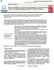
Limbs: The thoracic and pelvic limbs present marked micromelia, with hypoplastic joints and with no mineralized tissue [14]. The shortening is greater in the proximal bones, also affecting the scapulae and the pelvis [11]. Tegument: Depending on the gestational age of the affected fetus, the body may have no hair, or have hair only in some areas or be totally covered in hair [6,7,14]. Figure 1 shows several of the abnormalities described above in an affected Nellore calf with a gestational age of approximately eight months.
Figure 1: Clinical phenotype of a female Nellore calf with prenatal lethal chondrodysplasia (bulldog calf). Most of the pathoanatomical abnormalities described in the text are present in this affected calf. A) Macrocephalia, frontal bossing, flattened face and protruding tongue; B) Severe micromelia and bulky abdomen.
Histopathology The histological alterations of BPLC are evident on the growth plates and indicate a defect of the endochondral ossification [6]. The epiphysis are disorganized, have hyaline cartilage with numerous hypertrophied chondrocyte and show no differentiation between their zones [6,7]. The metaphysis are short and have thick trabeculae, showing irregular and deficient calcification [14,17]. The diaphysis may be thin and mainly constituted of cancelous osseous tissue and some compact tissue [6], or have a well-developed and normal looking cortical bone [7].
Genetic Features Although cases of BPLC in different breeds have had essentially the same clinical phenotype, the data currently available do not make it possible to affirm that they have the same cause. It is likely that they are etiologically different, i.e., they may be caused by different genes (locus heterogeneity) [6,7]. For this reason, the Mendelian and molecular aspects of the Dexter breed will be explained first, followed by considerations on the other breeds.
Inheritance pattern of BPLC in the Dexter breed The origins of the Dexter breed can be traced back to the nineteenth century from the Kerry breed, when there were individuals with short legs (Dexter type) and long legs (Kerry type). The Dexter type has a mild form of chondrodysplasia and, for this reason, its legs are short [6,8].
Hereditary Genet ISSN:2161-1041 HGCR, an open access journal
BPLC in the Dexter is caused by incompletely dominant mutations in the aggrecan gene (ACAN gene). If we represent the ACAN gene as “a” and the mutant allele as “A”, the long-legged Dexter has an “aa” genotype (homozygote), the short-legged Dexter has an “Aa” genotype (heterozygote), and the bulldog Dexter has an “AA” genotype (homozygote). Thus, the homozygosity of the “A” allele causes a defect in the endochondral ossification that is so severe that it is incompatible with life [6,8]. Therefore, based on the principle of Mendelian genetics, if two heterozygote Dexters are crossed, there is a 25% chance of the offspring having long legs, a 50% chance that it will have short legs, and a 25% chance that it will be affected by BPLC (bulldog calf), as shown in Figure 2. The inheritance pattern that we have described is classically known as incomplete dominance (semidominance). In human medical genetics, it has become the custom to refer to the phenotype that is manifested in heterozygotes to any degree as being dominant, as is the case of human achondroplasia. However, homozygotes for the human achondroplasia allele have a more severe clinical phenotype. Therefore, their inheritance mechanism corresponds to the concept of incomplete dominance of classical genetics [19]. If we adopt the language of medical genetics, BPLC in the Dexter breed has a dominant autosomal inheritance pattern with prenatal lethality.
Molecular aspects of BPLC in the Dexter breed In 2007, Cavanagh et al. [8] discovered that mutations in the ACAN gene are the cause of BPLC in the Dexter breed. The ACAN gene is located in band 5 of region 1 of the long arm of bovine chromosome 21 (BTA21q15). It encodes the aggrecan, a protein present in the
Volume 3 • Issue 3 • 1000132
Citation:
Moura E, Prado AMRB, Pimpão CT, Murakami CT, Ribeiro DR (2014) Genetic and Pathoanatomical Features of the Bovine Prenatal Lethal Chondrodysplasia . Hereditary Genet 3: 132. doi:10.4172/2161-1041.1000132
Page 3 of 5 extracellular matrix of the cartilaginous tissue and is necessary for its normal formation. It was formerly known as chondroitin sulfate proteoglycan core protein 1 (CSPGCP or CSPG1). Cavanagh et al. [8] found two mutations of the ACAN gene responsible for BPLC in all the cases that they tested. The most frequent was an insertion of 4 bp in the Exon 11
(2266_2267insGGCA), creating a reading frame and a premature termination codon. The most rare was a transition in the exon 1 (– 198C>T), creating a new initiation codon situated 199 bp before the normal initiation codon. Both the mutations cosegregate with the chondrodysplasia of the Dexter. Currently, diagnostic tests are available for both these mutations in several countries.
Figure 2: Risk of prenatal lethal chondrodysplasia in a mating of short-legged Dexters.
Considerations on BPLC in other breeds In all the cases of BPLC described in the Holstein breed, animals of both sexes were affected and the parents were phenotypically normal, leading to the assumption of autosomal recessive inheritance [7]. The existence of common ancestors in one of the reports strengthens this hypothesis [7]. However, there is a need for further studies in order to determine the inheritance pattern. The cases described in the Jersey breed have no detailed information regarding the parents, but it may be assumed through by the context that they were phenotypically normal. There is also no information regarding the sex of the affected animals [14,17]. The same is also true in the case that describes an affected Hostein-Jersey crossbred. If we accept that the parents were normal, it could be said that there was recessive inheritance in the cases of BPLC in the Jersey breed. However, it would not be safe to make any definite statement to this effect.
Hereditary Genet ISSN:2161-1041 HGCR, an open access journal
The family history of the affected Nellore calf that is illustrated in this paper is unknown, enabling no conclusion to be reached regarding the inheritance mechanism. It should also be borne in mind that Dexters have been used for crossbreeding or to form other breeds. This means that mutations of the ACAN gene responsible for the “bulldog” phenotype could be present in different bovine populations. Likewise, mutations in other genes that might cause BPLC may be found in the most different breeds, since there is essentially one bovine genome. There could be a variation in the genotype frequency of different breeds or populations [20].
Phenotypical Similarities between BPLC and Human Chondrodysplasias In the human species, some types of disproportionate dwarfism show phenotypic similarities with BPLC and have a known molecular base. Considering that the genes that control the development of
Volume 3 • Issue 3 • 1000132
Citation:
Moura E, Prado AMRB, Pimpão CT, Murakami CT, Ribeiro DR (2014) Genetic and Pathoanatomical Features of the Bovine Prenatal Lethal Chondrodysplasia . Hereditary Genet 3: 132. doi:10.4172/2161-1041.1000132
Page 4 of 5 vertebrates, and the epigenetic mechanisms that control their expression, are highly conserved [21], the knowledge of the molecular base of human chondrodysplasias may be a starting point for clarifying the etiology of cases in animals. Table 1 shows the main aspects of human chondrodysplasias whose major characteristic is lethal micromelic dwarfism. It should also be considered that different mutations in the same gene may cause phenotypic variations (allelic heterogeneity) or even different phenotypes (phenotypic heterogeneity) [19]. Therefore, bovine chondrodysplasias phenotypically distinct from BPLC could end up being the expression of alleles from the same gene. For instance, in humans, spondyloepiphyseal dysplasia, Kimberly type, is Chondrodysplasia
OMIM number Clinical phenotype (*) (main features)
Achondrogenesis type (Houston-Harris type)
IA 200600
Achondrogenesis (Fraccaro type)
IB 600972
type
caused by a non-lethal dominant mutation in the ACAN gene, causing a short proportionate stature [22], whereas spondyloepiphyseal dysplasia, aggrecan type, is caused by another non-lethal mutation of the ACAN gene, but is recessive and causes a severe form of disproportionate dwarfism [23,24].
Acknowledgements The authors would like to thank Dr. Valter da Silva Queiroz (Pontifícia Universidade Católica do Paraná) who referred to us the Nellore bulldog calf.
Lethality
Severe micromelia; Prenatal/perinatal macrocephaly; flattened nasal bridge; small thoracic cage; prominent abdomen.
Mutated gene
Inheritance pattern
TRIP11
Autosomal recessive (**)
[Thyroid hormone receptor interactor 11]
Severe micromelia; Prenatal/perinatal macrocephaly; flattened nasal bridge; small thoracic cage; prominent abdomen.
SLC26A2
Achondrogenesis type II (Langer- 200610 Saldino type)
Micromelia; macrocephaly; Prenatal/neonatal flattened nasal bridge; small thoracic cage; prominent abdomen; cleft palate.
COL2A1
Homozigous achondroplasia
100800
Micromelia; macrocephaly; flattened nasal bridge; small thoracic cage; prominent abdomen.
Thanatophoric dysplasia, type I
187600
Micromelia; macrocephaly; Usually perinatal flattened nasal bridge; small thoracic cage; prominent abdomen.
Thanatophoric dysplasia, type II
Thanatophoric Glasgow variant
187601
dysplasia, 273680
[Solute carrier family 26 (sulfate transporter), member 2]
[Collagen, type II, alpha-1]
Usually neonatal (only FGFR3 homozygotes). Heterozygous achondroplasia is nonlethal and is the most common form of human dwarfism.
Micromelia; macrocephaly; Usually perinatal flattened nasal bridge; small thoracic cage; prominent abdomen. Micromelia; macrocephaly; Neonatal flattened nasal bridge; small thoracic cage; prominent abdomen.
FGFR3 [Fibroblast receptor 3]
growth
factor
growth
factor
FGFR3 [Fibroblast receptor 3]
FGFR3 (likely)
Autosomal recessive (**)
Autosomal (**)
dominant
Autosomal (**)
dominant
Autosomal (**)
dominant
Autosomal (**)
dominant
Autosomal recessive (*)
(*) Reference 24; (**) Reference 2.
Table 1: Human lethal micromelic chondrodysplasias
References
4.
1.
5.
2. 3.
Spranger J, Benirschke K, Hall J G, Lenz W, Lowry R B et al. (1982) Errors of morphogenesis: Concepts and terms. Recommendations of an International Work Group. J Pediatr 100: 160-165. Krakow D, Rimoins DL (2010) The skeletal dysplasias. Genet Med 12: 327-341. Moura E, Pimpão C T (2012) A bird's-eye view of veterinary medicine. Veterinary dysmorphology. In: Perez-Marin, C C InTech, Rijeka.
Hereditary Genet ISSN:2161-1041 HGCR, an open access journal
6. 7.
White P J, Windsor P A (2012) Congenital chondrodystrophy of unknown origin in beef herds. Vet J 193: 336–343. Agerholm J S (2007) Inherited Disorders in Danish Cattle. APMIS 115: 19-20. Harper P A W, Latter M R, Nicholas F W, Cook R W, Gill P A (1998) Chondrodysplasia in Australian Dexter cattle. Aust Vet J 76: 199-202. Agerholm J S, Arnbjerg J, Andersen O (2004) Familial chondrodysplasia in Holstein calves. J Vet Diagn Invest 16: 293-298.
Volume 3 • Issue 3 • 1000132
Citation:
Moura E, Prado AMRB, Pimpão CT, Murakami CT, Ribeiro DR (2014) Genetic and Pathoanatomical Features of the Bovine Prenatal Lethal Chondrodysplasia . Hereditary Genet 3: 132. doi:10.4172/2161-1041.1000132
Page 5 of 5 8. 9. 10. 11. 12. 13. 14. 15. 16.
Cavanagh J A L, Tammen I, Windsor P A, Bateman J F, Savarirayan R, et al. (2007) Bulldog dwarfism in Dexter cattle is caused by mutations in ACAN. Mamm Genome 18: 808–814. Hogben L T, Crew F A E (1923) Studies on internal secretion II — Endocrine activity in fœtal and embryonic life. Br J Exp Biol 1: 1-13. Seligmann C G (1904) Cretinism in calves. J Pathol Bact 9: 311–22. Crew F A E (1924) The bull-dog Calf: A contribution to the study of achondroplasia. Proc R Soc Med 17: 39–58. Rousseau F, Bonaventure J, Legeai-Mallet L, Pelet A, Rozet JM, et al. (1994) Mutations in the gene encoding fibroblast growth factor receptor-3 in achondroplasia. Nature 371: 252-254. Shiang R, Thompson L M, Zhu YZ, Church D M, Fielder T J, et al. (1994) Mutations in the transmembrane domain of FGFR3 cause the most common genetic form of dwarfism, achondroplasia. Cell 78: 335-342. Coelho A C B, Marcolongo-Pereira C, Soares M P, Quevedo P S, RietCorrea F, et al. (2013) Condrodisplasia em bovinos no Sul do Rio Grande do Sul. Pesq Vet Bras 33: 1195-1200. Windsor P A, Agerholm J S (2009) Inherited diseases of Australian Holstein-Friesian cattle. Aust Vet J 87: 193–199. Wurster F, Bassuino D M, Juffo G D, Boos G S, Boabaid F M, et al. (2012) Condrodisplasia em type Dexter cattle aborted fetuses. Acta Sci Vet 40: 1-4.
Hereditary Genet ISSN:2161-1041 HGCR, an open access journal
17. 18. 19. 20. 21. 22.
23.
24.
Downs W M G Jr (1928) An american ‘dexter monster’. Anat Rec 37: 365-372. Windsor P A, Cavanagh J, Tammen I (2006) Hydrops fetalis associated with pulmonary hypoplasia in Dexter calves. Aust V J 84: 278-281. Nussbaum R L, McInnes R R, Willard H F (2007) Thompson & Thompson Genetics in medicine. Saunders, Philadelphia. Elsik CG, Tellam RL, Worley KC, Gibbs RA, Muzny DM, et al. (2009) The genome sequence of taurine cattle: a window to ruminant biology and evolution. Science 324: 522-528. Barbera B A, Rastegara M (2010) Epigenetic control of Hox genes during neurogenesis, development, and disease. Ann Anat 192: 261–274. Gleghorn L, Ramesar R, Beighton P, Gillian Wallis G (2005) A mutation in the variable repeat region of the aggrecan gene (AGC1) causes a form of spondyloepiphyseal dysplasia associated with severe, premature osteoarthritis. Am J Hum Genet 77: 484–490. Tompson S W, Merriman B, Funari V A, Fresquet M, Lachman R S, et al (2009) A recessive skeletal dysplasia, SEMD aggrecan type, results from a missense mutation affecting the C-type lectin domain of aggrecan. Am J Hum Genet 84: 72-79. http://omim.org/
Volume 3 • Issue 3 • 1000132