Stem Cell Rev (2007) 3:183–191 DOI 10.1007/s12015-007-0008-4
hESC Adaptation, Selection and Stability C. Grandela & E. Wolvetang
Published online: 22 May 2007 # Humana Press Inc. 2007
Introduction Human Embryonic Stem Cells (hESC) are vigorously investigated as a source for cell replacement therapies. In order to deliver on their promise large amounts of high quality hESC will be needed that are genetically stable, free of animal products and manipulated to evade the immune system through either SCNT or other technologies. Here we will review several aspects of hESC biology that may directly or indirectly affect their genetic stability.
hESC Culture Conditions Human embryonic stem (hESC) cell lines are established from the inner cell mass (ICM) of the blastocyst and differentiate into all cell lineages of the body [29, 31]. They were initially derived in 1998 and have enormous potential
C. Grandela PDBEB – Programa Doutoral em Biologia Experimental e Biomedicina, Department of Zoology, Center for Neuroscience and Cell Biology, University of Coimbra, Coimbra, Portugal C. Grandela Monash Institute of Medical Research, Monash University, Clayton, Australia E. Wolvetang (*) Australian Stem Cell Centre, Level 3 North Building 75 (STRIP) Wellington Road, Clayton 3168 Victoria, Australia e-mail:
[email protected] E. Wolvetang Department of Anatomy and Cell Biology, School of Biomedical Sciences, Monash University, Clayton, Australia
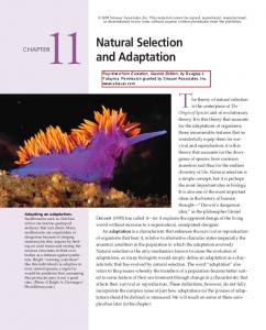
as a source of cells for cell replacement therapies and as a model for early human development [13]. Two culture conditions are widely used to propagate hESC. The standard method of culturing hESC stock cultures involves weekly mechanical passaging of morphologically undifferentiated appearing parts of a hESC colony onto full density (6×10⁴ cell/cm2) mouse or human fibroblast feeder layers in a medium containing 20% FCS [29, 31]. This protocol, albeit time, money and effort intensive, has proven to be a safe and reliable method for the long term propagation of hESC. This is highlighted by the fact that hESC thus cultured have never been reported to develop genetic abnormalities during culture. However, the major drawback of this method is the low number of hESC available for experimentation. Most laboratories therefore opt to maintain hESC stock cultures using this standard method and regularly establish fresh bulk cultures from these stocks for experimentation [2]. Bulk culture of hESC is performed according to a method developed by Amit et al. which involves passaging of enzymatically (collagenase, trypsin, etc) or non-enzymatically (cell dissociation solution, EDTA, etc) dissociated clumps of hESC on 1/3 density feeders (2×10⁴ cell/cm2) in KSOR with 20% serum replacement. This method indeed allows for dramatic expansion of hESC (Fig. 1). However, a number of reports have indicated that this is accompanied by an increase in genetic instability, methylation changes and mitochondrial mutations, abnormalities also seen in cancer and Embryo Carcinoma (EC) cells (the transformed counterpart of hESC) [5, 8, 24]. Since large amounts of undifferentiated stem cells of high quality will be needed for regenerative medicine approaches in the future, a deeper understanding of the molecular mechanisms that control the genetic stability of hESC is required. In addition culture methods that prevent the occurrence of genetic instability and/or
184
Stem Cell Rev (2007) 3:183–191
Fig. 1 Comparison of “Standard” culture method with “Bulk”-culture method. See text for detailed explanation
technologies to purge abnormal hESC from large scale hESC cultures will have to be developed.
Pre-existing versus Acquired Genetic Abnormalities? The fact that hESC cultured under standard conditions display such remarkable genetic stability (normal karyotypes for over 200 weekly passages) as compared to bulk cultured cells (frequent abnormal karyotypes between
passage 20 and 60) suggests fundamental differences between these two culture systems. One can envisage two broad possible explanations for the occurrence of genetic abnormalities in bulk cultured hESC. (1) Bulk culture directly or indirectly causes genetic instability (2) Genetic instability is an intrinsic feature of hESC in general but the phenotype of genetically abnormal cells only becomes apparent in bulk cultured hESC. If (1) is correct and the abnormalities occur as a consequence of the bulk culture conditions then bulk cultured hESC should be found to
Stem Cell Rev (2007) 3:183–191
exhibit a combination of increased mutagenesis, reduced apoptosis, reduced DNA-repair and enhanced growth/ survival properties as compared to hESC grown in standard conditions. If (2) is correct and hESC grown under standard conditions show similar intrinsic genetic instability as bulk cultured hESC then one would predict that standard grown hESC exhibit more efficient mechanisms to delete/suppress abnormal cells, display enhanced repair DNA or that the acquired genetic abnormalities do not confer sufficient growth/survival enhancement under standard conditions to take over the culture. In order to test these hypotheses described above CGH arrays, SNP arrays and high through put whole genome sequencing will have to be employed in the future because the method that is most widely used to assess genetic stability of hESC at present is G-banding karyotype analysis. This method is only a low resolution assay, as 5–10% of all cells analysed need to show an abnormal karyotype to call the hESC genetically abnormal and does not identify small deletions or mutations.
Can We Phenotypically Distinguish Genetically Normal and Abnormal hESC? Embryocarcinoma (EC) cells are the transformed genetically unstable counterpart of hESC and characteristically display very similar cytogenetic abnormalities to hESC with acquired genetic abnormalities such as trisomy 12, 17 and X [5, 8, 24]. How then does one define a genetically healthy hESC? Some would argue that it is the ability to form teratomas with representatives of all three germ lineages that identifies the stem cell. However, EC cells also exhibit this property with the difference that undifferentiated primitive cells are still present in the graft that can be transplanted to new recipients, identifying them as a teratocarcinoma. However, since this particular experiment is usually not performed with hESC teratomas it is difficult to determine whether the hESC were actually stem cells or EC cells. Indeed, in the majority of reports there are only high power images of differentiated hESC shown and no overview of what other (undifferentiated?) cell types are present in such grafts. Others would argue that the expression of stem cell markers is proof of the presence of hESC. However, EC cells also express pluripotency markers and hESC with genetic abnormalities, such as ones that are marked by CD30 (see section below), actually display higher marker expression (>95%) than genetically healthy hESC (>70–80%) which usually display 15–20% spontaneous background differentiation. The presence of a fraction of spontaneously differentiating hESC in culture may therefore be a better indication of a genetically healthy hESC culture than hESC that express unusually high levels of pluripotency markers. This phenomenon is exacerbated
185
by the high proliferative potential of high pluripotency marker expressing hESC. Indeed our data indicate that the more undifferentiated a hESC line is, the faster it grows (Filipczyk et al. pers. comm.) Unfortunately this also applies to hESC with abnormally high pluripotency marker expression that more closely resemble EC cells. Such cells would therefore rapidly take over the culture and without good assays to discriminate hESC from EC cells or from ES cells with genetic alterations that do not show up in Gbanding karyotype analyses one could easily interpret the properties of such genetically compromised cells for those of hESC cells.
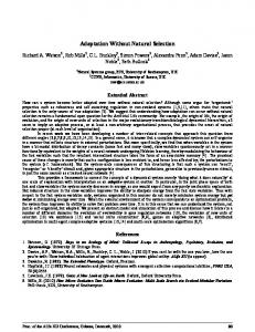
Selective Pressures During Establishment of HES Cell Lines Since we can not rule out the possibility that genetically compromised hESC are present from the inception of a hESC culture and because genetically abnormal hESC have a growth/survival/differentiation advantage over genetically “healthy” hESC it may be informative to consider the history of the cells and the selective pressures placed upon hESC cultures during their establishment and subsequent propagation (Fig. 2). hESC are cells isolated from the inner cell mass of day 5 blastocysts procured from to be discarded excess IVF embryos [29, 31]. The first decision in the history of a hESC line is therefore made at the oocyte stage when good quality oocytes are selected for IVF. In general the best fertilized cells are selected for implantation while the left over eggs with a potentially lower quality score can then made available for stem cell research. Post fertilization only some of the excess zygotes subsequently progress to blastocyst stage for reasons that are not fully understood at present. The next step in the establishment of a hESC line involves the selection of blastocysts with a high standard scoring classification (i.e. symmetry and low levels of cell fragmentation (apoptosis). Apoptosis of ICM cells is thought to be part of a quality control program perhaps aimed at eliminating abnormal cells from the developing embryo. This is supported by the observation that the incidence of apoptosis in in vitro produced embryos is increased by suboptimal culture conditions. Indeed, 7– 8% of cells in pre-implantation embryos undergo apoptosis during this time and this occurs mainly in the ICM [11]. Nevertheless, early human development is accompanied by a high degree of genetic abnormality, with one third of first trimester miscarriages attributed to chromosomal defects [23]. To our knowledge to date there has been no systematic investigation into the potential correlation between the level of ICM apoptosis at the blastocyst stage and their subsequent ability to establish hESC lines or how this relates to their long term genetic stability. However,
186
Stem Cell Rev (2007) 3:183–191
Fig. 2 The sequence of potential selective pressures during the establishment of a hESC culture. Excess oocytes not selected for reproduction are made available for hESC establishment. A subset of fertilized embryos progresses to blastocyst stage. Inner cell mass cells are selected for low apoptotic index. A subset of hESC proceeds to form a cell line after immunosurgery. A subset of hESC responds to MEF secreted maintenance factors and media components. Further selection of hESC occurs during culture
several studies have correlated the occurrence of chromosome abnormalities in SCNT embryos with the frequency of abnormalities in the donor cell line [22]. Donor cell lines with a higher percentage of genetically abnormal cells were found to cause a higher rate of chromosomally abnormal blastomeres in the SCNT embryos. It would be interesting to determine whether donor nuclei with chromosomal amplifications also detected in hESC (such as trisomy 12, 17, or X) show an enhanced ability to establish ES-cell lines. Following immuno-surgery and isolation of the inner cell mass, the next stage of hES derivation involves the propagation of isolated clumps of inner cell mass cells after plating on full density mouse or human fibroblast feeder layers. Again only a subset of these clumps can be coaxed into becoming a hESC line with rather low frequency. It seems self-evident that during this step there is a selection for cells that can respond to the maintenance factors excreted by the feeder layers and the medium components that aid in the establishment of hESC lines. In addition one would select for hESC that exhibit low apoptosis and a low
propensity to differentiate since these are the cells that have the highest proliferative capacity. There is a large body of circumstantial evidence to suggest that mES lines adapt to their culture conditions and that various strains or substrains differ in their ability to generate ES cell lines or to contribute to the germline. Indeed, some mouse strains appear unable to establish mES lines, although the exact rationale for this phenomenon remains unknown. Although these parameters are not easy to assess in a human context it is not unreasonable to infer that a similar process occurs with hESCs. In addition there is a school of thought that argues that this selection is related to the occurrence of specific genetic and epigenetic changes that may aid in this adaptation to in vitro culture. As argued above hESC may have an innate ability to adapt to culture conditions or that certain culture conditions select for hESC with properties that allow propagation in a particular culture method. Since hESCs generally do not proliferate as single cells but rather as small clumps there is a possibility that hESC lines are intrinsically heterogeneous for this very reason from their
Stem Cell Rev (2007) 3:183–191
inception. Indeed, the vast majority of hESC lines have not been derived from a single clone. An in depth analysis of the genetic heterogeneity within hESC lines is further complicated by the fact that hESCs cultured under the standard most optimal culture conditions rapidly become a heterogeneous population of cells when analysed at the gene expression level. Transcriptome analysis of gene expression in hESC stratified on the basis of their expression of known pluripotency marker expression shows that hESCs are not only in a constant flux of spontaneous differentiation, as judged by the presence of 20% of differentiated cells under these conditions, but indeed display substantial differences in mRNA expression [19] as compared to hESC with lower, but still positive, expression of the pluripotency markers. Indeed, transcriptome analysis of FACS sorted hESC stratified on the basis of the highest expression of pluripotency markers indicates that that these ultra-stem cells already express supposedly lineage specific markers such as nestin and brachyury by array analysis and RT-PCR, albeit at low levels.
Chromosomal Abnormalities and Potential Significance Although the original reports on HESCs indicated that they can maintain a normal diploid karyotype, there is evidence that this depend on culture conditions. Different laboratories have reported that HESCs cultured for long periods of time tend to gain chromosomal abnormalities, resembling the ones observed in hEC cells. The most frequently observed karyotypic abnormalities are gain of chromosome 17q, 12 and X chromosome [5, 8, 24], while there also seems to be a high proportion of partial or total chromosome 1 duplication. It has been postulated that increased dosage of those chromosomes might give the cells some sort of selective advantage. Draper et al. [8] were the first to report gain of chromosome 17q and chromosome 12 in H7 and H14 cell lines cultured with Knockout SR replacer (Invitrogen/ GIBCO) instead of foetal calf serum and passaged with an enzymatic method (collagenase IV). They performed interphase fluorescence in situ hybridization (FISH) and showed that after 22 passages trisomy 17 was observed in 76% of the cells and after an additional 17 passages, in 95% of the cells, again indicating that such cells possess enhanced survival or proliferation properties. Buzzard and colleagues [6] cultured six NIH-registered HESC lines (hES1-6) between 34 and 140 passages using mechanical method of passaging cells rather than enzymatic or chemical methods of cell dissociation. They reported just one karyotype abnormality was detected in an early passage hES5, demonstrating that HESCs are not necessarily predisposed to karyotypic abnormalities even grown for
187
extended time. Another group studied 3 cell lines (BG01, BG02 and BG03) and showed that karyotypic changes also give rise to differences in expression of genes, especially those involved in pluripotency [24]. They reported that bulk passaging (nonenzymatic- Cell Dissociation Buffer- or enzymatic- colagenase, trypsin) methods can compromise the genetic stability of HESCs, in contrast with manual methods. The bulk passage methods can be used for shorter periods (