Poster No. CG17P-0612
High Angular Resolution Diffusion Imaging Correlates of Depression in Parkinson’s Disease: A Connectometry Study M. Mayeli1, S. Sobhani2 , F. Ghazi Sherbaf 2, S. Mohammadi Jooyandeh3, F. Rahmani2, M.H. Aarabi4 1. Neuropsychology Association, Students’ Scientific Research Center, Tehran University of Medical Sciences, Tehran, Iran 2. Neuroimaging Network, Universal Scientific and Education Network, Tehran, Iran 3. Department of Psychiatry and Psychotherapy, University of Regensburg, Regensburg, Germany 4. Basir Eye Health Research Center, Tehran, Iran
Introduction: Depression is a significant disabling feature in Parkinson's disease, however the neuropathology of this comorbidity is still unclear. To further investigate the relationship between brain microstructural alterations and depressive symptoms in PD, we employed a multiple regression study to survey the connectivity of whole white matter regions by using diffusion MRI connectometry, in regard to depressive symptoms assessed by Hospital Anxiety and Depression Scale (HADS) in 27 patients with confirmed diagnosis of PD. A significant negative association (FDR < 0.05) was demonstrated between depressive symptoms in PD patients and left Cingulum, Genu and Splenium of the Corpus Callosum, anterior and posterior limbs of the right internal capsule. This finding may provide new potentials in better understanding the neural basis of depressive symptoms in Parkinson's disease.
Methods: We studied 27 patients clinically diagnosed with Parkinson's disease. Disease stage within the patient population was assessed in the “on” state using the Hoehn & Yahr scale. Twenty-four of the 27 patients were taking a combination of several classes of drugs: levodopa (immediate and controlled release), non-ergot-derived dopamine receptor agonists (Pramipexole, Ropinirole), and a monoamine oxidase B inhibitor (Rasagiline). The remaining 3 patients were not taking any antiParkinsonian medications at the time of scanning. Levodopa and dopamine agonist dosages were pooled and summarized as the levodopa equivalent daily dose.
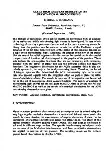
Results: The connectometry analysis was implemented to identify white matter pathways associated with depression. The analysis showed a significant negative association (False discovery rate (FDR) < 0.05) between HADS in PD patients and Genu, Splenium, right anterior limb of internal capsule, right posterior limb of internal capsule, and left cingulum (Figure 1).
Discussion: The principal finding of this study is that the higher scores of depression subscale of the HADS test is associated with the lower connectivity in specific white matter regions including the left cingulum, genu and splenium parts of the corpus callosum, and also the anterior and posterior limbs of the right internal capsule. In other words, more severe depressive symptoms are contributed to lower white matter integrity of fiber tracts in the mentioned regions of the brain. The effect of confounders such as anxiety, cognitive disturbances, duration and severity of the disease and the treatment dosages were controlled.
Figure 1. White matter pathways with significantly associated in PD patients (a) genu of corpus callosum (b) Splenium of corpus callosum, (c) right anterior limb of internal capsule, (d) right posterior limb of internal capsule, and (e) left cingulum
Conclusion: In sum, disturbed tissue organization in cingulum, corpus callosum and internal capsule supports the hypothesis that diminished connectivity between cortical and subcortical regions of brain, involving complicated neural circuits, manifests mood deregulations. The principal limitation of this study is not specifically probing the tracts in the mentioned regions, each known to have extensive connections and multiple circuits involved in different tasks. Lack of follow-up comparison and small sample of patients highlights the dire need of further studies with follow-up data of PD patients with depressive symptoms. It is worthy to note that caution should be exercised when discussing our results due to lack of HADS validity to diagnose depression in the clinical setting.
Conflict of Interests The authors declare no conflict of interests. References Reijnders JS, Ehrt U, Weber WE, Aarsland D, Leentjens AF. A systematic review of prevalence studies of depression in Parkinson's disease. Movement Disorders 2008; 23: 183-189. van Mierlo TJ, Chung C, Foncke EM, Berendse HW, van den Heuvel OA. Depressive symptoms in Parkinson's disease are related to decreased hippocampus and amygdala volume. Mov Disord 2015; 30: 245-52. Gutman DA, Holtzheimer PE, Behrens TE, Johansen-Berg H, Mayberg HS. A tractography analysis of two deep brain stimulation white matter targets for depression. Biological psychiatry 2009; 65: 276-282.
Mahsa Mayeli et al. 2017
[email protected]