B American Society for Mass Spectrometry, 2012
J. Am. Soc. Mass Spectrom. (2012) 23:952Y963 DOI: 10.1007/s13361-012-0345-7
RESEARCH ARTICLE
High Resolution LC-MSn Fragmentation Pattern of Palytoxin as Template to Gain New Insights into Ovatoxin-a Structure. The Key Role of Calcium in MS Behavior of Palytoxins Patrizia Ciminiello, Carmela Dell’Aversano, Emma Dello Iacovo, Ernesto Fattorusso, Martino Forino, Laura Grauso, Luciana Tartaglione Dipartimento di Chimica delle Sostanze Naturali, Università degli Studi di Napoli Federico II, via D. Montesano 49, 80131 Napoli, Italy
Abstract Palytoxin is a potent marine toxin and one of the most complex natural compounds ever described. A number of compounds identified as palytoxin congeners (e.g., ovatoxins, mascarenotoxins, ostreocins, etc.) have not been yet structurally elucidated due to lack of pure material in quantities sufficient to an NMR-based structural investigation. In this study, the complex fragmentation pattern of palytoxin in its positive high resolution liquid chromatography tandem mass spectra (HR LC-MSn) was interpreted. Under the used conditions, the molecule underwent fragmentation at many sites of its backbone, and a large number of diagnostic fragment ions were identified. The natural product itself was used with no need for derivatization. Interestingly, most of the fragments contained calcium in their elemental formula. Evidence for palytoxin tendency to form adduct ions with calcium and other divalent cations in its mass spectra was obtained. Fragmentation pattern of palytoxin was used as template to gain detailed structural information on ovatoxin-a, the main toxin produced by Ostreopsis ovata, (observe correct font) a benthic dinoflagellate that currently represents the major harmful algal bloom threat in the Mediterranean area. Either the regions or the specific sites where ovatoxin-a and palytoxin structurally differ have been identified. Key words: HR LC-MSn, Palytoxin, Ovatoxin-a, Calcium adducts
Introduction
P
alytoxin (Figure 1) is one of the most potent non-protein marine toxins so far known as well as one of the most complex non-polymeric natural compounds ever described: it presents a long and highly functionalized alkyl chain containing, among others, 42 hydroxyl groups and seven Electronic supplementary material The online version of this article (doi:10.1007/s13361-012-0345-7) contains supplementary material, which is available to authorized users. Correspondence to: Carmela Dell’Aversano; e-mail:
[email protected]
ether rings, with no repeating units [1–4]. A number of palytoxin analogues have been so far isolated either from soft corals (Palythoa genus) or dinoflagellates (Ostreopsis spp.): homo, bishomo-, neo-, and 73-deoxy-palytoxin [5], ostreocin-D (namely 42-hydroxy-3,26-didemethyl-19,44dideoxypalytoxin) [6], and 42-hydroxy-palytoxin [7]. Structure elucidation of these compounds was carried out by extensive NMR analyses and in most cases through the use of degradation/derivatization reactions [1, 5, 8]. This kind of study requires relatively high amounts of pure material and, therefore, structures of a number of minor congeners of palytoxin so far identified were not elucidated due to lack of Received: 11 November 2011 Revised: 29 December 2011 Accepted: 21 January 2012 Published online: 22 February 2012
P. Ciminiello et al.: HR LC-MSn of Palytoxin and Ovatoxin-a
953
Cleavage #27
#28
#26
#25 #24
#23
#22 #21
#20
#18
#19
#17
#15
#16
#14
#11
#13 #12
#9+#12
#10+#12
#8+#12
#7+#12
#6+#12
#5+#12 #4+#16
2648.5 1315.7 896.2
#4+#15
2
MS 2 MS 2 MS 3 MS 3 MS 3 MS 4 MS
#4+#13 #4+#12
Ovatoxin-a
2680.4 1331.7 906.8
#4
Palytoxin
MS 2 MS 2 MS 3 MS 3 MS 3 MS 4 MS
#3 #2
2
Precursor ion
#1+#4
CID
743.4 507.3 327.1 309.1
727.4 507.3 327.1 309.1
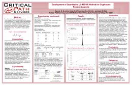
Figure 1. Structure of palytoxin and HR LC-MSn-based structural hypothesis for ovatoxin-a. An NMR study [20] on pure ovatoxin-a demonstrated that ovatoxin-a is the 42-hydroxy-17,44,64-trideoxy-palytoxin. Cleavages resulting from various HR CID MSn spectra are reported in the structure and in the scheme below. All cleavages in palytoxin occurred also in ovatoxin-a apart from those marked as white spots in black cells
pure material in quantities sufficient to structural investigation. For this purpose, mass spectrometry (MS) could be a useful tool as it is both sensitive and selective even when analytes are contained at trace levels in quite complex matrices. However, structural complexity of palytoxins is reflected in equally complex mass spectra, and this hampers a straightaway interpretation of their fragmentation patterns [9]. Significant efforts in this direction were made by Ukena et al. [10], who used negative ion fast atom bombardment (FAB) collision induced dissociation (CID) tandem MS to confirm the structure of ostreocin-D. However, this study
was not carried out on ostreocin-D itself but on a derivatized sample of ostreocin-D obtained by reaction with 2-sulfobenzoic acid cyclic anhydride; this resulted in introduction of a negative charge in the molecule that facilitated the charge-remote fragmentations. In the present study, we fully interpreted the positive ion high resolution (HR) electrospray (ESI) MSn spectra of palytoxin; the natural product itself was used with no need for derivatization. In the spectra obtained, besides the abundant and uninformative ions due to water losses, a large number of diagnostic fragment ions were present;
954
P. Ciminiello et al.: HR LC-MSn of Palytoxin and Ovatoxin-a
interestingly, most of them contained calcium in their elemental formula. Evidence for palytoxin tendency to form adduct ions with calcium and other divalent cations in its ESI MS and MSn spectra was obtained. Fragmentation pattern of palytoxin, full of informative cleavages all along the entire backbone of the molecule, could provide a direct strategy to get structural information on uncharacterized palytoxin congeners, available in quantities too small to be studied by NMR. So, the results obtained were used for structural investigation of the main toxin produced by Ostreopsis ovata. This benthic dinoflagellate currently represents the major threat in the Mediterranean area both from an environmental and a public health perspective [11–13]. The most relevant episode associated to O. ovata occurred in 2005 along the Ligurian coasts (Italy) when hundreds of people required medical attention after exposure to marine aerosols [14, 15]. O. ovata blooms have repeatedly occurred in subsequent years in Italy as well as along the Mediterranean coasts of Spain, France, and Greece [11]. Putative palytoxin and several new palytoxin-like compounds, named ovatoxin-a, -b, -c, -d, and -e, were identified by our group in both natural and cultured O. ovata samples based on comparison of their HR LC-MS and MS2 behavior with that of palytoxin [9, 16–18]. In these early studies, elemental composition of ovatoxins was provided together with preliminary information on their structures, which allowed only including ovatoxins within the palytoxin group of toxins. In the present study, interpretation of the fragmentation pattern of palytoxin was used as rational basis for structural investigation of ovatoxin-a, the major component of O. ovata toxin profile [17, 18], and this allowed to identify the regions or even the specific sites of difference between palytoxin and ovatoxin-a.
Materials and Methods Chemicals and Materials All organic solvents and glacial acetic acid (Laboratory grade) were by Carlo Erba (Milan, Italy). Calcium, magnesium, strontium, zinc, potassium, sodium, lithium acetate, and disodium EDTA were purchased from Sigma Aldrich (Milan, Italy). Palytoxin was purchased from Wako Chemicals GmbH (Neuss, Germany) and dissolved in methanol/water (1:1, vol/vol) to a concentration of 10 μg/mL. A crude extract of O. ovata [18] containing ovatoxin-a at levels of 6 μg/mL was used.
HR LC-MS and MSn All analyses were performed on an Agilent 1100 LC binary system (Palo Alto, CA, USA) coupled to a hybrid linear ion trap LTQ Orbitrap XL Fourier transform mass spectrometer (FTMS) equipped with an ESI ION MAX source (ThermoFisher, San Josè, CA, USA). A 3 μm Gemini C18 column
(150×2.00 mm; Phenomenex, Torrance, CA, USA) was eluted at 0.2 mL/min with water (eluent A) and 95% acetonitrile/water (eluent B), both containing 30 mM acetic acid (control conditions). A gradient elution (20%–50% B over 20 min, 50%–80% B over 10 min, 80%–100% B in 1 min, and hold 5 min) was used. Injection volume was 5 μL. In adduct formation experiments, 100 nM calcium, magnesium, strontium, zinc, potassium, sodium, and lithium acetate were separately added to mobile phase. Disodium EDTA was added to mobile phase to a final concentration of 0.1, 1, 2, and 4 mM. HR full MS experiments (positive ions) were acquired either in the range m/z 800–1400 or m/z 2000–3000 at resolution settings 15,000 to 100,000. The following source settings were used: spray voltage = 4 kV, capillary temperature = 290 °C, capillary voltage = 45 V, sheath gas = 35, and auxiliary gas = 1 (arbitrary units), tube lens voltage = 165 V (m/z 800–1400) or 250 V (m/z 2000–3000). HR collision induced dissociation (CID) MS2 experiments were acquired on [M + H + Ca]3+, [M + 2H – H2O]2+, and [M + H]+ ions of palytoxin (m/z 906.8, 1331.7, and 2680.4, respectively) and ovatoxin-a (m/z 896.2, 1315.7, and 2648.5, respectively) at collision energies (CE) = 25%, 21%, and 65%, respectively. HR CID MS3 experiments were carried out on palytoxin (m/z 906.89743.4, 906.89507.3, and 906.8 9327.1) and ovatoxin-a (m/z 896.2 9727.4, 896.29507.3, and 896.29327.1) at CE = 25%. HR CID MS4 experiments were carried out on palytoxin (m/z 906.89 327.19309.1) and ovatoxin-a (m/z 896.29327.19309.1) at CE = 20%. HR pulsed Q collision induced dissociation (PQD) MS2 experiments were acquired on m/z 906.8 and 1331.7 of palytoxin at CE = 37% and 25%, respectively. HR high energy collision dissociation (HCD) MS2 experiments were acquired on m/z 906.8, 1331.7 and 2680.4 of palytoxin at CE = 48%, 25%, and 48%, respectively. A 60,000 resolving power, an activation Q of 0.250, and an activation time of 30 ms were used in all cases. Calculation of elemental formulae was performed by using the monoisotopic ion peak of each ion cluster. A mass tolerance of 5 ppm was used and the isotopic pattern of each ion cluster was considered.
Results and Discussion HR LC-MSn spectra of palytoxin were obtained under various fragmentation modes (namely CID, PQD, and HCD) using as precursors the most abundant triply-, doubly-, and mono-charged ions of the full MS spectra (SI Figure S1a, b) of m/z 906.8, 1331.7, and 2680.4, respectively. The most informative results were obtained by interpretation of MSn spectra of the triply-charged ion of m/z 906.8; this ion, previously assigned to [M + 2H + K]3+ [18], had to be reassigned since most of the fragments contained in its MSn spectra were not consistent with any reasonable cleavage of palytoxin molecule, including the precursor formula C129H225KN3O54 in the element con-
P. Ciminiello et al.: HR LC-MSn of Palytoxin and Ovatoxin-a
strains (errors 95 ppm in most cases) [16–18]. A more thorough examination revealed that the alternative formula C129H224CaN3O54 could be assigned to the precursor, that is to say the ion of m/z 906.8 could be a palytoxin calcium adduct in place of a potassium adduct; in this way, the unassigned ions of the MSn spectra could all be interpreted as calcium-containing fragments deriving from reasonable cleavages of palytoxin molecule. Confident option between the two alternative formulae for the precursor was not immediate as the monoisotopic ion of m/z 906.4851 could be assigned either to [M + 2H + K]3+ or [M + H + Ca]3+ ion (Δ=– 0.737 ppm and 2.551 ppm, respectively), which have undistinguishable m/z values (at resolution 100,000) and the same isotopic pattern (SI Figure S1c). Definitive support for the calcium adduct option was gained by investigating formation of adduct ions of palytoxin in its MS spectra in the presence of various mono- and divalent cations, as described below.
Palytoxin Adducts with Mono- and Divalent Cations HR full MS spectra of palytoxin were acquired in the mass range m/z 800–1400 after addition of various monovalent (K+, Na+, Li+) or divalent (Ca2+, Mg2+, Sr2+, Zn2+) cations to the mobile phase, and they were compared with the spectrum of palytoxin obtained under control conditions (Figure 2a). Addition of K+ acetate to mobile phase (Figure 2b) resulted in a significant increase in intensity of the doublycharged [M + H + K]2+ ion of palytoxin of m/z 1359.7 (monoisotopic m/z 1359.2259, C 129 H 224 KN 3 O 54 , Δ = 0.674 ppm), whereas the appearance of the mass region containing triply-charged ions (m/z 850–920) remained almost unchanged with respect to control, with the ion of m/z 906.8 still dominating the spectrum. Conversely, addition of Ca2+ acetate to mobile phase (Figure 2c) dramatically changed the appearance of palytoxin full MS spectrum, which contained only the triply-charged ion of m/z 906.8, while doubly-charged ions of palytoxin in the mass range m/z 1300–1370 were barely detectable. Such results supported involvement of calcium in the formation of the ion of m/z 906.8. To unravel any doubt, increasing concentrations of the divalent ion chelating agent EDTA [19] (0.1– 4 mM) were added to the mobile phase: intensity of the ion of m/z 906.8, as well as of all the triply-charged ions of palytoxin, decreased as EDTA concentration increased, with no triply-charged ion of palytoxin being present in the spectrum at EDTA concentrations≥4 mM (Figure 2d). These results clearly indicated that in full MS spectrum of palytoxin, the ion of m/z 906.8 was actually a calcium adduct, namely [M + H + Ca]3+. Figure 2. HR full MS spectra of palytoxin (m/z 800–1400) obtained under control conditions (a) and after addition of various monovalent (b), (e), (g) and divalent cations (c), (f), (h), (i), (j) or disodium EDTA (d) to mobile phase. The most abundant peak of each ion cluster is shown
b
955
956
P. Ciminiello et al.: HR LC-MSn of Palytoxin and Ovatoxin-a
A further interesting result of the EDTA experiment was that the triply-charged ion of palytoxin of m/z 901.5 (Figure 2a), previously assigned to a palytoxin adduct with a monovalent cation (sodium), was actually a palytoxin adduct with a divalent cation, as it disappeared after EDTA addition. The most appropriate candidate was magnesium as the monoisotopic ion peak of m/z 901.1581 could be assigned either to [M + 2H + Na]3+ or [M + H + Mg]3+ ions (Δ=–3.354 and 1.290 ppm, respectively) which, once again, present undistinguishable m/z values and the same isotopic pattern under the experimental resolution setting (SI Figure S1d). The Mg adduct option was definitively proved by HR full MS spectra of palytoxin acquired adding Na+ and Mg2+ salts separately to the mobile phase; the results obtained (Figure 2e, f) paralleled those obtained in the K+ and Ca2+ experiments, respectively; particularly, the doublycharged ion of m/z 1351.7 (monoisotopic m/z 1351.2368, C129H224NaN3O54, Δ=–0.921 ppm) increased after addition of the monovalent cation (Na+), while the triply-charged ion of m/z 901.5 was the base peak after addition of the divalent cation (Mg2+). In order to have a wider view of palytoxin behavior in the presence of mono- and di-valent cations, individual addition of Li+, Sr2+ and Zn2+ acetate to the mobile phase was also carried out (Figure 2g, h, i). As expected, addition of Sr2+ and Zn2+ resulted into abundant triply-charged ions of m/z 922.7977 (monoisotopic m/z 922.4641, C 129 H 224 N 3 O 54 Sr, Δ = 0.330 ppm) and m/z 914.8047 (monoisotopic m/z 914.4716, C129H224N3O54Zn, Δ=–0.043 ppm), respectively, and into barely detectable doubly-charged ions. On the contrary Li+ addition resulted into a quite abundant [M + H + Li]2+ ion of m/z 1343.7505 (monoisotopic m/z 1343.2499, C129H225LiN3O54, Δ=–0.940 ppm) while no triply-charged lithium adduct was observed. Finally, when equimolar amounts of Ca2+, Mg2+, and Sr2+ acetate were concurrently added to mobile phase (Figure 2j), some selectivity among the tested divalent cations in formation of palytoxin triply-charged adducts emerged, with the base peak of m/z 906.8 being the palytoxin calcium adduct. In conclusion, the above results indicated that in the presence of divalent cations, triply-charged adduct ions of palytoxin dominate its full MS spectrum, while, in the presence of monovalent cations, formation of doublycharged adducts of palytoxin is favored, and that palytoxin presents the highest affinity for calcium among the tested divalent cations.
Fragmentation Pattern of Palytoxin HR CID MS2 of m/z 906.8 Following reassignment of the triply-charged ion of m/z 906.8 as [M + H + Ca]3+, the whole fragmentation pattern of palytoxin contained in its HR CID MS2 spectrum (Figure 3) was interpreted, including calcium in the element constraints. It has to be noted that correct ion assignment and
cleavage identification was complicated by two factors: (1) the ions representative of each cleavage were always followed by a number of ions due to subsequent water losses; this resulted in a very crowded spectrum and, in some cases, in extensive overlap of ion clusters; (2) a number of elemental formulae were possible for each ion within a mass tolerance of 5 ppm; fortunately, some alternative formulae could be ruled out on the basis of structural features of palytoxin, including degree of instaurations (RDB) and location of the three nitrogen atoms in the molecule; 2 N atoms are at the A-side terminal and 1 N atom is at the Bside terminal. HR CID MS2 spectrum of m/z 906.8 contained a number of diagnostic ions; a few of them, namely the mono-charged ion of m/z 327.1908 and the doubly-charged ion of m/z 1187.6228 with associated water losses, derived from cleavage between C-8 and C-9 of palytoxin molecule (Table 1, Figure 1) and they had been assigned to A- and B-side fragments of cleavage #4, respectively [18]. A very limited number of mono-charged ions could be assigned to protonated B-side fragments deriving from cleavage #15, #16, #17, #19, and #21 (Table 1, Figure 1), whereas all the remaining ions were calcium-containing fragments. Only A-side fragments were observed arising from cleavages #11, #22, #23, #24, #25, #26, #27, and #28, while cleavage #18 led to only B-side fragment (Table 1, Figure 1). On the contrary, several other cleavages (#4, #12, #13, #14, #15, #16, #17, #19, and #21) generated both Aside and B-side fragments (Table 1, Figure 1). A few ions could not be assigned to any fragment containing either the A-side or the B-side terminal of palytoxin molecule. All of them were found to be internal fragments due to a combination of two cleavages; they contained no nitrogen atom in their elemental formula and originated from the inner portion of the molecule stretching from C-9 to C-48 (Table 2, Figure 1). Such fragments can be divided in two groups: the first includes fragments resulting from the B-side fragment of cleavage #4 after further fragmentation at cleavage sites #12, #13, #15, and #16; the second group includes fragments resulting from the A-side fragment of cleavage #12 after further fragmentation at new cleavage sites labeled as #5, #7, #8, and #9 (Table 2, Figure 1).
HR CID MS3 and MS4 Some HR CID MS3 and MS4 experiments were carried out by fragmenting the most abundant ions of the HR CID MS2 spectrum of m/z 906.8. Useful information was obtained through fragmentation of the internal fragment ions of m/z 743.4 and 507.3, relevant to part structures C-9 to C-41 (cleavage #4+#12) and C-18 to C-41 (cleavage #8+#12), respectively. Most of the ions contained in the MS3 of m/z 743.4 were relevant to internal fragments already assigned in the MS2 of m/z 906.8 (cleavages #5+#12; #7+#12, and #8+#12) (Table 2, Figure 1); in addition, two further fragment ions of m/z 567.3 and 477.3 were contained in the spectrum, relevant to
300
400
500
600
700
800
900 m/z
1000
1100
1200
1300
1400
1526.8081 #16
1450
1500
1526.8081 #15
1508.7979
1490.7873
1472.7772
1455.7748
1236.1504 #26 1237.1370 #27
1187.6228 #4
1175.1131 #23 1178.6182
1166.1080 1169.6131
1160.6080
1142.5993 1145.1038 #22 1148.1011 1151.6033 1157.1031
1150
1227.1446 1228.1324
m/z
1223.1432 #25
1214.1362 1216.1341 #24 1218.1392 1219.1257
1118.0879 1121.0922 1124.5884 1127.0932 1130.0976 #21 1133.5935 1136.0980
900 m/z
1452.7717 #17
1406.7657 #18
1400 1452.7717 #16
1434.7612
1209.1337
1100
1322.1864 #28
m/z
1236.1504 #26 1237.1370 #27
1350
1205.1311
1200.1288
m/z
1223.1432 #25
1300
1112.0874
1103.0818
1094.0758
647.3343 #17
633.3363 #16 638.3277
850
1175.1131 #23 1187.6228 #4 1216.1341 #24
800 1196.1259
1187.1213
650
1370.7437 1380.7299 1388.7558 1398.7403 1406.7657 #17 1416.7506
750
1145.1038 #22
700 m/z 894.8087
743.3878 #4+#12 745.8630 #16
736.8576
624.3310
615.3259
500
1130.0976 #21
1313.1821 1322.1864 #28
807.8892 C#12
600
precursor ion [M+H+Ca]3+
792.0846 #4 797.8864 #14 804.3846 #13 804.4369 #19
743.3878 #4+#12
727.8523
606.3206
596.3183 #15
450
948.4986 #19
807.8892 #12
782.8815 #15
745.8630 #16
804.3876 C#13 804.4369 C#19
718.8470
587.3127
578.3075
572.3074 #14 400 m/z
694.8289 #18
750 797.8864 #14 798.8831
709.8420
550
633.3363 #16 647.3343 #17 657.3514 #5+#12
572.3074 #14 596.3183 #15
650 700.8362
694.8289 #18
685.8240
557.3022 563.3016 566.3074 #13
535.2891 537.3075 #7+ #12 544.2943 #12 548.2973
526.2836
350
507.2982 #8+#12 537.3075 #7+ #12 544.2943 #12 566.3074 #13
779.8750 780.0774 782.8815 #15 786.0811 786.0811 788.8809 792.0846 #4 795.3826
770.8705 774.0740 773.8760
764.8708
657.3514 #5+#12
300
372.1975 #4+#12 394.2105 #4+#13 406.2218 #21 424.2211 #4+#15 446.2214 #11 461.2389 #4+#16 477.2878 #9+#12
327.1908 #4
755.8654
327.1908 #4
948.4986 #19
939.4923
930.9880
894.8087
888.8055
882.8021
864.7919 870.7953 876.7987
858.7883
507.2982 #8+#12
477.2878 #9+#12
437.2165 443.2286 446.2214 #11 452.2339 461.2389 #4+#16
415.2160 424.2211 #4+#15
406.2218 #21
394.2105 #4+#13
372.1975 #4+#12
309.1805
P. Ciminiello et al.: HR LC-MSn of Palytoxin and Ovatoxin-a 957
x 20
950
1200 m/z
m/z 1500 1550
1600
Figure 3. HR CID MS2 spectrum of the [M + H + Ca]3+ ion of palytoxin of m/z 906.8. The most abundant peak of each ion cluster is shown together with cleavage numbers (#1–28) each fragment is relevant to. Ion assignment is reported in Tables 1 and 2, while cleavages are shown in Figure 1. In mass expansions of selected regions of the spectrum, ions due to different cleavages are displayed in different colors. It has to be noted that in most cases, each fragment ion was followed by several ions because of subsequent water losses; their assignment is reported in Table S1
958
P. Ciminiello et al.: HR LC-MSn of Palytoxin and Ovatoxin-a
part structures ranging from C-16 to C-41 (cleavage #6+#12) and from C-19 to C-41 (cleavage #9+#12), respectively. The latter ion was also contained in the MS3 spectrum of m/z 507.3 together with an additional fragment of m/z 447.3 relevant to part structure C-20 to C-41, due to cleavage #10+#12. Such results, confirmed assignment of the internal fragment ions contained in MS2 of m/z 906.8 to the inner portion of palytoxin molecule ranging from C-9 to C-41 (Table 2), and provided further structural details on this region of the molecule, where some palytoxin-like compounds (ostreocin-D) present structural differences with palytoxin. The A-side fragment of cleavage #4 was also fragmented within MS3 (precursor m/z 906.89327.1) and MS4 (precursor m/z 906.8 9327.1 9309.1) experiments. The fragments obtained due to cleavages #1+#4, #2, and #3 (Table 2, Figure 1) provided information on the part structure ranging from C-8 to the A-side terminal of palytoxin which is a further region of the molecule where structural differences among palytoxin and most of its congeners (ostreocin-D, homo, bishomo-, neopalytoxin, ovatoxin-b, -c, and -e) occur [5, 6, 10, 18].
HR CID MS2 of m/z 2680.4 and 1331.7 HR CID MS2 spectra of both [M + H]+ of m/z 2680.4 and [M + 2H – H2O]2+ of m/z 1331.7 were acquired; while the spectrum of the former precursor was dominated by ions due to water losses and contained only two diagnostic fragments due to cleavage #16 and #19, the spectrum of the m/z 1331.7 confirmed several already observed cleavages and contained the additional cleavage #20 (Table 1, Figure 1).
HR PQD and HCD MSn of m/z 2680.4, 1331.7 and 906.8 Alternative fragmentation modes, namely pulsed Q collision induced dissociation (PQD) and high energy collision dissociation (HCD) were also investigated. None of these experiments were as informative as HR CID MS2 spectra reported above since they contained only some ions due to the observed cleavages (SI Table S2).
Fragmentation Pattern of Ovatoxin-a Previous studies on ovatoxin-a allowed to assign its elemental formula C129H223N3O52, thus ascertaining that it presents two oxygen atoms less than palytoxin (C129H223N3O54). The elemental composition difference between ovatoxin-a and palytoxin was found to occur in the wide region ranging from position 9 to 115, on the basis of elemental formulae of A- and B-side fragments of cleavage #4 [17, 18]. So it was suggested that ovatoxin-a and palytoxin share the region ranging from position 8 to the A-side terminal of the molecule, at least in its elemental composition. The approach proposed in the present study for palytoxin was used for ion assignment of all mono-, doubly-, and
triply-charged ions (Tables 1 and 2) contained in HR CID MSn spectra of ovatoxin-a. Fragmentation pattern of ovatoxin-a closely paralleled that of palytoxin: all fragments contained in its HR CID MS2 (Figure 4), MS3, and MS4 spectra were due to the same cleavages observed for palytoxin (Figure 1) with the exception of only four out of the 32 cleavages (cleavage #14, #20, #28, and #6+#12) that were lacking in ovatoxin-a. This pointed to a close structural similarity between the molecules, suggesting that they share the same backbone; however, there were some clues in the spectra that indicated the regions and, in some cases, the specific sites where structural differences between the two compounds occur. Such structural information could be drawn by comparison between elemental formulae of fragments of palytoxin and ovatoxin-a for each cleavage (Tables 1 and 2), the most salient details being reported below. The regions near the A- and the B-side terminal were deduced the same in both palytoxin and ovatoxin-a, whereas structural differences between palytoxin and ovatoxin-a had to be comprised in the core of the molecules ranging from C9 to C-78, where cleavages generated fragments with elemental composition having either 1 or 2 oxygen atoms less in ovatoxin-a than in palytoxin. Particularly, fragmentation of ovatoxin-a between C-78 and C-79 (cleavage #19) generated a B-side fragment with the same elemental composition as the relevant fragment in palytoxin spectrum suggesting that the two molecules share the part of the molecule stretching from C-79 to C-115. This was further corroborated by the A-side fragments generated by cleavage #19, #21, #22, #23, #24, #25, #26, and #27 in ovatoxin-a: all of them presented two oxygen atoms less than the relevant fragments in palytoxin. Absence of one of the two oxygens was found to occur in the region ranging from C-53 to C-78, as the B-side fragment generated by cleavage between C-52 and C-53 (cleavage #18) in ovatoxin-a presented only one oxygen atom less than the relevant fragment in palytoxin. Unfortunately, no fragmentation occurred in the region from C-53 to C-78, so the exact site of difference between the molecules could not be identified. Moving toward A-side terminal up to C-46, all A- and Bside fragments generated by cleavage #17, #16, and #15 in ovatoxin-a still contained one oxygen atom less than the relevant fragments in palytoxin. This suggested that the lack of a further oxygen atom should occur in the region stretching from C-45 to the A-side terminal and, thus, ovatoxin-a should parallel palytoxin structure in the region from position 46 to 52. Cleavage #13 in ovatoxin-a originated a B-side fragment containing two oxygen atoms less than the relevant fragment in palytoxin and an A-side fragment with the same elemental composition as in palytoxin. This suggested that compared with palytoxin, ovatoxin-a lacks of an hydroxyl group either at C-44 or C-45; no choice could be made between such positions, as cleavage #14 did not occur in ovatoxin-a.
P. Ciminiello et al.: HR LC-MSn of Palytoxin and Ovatoxin-a
959
Table 1. Assignment of Fragments Contained in HR CID MS2 Spectra of Palytoxin and Ovatoxin-a to Relevant Cleavages (Clv). Elemental Formulae of the Mono-Isotopic Ion Peaks (m/z) are Reported with Ion Charge State (1+, 2+, or 3+), Relative Double Bonds (RDB), and Errors (Δ, ppm)
a b c
Ions in the HR CID MS2 spectra of the [M + H + Ca]3+ ion of palytoxin (m/z 906.8) and ovatoxin-a (m/z 896.2), respectively. Ions in the HR CID MS2 spectra of the [M + 2H – H2O]2+ ion of palytoxin (m/z 1331.7) and ovatoxin-a (m/z 1315.7), respectively. nd = not detected.
960
P. Ciminiello et al.: HR LC-MSn of Palytoxin and Ovatoxin-a
Table 2. Assignment of the internal Fragments Contained in HR CID MS2 and of the Fragments Contained in HR CID MS3 and MS4 Spectra of Palytoxin and Ovatoxin-a to Relevant Cleavages (Clv). Elemental Formulae of the Monoisotopic Ion Peaks (m/z) are Reported Together with Ion Charge State (1+, 2+, or 3+), Relative Double Bonds (RDB), and Error (Δ, ppm)
Δ
a
Ions in the HR CID MS3 spectra of palytoxin (m/z 906.89327.1) and ovatoxin-a (m/z 896.29327.1). Ions in the HR CID MS4 spectra of palytoxin (m/z 906.89327.19309.1) and ovatoxin-a (m/z 896.29327.19309.1). c Ions in the HR CID MS2 spectra of the [M + H + Ca]3+ ion of palytoxin (m/z 906.8) and ovatoxin-a (m/z 896.2). d Ions in the HR CID MS3 spectra of palytoxin (m/z 906.89743.4) and ovatoxin-a (m/z 896.29727.4). e Ions in the HR CID MS3 spectra of palytoxin (m/z 906.89507.3) and ovatoxin-a (m/z 896.29507.3). b
Δ
300
400
500
600
700
800
900 m/z
1000
1100
1200.1383 #24
1400 m/z
1200
1300
1400 1450
1500
1510.8129 #15
1220.1546 #26 1221.1410 #27
1211.1492 1212.1373
1207.1469 #25
1171.6273 #4
1159.1197 #23 1162.6227
1102.0929 1105.0973 1111.0974 1108.5845 1114.1024 #21 1117.5972 1120.1032 1123.1021 1126.6026 1129.1085 #22 1132.1020 1135.6084 1141.1065 1144.6130 1150.1130 1153.6178
1087.0874 1093.0819 1096.0914
900 m/z
1492.8023
1474.7919
1456.7814
1198.1417 1200.1383 #24 1202.1442
1100
1510.8129 #15
1350
1436.7765 #16
750
1436.7765 #16 1438.7758
m/z 1189.1360 1191.1342 1193.1381
1184.1343
1180.1310
1175.1267
m/z
1418.7659
1346.7232 1354.7502 1364.7341 1372.7605 1382.7446 1390.7711 #17 1400.7555
1078.0815
850
1390.7711 #17
1300
1159.1197 #23
650
1207.1469 #25 1220.1546 #26 1221.1410 #27
800 1171.1261
639.3361 #17 641.3559 #5+#12
500
1171.6273 #4
m/z 1328.7132
700
1114.1024 #21 1129.1085 #22
884.1451
737.8650 #16
600
precursor ion [M+H+Ca]3+
804.4365 #19
800.3929 #12
616.3335 621.3256 625.3387 #16 630.3310
607.3279
588.3204 #15
450
932.5036 #19
750
774.8833 #15 781.4213 #4 788.3925 #13 804.4365 #19 800.3929 #12
727.3927 #4+#12 728.8596
719.8543
579.3151
566.3074 #13 570.3100
557.3021
548.2969
536.2967 #12
400 m/z
727.3927 #4+#12 737.8650 #16
788.3925 #13 790.8860
781.4213 #4
710.8491
701.8436
692.8384
550
625.3387 #16 639.3361 #17 641.3559 #5+#12 686.8319 #18
588.3204 #15
757.4037
763.4105 765.8781 769.4141 770.3828 774.8833 #15 775.4173 779.3875
756.8727
683.8331 686.8319 #18 350
507.2981 #8+#12 521.3137 #7+#12 536.2967 #12 566.3074 #13
394.2104 #4+#13 406.2215 #21 416.2233 #4+#15 438.2239 #11 447.2762 #10+#12 453.2417 #4+#16 477.2868 #9+#12
747.8673 751.4035
518.2858 521.3137 #7+#12 527.2914
300
364.2001 #4+#12
327.1908 #4
674.8286 327.1908 #4
932.5036 #19
923.4983
914.4923
884.1451
866.1351 872.1385 878.1419
848.1250 854.1283 860.1316
507.2981 #8+#12
477.2868 #9+#12
429.2194 438.2239 #11 447.2762 #10+#12 453.2417 #4+#16
416.2233 #4+#15
406.2215 #21
394.2104 #4+#13
364.2001 #4+#12
309.1804
P. Ciminiello et al.: HR LC-MSn of Palytoxin and Ovatoxin-a 961
x 10
950
m/z 1150
m/z 1200
1500 1550
1600
Figure 4. HR CID MS2 spectrum of the [M + H + Ca]3+ ion of ovatoxin-a of m/z 896.2. The most abundant peak of each ion cluster is shown together with cleavage numbers (#1–28) each fragment is relevant to. Ion assignment is reported in Tables 1 and 2, while cleavages are shown in Figure 1. In mass expansions of selected regions of the spectrum, ions due to different cleavages are displayed in different colors. It has to be noted that in most cases, each fragment ion was followed by several ions because of subsequent water losses; their assignment is reported in Table S1
962
P. Ciminiello et al.: HR LC-MSn of Palytoxin and Ovatoxin-a
Although elemental formula of the A-side fragment of cleavage #13 was the same for both ovatoxin-a and palytoxin, structural details in the region stretching from C43 to A-side terminal cannot be assumed to be the same for the two molecules, as fragment ions obtained by cleavages within this region were different between ovatoxin-a and palytoxin. Cleavage #12, between C-41 and C-42, unlike cleavage #13, generated A- and B-side fragments, both lacking one oxygen atom in ovatoxin-a compared with palytoxin. This indicated that in ovatoxin-a, an additional hydroxyl group was likely present at C-42 and that a further oxygen atom must be missing in the region from C-41 to the A-side terminal. The A-side fragment of cleavage #11, containing one oxygen atom less in ovatoxin-a than in palytoxin, allowed locating the missing oxygen in the more restricted region from C-30 to the A-side terminal, and extended structural similarity between palytoxin and ovatoxin-a to the region from C-41 to C-31. A comparative analysis of the internal fragments due to double cleavages (Table 2) in ovatoxin-a and palytoxin suggested that the former lacks the hydroxyl group at C-17. This was deduced by comparing fragments generated by two subsequent double cleavages, namely cleavage #8+#12 and #7+#12, relevant to part structures ranging from C-18 to C41 and from C-17 to C-41, respectively; while the fragment due to cleavage #8+#12 had the same elemental composition in both molecules, the fragment due to cleavage #7+ #12 presented one oxygen atom less in ovatoxin-a than in palytoxin. Finally, comparison between ovatoxin-a and palytoxin of fragments due to cleavages #10+#12, #9+#12, #5+#12, #4+ #12, #3, #2, and #1+#4 (Table 2, Figure 1) suggested that the regions ranging from C-30 to C-18 and from C-16 to the A-side terminal were the same in both molecules. Summing-up, on the basis of the above results, ovatoxina, compared with palytoxin, (1) lacks three hydroxyl groups, one at C-17, one at C-44 or C-45, and one in the region C-53 to C-78, respectively; (2) presents an additional hydroxyl group at C-42, similarly to 42-hydroxy palytoxin and ostreocin-D [6, 7]; (3) presents the same structural features as palytoxin in the regions ranging from the A-side terminal to C-16, from C-18 to C-41, from C-46 to C-52, and from C79 to the B-side terminal. A concurrent NMR study we carried out on pure ovatoxin-a [20] confirmed the presence in the molecule of the hydroxyl group at C-42, the absence of the hydroxyl group at C-17, and most importantly allowed to exactly locate the missing hydroxyl groups at C-44 and C-64, thus demonstrating that ovatoxin-a is the 42-hydroxy-17,44,64trideoxy derivative of palytoxin.
Conclusion Interpretation of the complex fragmentation pattern of palytoxin demonstrated that much structural information can be obtained from the diagnostic fragment ions contained
in positive HR MSn spectra of palytoxin, as the molecule undergoes fragmentation at many sites of its backbone with the only exception of the regions containing methylene chains and the region ranging from C-53 to C-78. The fragmentation pattern of palytoxin was used as a template to gain detailed structural information on the major component of O. ovata toxin profile, ovatoxin-a, directly in the algal extract. A concurrent NMR study that fully elucidated the structure of ovatoxin-a [20] proved validity of the HR LC-MSn-based approach in confirming all the structural hypotheses made. Although the observed fragmentation was not as complete as that reported by Ukena et al. [10] for the 2sulfobenzoic acid derivative of ostreocin-D, the present approach required no derivatization. Therefore, HR LCMSn spectra of palytoxin and ovatoxin-a could be used as fingerprints for a confident identification of these molecules in crude extracts. Since natural extracts are usually contaminated by a complex mixture of palytoxin-like compounds (mascarenotoxins, ovatoxins, ostreocins, etc.) [10, 17, 18, 21], this approach could also be used for a preliminary structural characterization of the unknown minor components contained in the extracts at levels too low for NMR studies. The present study also showed the key role played by divalent cations, and particularly by calcium, to form adduct ions of palytoxin in ESI MS spectra. This is a quite peculiar behavior that should be taken into account in future MS studies on palytoxin congeners for correct assignment of triply-charged ions in their full MS spectra and for interpreting their MSn spectra. Palytoxin affinity for divalent cations could represent an interesting aspect to address studies on the molecular mechanism of action of palytoxinlike compounds [22].
Acknowledgments The authors acknowledge that this work is a result of a research supported by MURST PRIN 2009.
References 1. Ciminiello, P., Dell'Aversano, C., Fattorusso, E., Forino, M., Grauso, L., Tartaglione, L.: A 4-decade-long (and still ongoing) hunt for palytoxins chemical architecture. Toxicon 57, 362–367 (2011) 2. Moore, R.E., Scheuer, P.J.: Palytoxin: A new marine toxin from a coelenterate. Science 172, 495–498 (1971) 3. Moore, R.E., Bartolini, G.: Structure of palytoxin. J. Am. Chem. Soc. 103, 2491–2494 (1981) 4. Uemura, D., Ueda, K., Hirata, Y., Naoki, H., Iwashita, T.: Further studies on palytoxin. II. Structure of palytoxin. Tetrahedron Lett. 22, 2781–2784 (1981) 5. Uemura, D., Hirata, Y., Iwashita, T., Naoki, H.: Studies on palytoxins. Tetrahedron 41, 1007–1017 (1985) 6. Ukena, T., Satake, M., Usami, M., Oshima, Y., Naoki, H., Fujita, T., Kan, Y., Yasumoto, T.: Structure elucidation of ostreocin D, a palytoxin analog isolated from the dinoflagellate Ostreopsis siamensis. Biosci. Biotechnol. Biochem. 65, 2585–2588 (2001) 7. Ciminiello, P., Dell’Aversano, C., Dello Iacovo, E., Fattorusso, E., Forino, M., Grauso, L., Tartaglione, L., Florio, C., Lorenzon, P., De Bortoli, M., Tubaro, A., Poli, M., Bignami, G.: Stereostructure and biological activity of
P. Ciminiello et al.: HR LC-MSn of Palytoxin and Ovatoxin-a
8.
9. 10.
11.
12.
13.
14.
15.
42-hydroxy-palytoxin: A new palytoxin analogue from Hawaiian Palythoa subspecies. Chem. Res. Toxicol. 22, 1851–1859 (2009) Armstrong, R.W., Beau, J.M., Cheon, S.H., Christ, W.J., Fujioka, H., Ham, W.H., Hawkins, L.D., Jin, H., Kang, S.H., et al.: Total synthesis of a fully protected palytoxin carboxylic acid. J. Am. Chem. Soc. 111 (19), 7525–7530 (1989) Ciminiello, P., Dell’Aversano, C., Dello Iacovo, E., Fattorusso, E., Forino, M., Tartaglione, L.: LC-MS of palytoxin and its analogues: State of the art and future perspectives. Toxicon 57(3), 376–389 (2011) Ukena, T., Satake, M., Usami, M., Oshima, Y., Fujita, T., Naoki, H., Yasumoto, T.: Structural confirmation of ostreocin-D by application of negative ion fast-atom bombardment collision-induced dissociation tandem mass spectrometric methods. Rapid Commun. Mass Spectrom. 16, 2387–2393 (2002) Ciminiello, P., Dell’Aversano, C., Fattorusso, E., Forino, M.: Recent developments in Mediterranean harmful algal events. In: Fishbein, J.C. (ed.) Advances in Molecular Toxicology, vol. III, p. 1. Elsevier V, Amsterdam (2009) Accoroni, S., Romagnoli, T., Colombo, F., Pennesi, C., Di Camillo, C. G., Marini, M., Battocchi, C., Ciminiello, P., Dell'Aversano, C., Dello Iacovo, E., Fattorusso, E., Tartaglione, L., Penna, A., Totti, C.: Ostreopsis cf. ovata bloom in the northern Adriatic Sea during summer 2009: Ecology, molecular characterization, and toxin profile. Mar. Pollut. Bull. 62, 2512–2519 (2011) Honsell, G., De Bortoli, M., Boscolo, S., Dell’Aversano, C., Battocchi, C., Fontanive, G., Penna, A., Berti, F., Sosa, S., Yasumoto, T., Ciminiello, P., Poli, M., Tubaro, A.: Harmful dinoflagellate Ostreopsis cf. ovata Fukuyo: detection of ovatoxins in field samples and cell immunolocalization using antipalytoxin antibodies. Environ. Sci. Technol. 45, 7051–7059 (2011) Durando, P., Ansaldi, F., Oreste, P., Moscatelli, P., Gasparini, L.M., Icardi, G.: Ostreopsis ovata and human health: Epidemiological and clinical features of respiratory syndrome outbreaks from a 2-year syndromic surveillance, 2005–2006, in north-west Italy. Eur. Surveill. 12 (23) (2007) Mangialajo, L., Bertolotto, R., Cattaneo-Vietti, R., Chiantore, M., Grillo, C., Lemee, R., Melchiorre, N., Moretto, P., Povero, P., Ruggieri,
16.
17.
18.
19.
20.
21.
22.
963
N.: The toxic benthic dinoflagellate Ostreopsis ovata: Quantification of proliferation along the coastline of Genoa, Italy. Mar. Pollut. Bull. 56, 1209–1214 (2008) Ciminiello, P., Dell’Aversano, C., Fattorusso, E., Forino, M., Magno, G.S., Tartaglione, L., Grillo, C., Melchiorre, N.: The Genoa 2005 outbreak. Determination of putative palytoxin in Mediterranean Ostreopsis ovata by a new liquid chromatography tandem mass spectrometry method. Anal. Chem. 78, 6153–6159 (2006) Ciminiello, P., Dell’Aversano, C., Fattorusso, E., Forino, M., Tartaglione, L., Grillo, C., Melchiorre, N.: Putative palytoxin and its new analogue, ovatoxin-a, in Ostreopsis ovata collected along the Ligurian coast during the 2006 toxic outbreak. J. Am. Soc. Mass Spectrom. 19, 111–120 (2008) Ciminiello, P., Dell’Aversano, C., Dello Iacovo, E., Fattorusso, E., Forino, M., Grauso, L., Tartaglione, L., Guerrini, F., Pistocchi, R.: Complex palytoxin-like profile of Ostreopsis ovata. Identification of four new ovatoxins by high-resolution liquid chromatography/mass spectrometry. Rapid Commun. Mass Spectrom. 24, 2735–2744 (2010) Ke, J., Yancey, M., Zhang, S., Lowes, S., Henion, J.: Quantitative liquid chromatographic-tandem mass spectrometric determination of reserpine in FVB/N mouse plasma using a “chelating” agent (disodium EDTA) for releasing protein-bound analytes during 96-well liquidliquid extraction. J. Chromatogr. B 742(2), 369–380 (2000) Ciminiello, P., Dell’Aversano, C., Dello Iacovo, E., Fattorusso, E., Forino, M., Grauso, L., Tartaglione, L., Guerrini, F., Pezzolesi, L., Pistocchi, R., Vanucci, S.: Isolation and structure elucidation of Ovatoxin-a, the major toxin produced by Ostreopsis ovata. J. Am. Chem. Soc. 134, 1869–1875 (2012) Lenoir, S., Ten-Hage, L., Turquet, J., Quod, J.P., Bernard, C., Hennion, M.C.: First evidence of palytoxin analogues from an Ostreopsis mascarenensis (Dinophyceae) benthic bloom in southwestern Indian Ocean. J. Phycol. 40, 1042–1051 (2004) Rossini, G.P., Bigiani, A.: Palytoxin action on the Na+/K+-ATPase and the disruption of ion equilibria in biological systems. Toxicon 57, 429– 439 (2011)