Journal of
Cancer Therapeutics & Research ISSN 2049-7962
Research
Open Access
High SIRT1 expression and low DBC1 expression are associated with poor prognosis in colorectal cancer Keiji Kikuchi1*, Akira Noguchi1, Hiroyuki Takahashi2, Hauchaun Zheng3, Yoichi Kameda4, Hironobu Sekiguchi5, Makoto Akaike6, Yohei Miyagi1 and Yasuo Takano1 *Correspondence:
[email protected] 1 Molecular Cell Biology Division, Kanagawa Cancer Center Research Institute, Yokohama, Kanagawa, Japan. 2 Department of Pathology, School of Medicine, Kitasato University, Sagamihara, Kanagawa, Japan. 3 Department of Biochemistry and Molecular Biology, College of Basic Medicine, China Medical University, Shenyang, Liaoning, China. 4 Department of Pathology, Kanagawa Cancer Center, Yokohama, Kanagawa, Japan. 5 Department of Laboratory Medicine, Kanagawa Cancer Center, Yokohama, Kanagawa, Japan. 6 Department of Surgery, Kanagawa Cancer Center, Yokohama, Kanagawa, Japan.
Abstract
Background: SIRT1 deacetylates various cellular proteins in addition to histones and functions as an either tumor-promoter or tumor-suppressor. The participation of SIRT1 in colorectal cancer onset and progression remains controversial. SIRT1 activity is regulated among the others by deleted in breast cancer 1 (DBC1). We asked if the expression of the SIRT1-inhibitor DBC1 affects contribution of SIRT1 to colorectal cancer. Methods: We examined the expression of SIRT1 and DBC1 in 114 cancers by immunohistochemical staining (IHC) and Western blot analysis (WB) of proteins extracted from tissues. In 55 cancers where the results of IHC and WB are consistent, we analyzed correlations with clinico-pathological features and prognosis of the patients. Results: High expression of SIRT1 and DBC1 was observed in 8 and 12 cases, respectively. High SIRT1 expression expression was significantly associated with shorter patients survival specifically in cases with low DBC1 expression. Conclusions: High expression of SIRT1 and low expression of its inhibitor DBC1 associate with poor prognosis in colorectal cancer patients.tended to positively correlated with poor patient prognosis, while high DBC1 expression correlated with poor differentiation. Multivariate analysis showed high SIRT1 expression can be an independent marker for poor patient prognosis. While expressions of SIRT1 and DBC1 were coordinatedly elevated in cancers, high SIRT1 Keywords: colorectal cancer, SIRT1, DBC1, prognosis
Background
SIRT1 (Silent information regulation 2 (Sir2) homolog 1) is the closest mammalian homolog of yeast Sir2 that encodes nicotinamide adenine dinucleotide (NAD+)dependent, trichostatin A-insensitive class III histone deacetylase [1]. In addition to histones, SIRT1 deacetylates various cellular proteins and participates to a broad range of physiological or pathological processes in response to metabolic conditions and stresses [2]. SIRT1 either promotes or suppresses carcinogenesis [2,3-5]. SIRT1 has been found in the promoters of densely hypermethylated tumor suppressor genes such as E-cadherin, MLH1, and p27 in tumor cells and may contribute to their transcriptionally inactive state [6,7]. SIRT1 deacetylates and inactivates a number of tumor suppressor proteins including p53, Rb and E2F [8] and pharmacological inhibition or siRNAmediated down-regulation of SIRT1 induces growth arrest and apoptosis in cancer cells [9 -11]. In agreement with these observations implying oncogenic functions of SIRT1 in vitro, elevated levels of SIRT1 expression have been
reported in various malignant tumors (reviewed in ref. 12). However, several lines of evidence indicate that SIRT1 can also function as a tumor suppressor. SIRT1 deacetylates and inactivates possible tumor promoter proteins including NF-kB and survivin [8]. Sirt1-/- mice have an impaired DNA damage response and genome instability, and activation of SIRT1 by resveratrol suppresses tumor formation in Sirt1+/-p53+/- mice [13]. In addition, SIRT1 overexpression reduces colon tumor formation in ApcMin/+ mice, possibly through deacetylation of β-catenin [14]. Thus SIRT1 shows dual functions as tumor promoter and tumor suppressor, depending on the cellular genetic background and/ or experimental conditions [3-5]. Correlations of SIRT1 expression with clinico-pathological features in different cancer types and with patients prognosis also suggest different contribution of SIRT1 to cancer outcome, including responses to therapy. SIRT1 overexpression positively correlates with poor prognosis in B cell lymphoma [15], gastric cancer [16] and breast cancer [17], while it is related to favorable outcome in ovarian epithelial cancer [18].
© 2013 Kikuchi et al; licensee Herbert Publications Ltd. This is an Open Access article distributed under the terms of Creative Commons Attribution License (http://creativecommons.org/licenses/by/3.0). This permits unrestricted use, distribution, and reproduction in any medium, provided the original work is properly cited.
Kikuchi et al. Journal of Cancer Therapeutics & Research 2013, http://www.hoajonline.com/journals/pdf/2049-7962-2-1.pdf
Currently, the molecular background that determines participation of SIRT1 to tumor development and responses to the therapy is yet to be clarified. One of the regulators of SIRT1 for tumorigenesis may be the protein encoded by the Deleted in Breast Cancer gene 1/KIAA1967 (DBC1) [19]. The DBC1 gene was originally identified during a cloning of tumor suppressor genes on a human chromosome 8p21 region that is frequently deleted in breast cancers [20]. Although the role of DBC1 in tumorigenesis remains largely undefined [19], DBC1 is shown to bind with SIRT1 and negatively regulate its deacetylase activity [21-24]. Therefore, not only the levels of SIRT1 expression but also a relative abundance of SIRT1 and DBC1 could be important in tumor development. So far, an increase of DBC1 protein levels in cancers when compared to normal tissues is observed in breast cancers and gastric cancers and it is associated with poor prognosis of the patients [16,17,25]. However, the relative abundance of SIRT1 and DBC1 in cancers and its relationship to tumor development has not been investigated except one report in breast cancer, where correlation between expression of SIRT1 and DBC1 is lost [25]. In colorectal cancers, participation of SIRT1 is still controversial. Increase of SIRT1 expression is noted in at least some of colorectal cancers [26,27]. It appears to be tumor-promotive because it associates with unfavorable phenotypes such as CpG island methylation of several genes including the tumor-suppressive RUNX3 as well as high tumor grade, but at the same time it also associates with microsatellite instability, a favorable phenotype [27]. Observations in the animal models suggest that SIRT1 is somewhat suppressive for colorectal cancers [14,26]. To evaluate the significance of SIRT1 expression in colorectal cancer, we have measured expression levels of SIRT1 and DBC1 by Western blot analysis of proteins extracted from fresh normal- and cancerous- tissues of 96 colorectal cancer patients. We analyzed the correlations between expressions of SIRT1 and DBC1, progression of cancer and prognosis of the patients.
Methods
Patients and samples
A total of 114 colorectal cancer patients who had received radical colorectomy at the Kanagawa Cancer Center Hospital between May 2006 and December 2007 were included. Cancerous and corresponding noncancerous tissues were stored at -90ºC. All cases were reviewed and the histological typing was reconfirmed by at least three pathologists (AN, HT, YT, YK and YT) according to the World Health Organization classification guidelines [28]. Adenocarcinomas were subdivided into well differentiated, moderately differentiated, and poorly differentiated. Other clinicopathological variables were also reconfirmed, such as lymphatic and vessel invasion and depth of invasion. Depth of invasion was divided into 4 groups (excluding
doi: 10.7243/2049-7962-2-1 carcinoma in situ, Tis): tumor invading submucosa, T1; tumor invading muscularis propria, T2; tumor invading subserosa or non-peritonealized pericolic or perirectal tissues, T3; and tumor directly invading other organs or structures and/or perforating visceral peritoneum, T4. Pathologic staging was reviewed based on the tumor node metastasis (TNM) staging system of the UICC [29]. The Institutional Review Board of the Kanagawa Cancer Center approved this study. All patients provided informed consent according to the Declaration of Helsinki. Immunohistochemical detection of accumulation of
SIRT1, DBC1, TP53, proliferating cells and apoptotic cells
The tissue microarrays containing the colorectal cancer and corresponding normal tissues of the 114 cases were constructed and stained with anti-SIRT1 rabbit monoclonal antibody (clone E104; Epitomics, Burlingame, CA), anti-DBC1 rabbit polyclonal antibody (IHC-00135; Bethyl Laboratories, Montgomery, TX), anti-p53 mouse monoclonal antibody (clone DO7; DAKO, Copenhagen, Denmark) or anti-Ki-67 monoclonal antibody (MIB-1; Santa Cruz Biotechnology, Santa Cruz, CA) essentially as described previously [30] except that antigen retrieval for the E104 antibody was done at pH 9.0 and it was detected by Epitomics goat antirabbit IgG. A Ki-67/MIB-1 labeling index was calculated as the percent of 200 tumor cells that were positive. Terminal deoxynucleotidyl transferase-mediated dUTP-biotin nick end-labeling (TUNEL) was made on the tissue microarrays using the ApopTag Plus Peroxidase In Situ Apoptosis Detection Kit (Chemicon, Temecula, CA) according to the manufacturer’s instructions. Apoptotic indices were calculated after counting apoptotic cells in 10 randomly selected cancerous fields at a magnification of × 400.
Western blot anayisis
Frozen tissues were weighed and homogenized in 10 to 20 times volume of the Tissue Protein Extraction reagent (Thermo Scientific; Rockford, IL) supplemented with protease inhibitor cocktail (Roche; Mannheim, Germany) using Polytron-type homogenizer. The lysates were centrifuged at 15,000 x g for 10 min and the supernatant was recovered. Protein concentrations were determined using Bradford reagent (BioRad Laboratories; Hercules, CA). Thirty mg (for SIRT1) or 20 mg (for DBC1 and b-actin) of protein was resolved by electrophoresis in an SDS 10% polyacrylamide gel and blotted onto a polyvinylidene difluoride membrane (Millipore; Bedford, MA). The membranes were incubated with the first antibodies (anti-SIRT1 rabbit monoclonal antibody (clone E104; Epitomics, Burlingame, CA); anti-DBC1 rabbit polyclonal antibody (IHC-00135; Bethyl Laboratories, Montgomery, TX); anti-b-actin mouse monoclonal antibody (clone AC74; Sigma, St. Louis, MO) overnight at 4 °C and they were detected using an enhanced chemiluminescence system (GE Healthcare, Buckinghamshire, UK) and LAS 4000 2
Kikuchi et al. Journal of Cancer Therapeutics & Research 2013, http://www.hoajonline.com/journals/pdf/2049-7962-2-1.pdf
doi: 10.7243/2049-7962-2-1 Table 1. Correlati of the results of immunohistochemical staining (IHC) and Western blot anaysis for expression of SIRT1 or DBC1. SIRT1 IHC low WB
high
total
low
69
11
high
19
11
30
88
22
110
total
R=0.299 p=0.020
80
DBC1
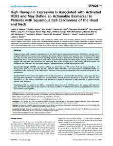
Figure 1. Immunohistochemical staining for SIRT1 (top panels) or DBC1 (bottom panels) in colorectal cancers. From left to right, representative panels of cancers where expression of the proteins is judged to be negative, low or high are shown. Magnification, x200.
Imager (GE Healthcare). The signals of SIRT1 and DBC1 were measured quantitatively using ImageQuant TL software (GE Healthcare) and normalized with that of β-actin.
IHC
WB total
total
low
high
low
49
26
high
13
19
32
62
45
107
R=0.229 p=0.020
75
analyses were done using Cox’s proportional hazard analysis.
Results
Expressions of SIRT1 and DBC1 determined by immunohistochemical staining and Western blot Mutation of TP53 was judged by combination of direct analysis Determination of the TP53 gene mutation
sequencing of the gene, accumulation of TP53 protein in Western blot analysis and that in immunohistochemistry (IHC). For direct sequencing of the gene, DNA was extracted from tissue samples as previously described and 96 cases were subjected to direct sequencing of 1,642 bp region spanning exons 5 to 8. Primers used for the amplification were : a forward primer, 5¢-CTG TTC ACT TGT GCC CTG ACT TTC AAC-3¢; a reverse primer, 5¢-TCT GAG GCA TAA CTG CAC CCT TGG TCT-3¢. Sequencing primers were 5¢-CTG TTC ACT TGT GCC CTG ACT TTC AAC-3¢ for exon 5, 5¢-AGG GCC ACT GAC AAC CAC CCT TAA C-3¢ for exon 6, 5¢-ACA GGT CTC CCC AAG GCG CAC TGG-3¢ for exon 7, and 5¢-TCT GAG GCA TAA CTG CAC CCT TGG TCT-3' for exon 8. In Western blots, p53 were detected using anti-p53 mouse monoclonal antibody (clone DO7; DAKO, Copenhagen, Denmark). TP53 was judged to carry mutation when the mutation was present in the sequence or, in cases where sequence data was unavailable or no mutation was detected, overexpression of the protein was observed both in Western blot analysis and in IHC. TP53 was judged to be wild type when no mutation was present in the sequence and no accumulation of the protein was detectable both by Western blot analysis and IHC.
Statistical analysis
Statistical analyses were performed using the SPSS software (version 19.0, IBM SPSS). Correlations among SIRT1 expression, DBC1 expression and clinicopathological variables were determined using the χ2 test (2-tailed). Cancer specific survival plots were generated using the Kaplan–Meier method and differences between the patient groups were determined by a log-rank test. Multivariate
We evaluated expressions of SIRT1 and DBC1 in colon tissues from 114 patients by both immunohistochemical (IHC) staining of formalin-fixed, paraffin-embedded specimens and Western blot analysis. After confirmation of specificity of anti-SIRT1 or anti-DBC1 antibodies in IHC and Western blot analysis using RNAi-mediated reduction of the proteins in colon cancer cells (Supplement figure S1), we measured expression of SIRT1 and DBC1 in normal and cancerous tissues of colon. Evaluation by IHC (Figure 1) showed that high expressions of SIRT1 and DBC1 were observed in 22 out of 110 cancers (20%) and 45 out of 107 cancers (42%), respectively (Table 1). In Western blot analysis, we measured band densities of SIRT1 or DBC1 and scored expression levels with the highest expression as 100 (Figure 2A). Although cancer-to-normal ratios of expression levels of SIRT1 or DBC1 were variable depending on the cases, in average expression of SIRT1 and DBC1 were elevated in cancers comparing to normal tissues (3.0-fold increase with p < 0.001 for SIRT1 and 3.0-fold increase with p < 0.001 for DBC1) (Supplement figure S2). From the distributions of expression levels (Figure 2B), we defined high expression of SIRT1 with scores of over 30 and that of DBC1 with scores of over 30. With these criteria, high SIRT1 expression cases were 7 cases (6.1%) in normal tissues and 32 cases (28.1%) in cancers, and high DBC1 expression cases were 7 cases (6.1%) in normal tissues and 34 cases (29.8%) in cancers. When the results of IHC and Western blot analysis are compared, those of SIRT1 coincided in 80 out of 110 cases (73%) and those of DBC1 coincided with 68 out of 107 cases (64%) (Table 1). Fifty-five cases showed consistent results of IHC and Western blot analysis for both SIRT1 and DBC1 and were used for further analysis. 3
Kikuchi et al. Journal of Cancer Therapeutics & Research 2013, http://www.hoajonline.com/journals/pdf/2049-7962-2-1.pdf
doi: 10.7243/2049-7962-2-1 Table 2. Frequency of high expression of SIRT1 or DBC1 in colorectal cancer and its correlation with clinicopathological features.
P value * -
-
-