JOURNAL OF BACTERIOLOGY, Oct. 2008, p. 6392–6397 0021-9193/08/$08.00⫹0 doi:10.1128/JB.00777-08 Copyright © 2008, American Society for Microbiology. All Rights Reserved.
Vol. 190, No. 19
Highly Efficient In Vitro Site-Specific Recombination System Based on Streptomyces Phage BT1 Integrase䌤† Lin Zhang,1 Xijun Ou,1 Guoping Zhao,1,2,3* and Xiaoming Ding1* State Key Laboratory of Genetic Engineering, Department of Microbiology and Microbial Engineering, School of Life Sciences, Fudan University, Shanghai 200433, China1; Institute of Plant Physiology and Ecology, Shanghai Institutes for Biological Sciences, Chinese Academy of Sciences, Shanghai 200032, China2; and Shanghai-MOST Key Laboratory of Health and Disease Genomics, Chinese National Human Genome Center at Shanghai, Shanghai 201203, China3 Received 3 June 2008/Accepted 28 July 2008
The Streptomyces phage BT1 encodes a site-specific integrase of the large serine recombinase subfamily. In this report, the enzymatic activity of the BT1 integrase was characterized in vitro. We showed that this integrase has efficient integration activity with substrate DNAs containing attB and attP sites, independent of DNA supercoiling or cofactors. Both intra- and intermolecular recombinations proceed with rapid kinetics. The recombination is highly specific, and no reactions are observed between pairs of sites including attB and attL, attB and attR, attP and attL, or attP and attR or between two identical att sequences; however, a low but significant frequency of excision recombination between attL and attR is observed in the presence of the BT1 integrase alone. In addition, for efficient integration, the minimal sizes of attB and attP are 36 bp and 48 bp, respectively. This site-specific recombination system is efficient and simple to use; thus, it could have applications for the manipulation of DNA in vitro. ing and host cofactors (18). In addition, the attachment sites of the C31 integrase are smaller than those of most tyrosine recombinases, with minimal sizes of 34 bp for attB and 39 bp for attP (11, 18). Streptomyces phage BT1, a homoimmune relative of C31, is inserted into the chromosomes of a wide range of Streptomyces spp. for lysogeny and encodes a large serine recombinase (5, 8). Similar to those of the C31 site-specific recombination system, the attB and attP sites of BT1 are quite different. They share only a 9-bp common sequence, which contains GT core dinucleotides flanked by an imperfect inverted repeat, at which strand exchange and cleavage can occur (8). Multiple pseudo-attP and -attB sites in mouse genomes were also found (3), and three pseudo-attP and two pseudoattB sites were identified in the intergenic regions of five different human chromosomes (5). BT1 integrase-mediated site-specific recombinations in Streptomyces spp. (8), in mice (2–4, 19), and in the human genome (5) were studied; however, neither biochemical characterization nor application of this recombination system in vitro has been reported to date. Here, we purified the heterogeneously expressed BT1 integrase and studied its catalyzed site-specific recombinations in vitro. Our results show that the BT1 integrase can catalyze integration of attB and attP efficiently and excision of attL and attR with low frequency without the RDF, and this is the first report that a large serine recombinase can catalyze bidirectional reactions in vitro. In addition, the minimal sizes of attB and attP required for efficient integration were determined.
Site-specific recombinases have been widely used in genetic engineering: for example, in vitro cloning, plant and mammalian cell genome modification, and gene therapy (3, 11, 12, 15, 16). Nearly all site-specific recombinases can be classified as tyrosine recombinases, also known as the integrase family, or serine recombinases, also known as the resolvase/invertase family, based on comparisons of amino acid sequences and different mechanisms of catalysis; these two types use tyrosine or serine, respectively, to attack the DNA sugar-phosphate backbone (14, 18, 20). The process of strand exchange catalyzed by tyrosine recombinases involves a Holliday junction intermediate and cleavage and rejoining of the strands one by one. In contrast, serine-mediated recombination involves a process of double-strand breakage, followed by rotation and religation (9). The well-known tyrosine recombinase integrase recognizes two different attachment substrate sites, the 25-bp attB gene (in the bacterial chromosome) and the 240-bp attP gene (in the phage genome), which are inverted into attL and attR with the aid of the integration host factor (a chaperone). Furthermore, excision between attL and attR requires recombination directionality factor (RDF), and recombinations mediated by tyrosine recombinases require supercoiling of the substrate DNAs (10). In contrast, the extensively studied large serine recombinase from Streptomyces phage C31 is thought to catalyze unidirectional recombination between DNA substrates attB and attP, independent of DNA supercoil-
* Corresponding author. Mailing address: Department of Microbiology and Microbial Engineering, School of Life Sciences, Fudan University, Shanghai 200433, China. Phone for Xiaoming Ding: 86 21 65643616. Fax: 86 21 65650149. E-mail:
[email protected] .cn. Phone for Guoping Zhao: 86 21 50801919. Fax: 86 21 50801922. E-mail:
[email protected]. † Supplemental material for this article may be found at http://jb .asm.org/. 䌤 Published ahead of print on 8 August 2008.
MATERIALS AND METHODS Heterogenous expression, purification, and analysis of BT1 integrase. The BT1 int gene was amplified by PCR from pFDZ15 (see Table S1 in the supplemental material), using primers ZL16 (5⬘-CATATGTCGCCCTTCATCG CTCCCGA-3⬘) and ZL18 (5⬘-CTCGAGCTACAGCGCCGCAAGCTCCCGC T-3⬘). The resulting 1,790-bp PCR product was digested with NdeI-XhoI and cloned into pET-28b(⫹) (Novagen) to yield pZL5808, which carries a His tag-
6392
VOL. 190, 2008
BT1 INTEGRASE-BASED IN VITRO RECOMBINATION
6393
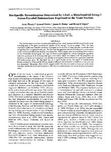
FIG. 1. In vitro recombination between attB and attP catalyzed by BT1 integrase. (A) Fractions from each purification step were obtained and analyzed by sodium dodecyl sulfate-polyacrylamide gel electrophoresis. BT1 integrase (68.2 kDa) displayed a band corresponding to the molecular mass marker of 66 kDa. Shown are total cell proteins from lysed BL21(DE3)/pZL5808 with (lane 1) or without (lane 2) IPTG induction, crude cell extract (lane 3), Ni-NTA column flowthrough (lane 4), four continuous fractions eluted from the column (lanes 5 to 8), gel filtration-purified protein (lane 9), and molecular markers (lane M). (B) Schematic diagrams of the substrates used; the expected recombination products are shown in panels C and E. (C) Recombination between supercoiling or linearized plasmids pZL5811 and pZL5812 (lanes 1 to 6). Substrates or the products were digested with Kpn⌱, followed by gel analysis. Supercoiled plasmid pZL5815 containing attB and attP was used as the substrate, and the recombinant products were digested with EcoRI, followed by gel electrophoresis (lanes 7 and 8). Multi-attR products in the assays were due to inter- and intramolecular reactions (see scheme in panel D for the mechanisms). (D) Schematic diagrams of the substrates used and the expected recombination products shown in panels C and F. (E) Time course of in vitro integration between two linearized att fragments, pZL5811 and pZL5812, predigested with Kpn⌱, illustrated as the scheme in panel B. The reaction times are indicated. (F) Time course of recombination with a substrate of linearized pZL5815 DNA containing attB and attP in direct orientation (see scheme in panel D). The reaction times are indicated.
fused N-terminal BT1 integrase gene. BL21(DE3) (Novagen), freshly transformed with pZL5808, was grown with 2⫻ YT (13) at 37°C for 3 to 4 h until late log phase (optical density at 600 nm, 0.8 to 1.0). Isopropyl -D-1-thiogalactopyranoside (IPTG) was added to give a final concentration of 0.1 mM to induce expression of the integrase, and growth at 20°C was continued overnight (10 to 16 h). After the cells were harvested by centrifugation, the pellets were resuspended, ultrasonicated, and subjected to Ni-nitrilotriacetic acid (NTA) purification according to the manufacturer’s instructions (Invitrogen Ni-NTA purification system). The fractions containing integrase were maintained in TEDS buffer (20 mM Tris-HCl, 2 mM EDTA, 1 mM dithiothreitol, 1 M NaCl, pH 8.0) and concentrated by ultrafiltration (Millipore) to give a final concentration of 3 mg/ml. After the addition of glycerol to give a final concentration of 50% (vol/vol), the purified protein was stored at ⫺30°C. For the further purity of the integrase, gel filtration was performed using a Superdex 200 10/300 GL column (Amersham Biosciences) connected to a fast protein liquid chromatography
system (GE AKTA purifier). The integrase peak fractions were pooled and concentrated by ultrafiltration. One unit of enzymatic activity was defined as the amount of enzyme necessary to produce 50% site-specific recombination of 100 ng linearized attB-attP DNA in a total reaction volume of 10 l in 30 min at 30°C, as described below. Construction of plasmids and preparation of DNA fragments for in vitro recombination reactions. The plasmids used in this study are listed in Table S1 in the supplemental material. Preparation of the Streptomyces coelicolor M145 genomic DNA was performed as described by Kieser et al. (13). The S. coelicolor chromosomal fragment containing attB (886 bp) was PCR amplified using primers ZL24 (5⬘-TGGACCCGGAGCCGATGC-3⬘) and ZL25 (5⬘-CGGAACAGA CGCCGCCAC-3⬘). The resulting PCR product was cloned into the Takara T-cloning vector pMD19 and designated pZL5812. Plasmid pZL5811 (int⫺) is a derivative of pRT802 (int⫹), in which the int gene is disrupted by deletion of an internal EcoRI fragment but the attP site is maintained. Plasmids pZL5813 (int⫹) and pZL5814 (int⫺), containing attL and attR, respectively, in direct orientations,
6394
ZHANG ET AL.
J. BACTERIOL.
TABLE 1. Purification of BT1 integrase No. of Sp act Amt of % integrase (no. of protein (mg) Recovery unitsa units/mg) 3 (10 )
Fraction source
Vol (ml)
Supernatant after ultrasonication Ni-NTA resin column flowthrough Gel filtrate
4.0
12.5
1.2
3.1
2.9
2.3
a
16
1,280
100
9.6
3,100
60
6.8
2,960
43
The unit is defined in Materials and Methods.
were obtained by BT1 integrase-mediated in vitro site-specific integration; plasmid pZL5812 (containing attB resistant to ampicillin) was recombined with plasmid pRT802 or pZL5811 (containing attP resistant to kanamycin), and transformants were then screened using kanamycin and ampicillin. Plasmid pZL5814 was digested by EcoRI and self-ligated to obtain plasmid pZL5816 or pZL5817, containing only attL or attR, respectively. Plasmid pZL5815, containing attB and attP in direct orientation, was constructed by inserting the 716-bp BoxI-KpnI fragment from pZL5812 into pZL5811. Plasmid pDZY101 was constructed for Streptomyces mutagenesis; this plasmid encodes a novel transposon and contains a BT1 attP site. All substrate DNA sequences used for determination of recombination directionality were linearized with restriction enzymes before the assays. The restriction enzymes and the plasmids and their products after digestion were as follows (schematic diagrams are shown in Fig. S2 in the supplemental material): plasmid pZL5812 was digested with Box⌱ to produce a 3,578-bp segment containing attB1, which was digested with BamH⌱ and Ssp⌱ to produce a 2,485-bp segment containing attB2; pZL5814 was digested with Box⌱ to produce a 2,611-bp segment containing attL1, which was digested with Nde⌱ and Nhe⌱ to produce a 2,957-bp segment containing attL2; pZL5816 was digested with EcoR⌱ and HindIII to produce a 1,623-bp segment containing attL3; pZL5813 was digested with Blp⌱ to produce a 3,624-bp segment containing attR1 and digested with Kpn⌱ to produce a 2,477-bp segment containing attR2, which was digested with Nhe⌱ and HindIII to produce an 8,188-bp segment containing attR3; pDZY101 was digested with BamH⌱ and Nru⌱ to produce a 4,947-bp segment containing attP1, and pZL5811 was digested with Sph⌱ and Kpn⌱ to produce a 2,612-bp segment containing attP2, which was digested with EcoR⌱ to produce a 4,434-bp segment containing attP3. For intramolecular excision assays, plasmid pZL5813 was digested with HindIII to produce an 8,724-bp segment containing attL and attR in direct orientations. Standard in vitro recombination assays. Standard recombination was carried out in a reaction mixture (10 l) containing 10 mM Tris-HCl, pH 8.0, 100 mM KCl, and integrase (4 U in 0.5 l). The reaction mixtures were incubated at 30°C for 30 min, and the reactions were terminated by heat inactivation at 75°C for 10 min; the products were separated by electrophoresis on agarose gels. Reaction mixtures using supercoiled plasmids as substrates were heat inactivated, precipitated with ethanol, and dissolved in distilled water for digestions and then separated by agarose gel electrophoresis. A linearized DNA fragment termed attB-attP was prepared by digestion of plasmid pZL5815 with EcoR⌱ for activity unit definition. The amount of DNA recombination was determined by agarose gel electrophoresis and optical-density-scanning analysis. Unless otherwise indicated, 4 U integrase was used for each reaction.
RESULTS AND DISCUSSION Isolation of the BT1 integrase. The 〉⌻1 integration system has been used for genomic manipulations in streptomycete, mouse, and human cells in vivo (4, 5, 8, 19). To characterize the enzymatic properties of the 〉⌻1 recombinase in vitro, we expressed the int gene product in Escherichia coli and purified the recombinant integrase to near homogeneity by using the procedures described in Materials and Methods. Figure 1A shows the isolated enzyme, with a purity of 90 to 95% as determined by optical-density-scanning analysis of the Coomassie blue-stained sodium dodecyl sulfate-polyacryl-
amide gel electrophoresis gel. Table 1 summarizes the isolation of the 〉⌻1 recombinase, which resulted in threefold purification overall and 43% recovery. To avoid precipitation of the enzyme, the 〉⌻1 recombinase was kept in buffer containing 1 M NaCl. The BT1 integrase catalyzes site-specific integration efficiently. We developed in vitro assays to monitor the activity of BT1 integrase (see Materials and Methods). To assess intermolecular integration, purified integrase was incubated with two supercoiled plasmids containing equimolar amounts of attB and attP. After digestion with restriction enzymes, integration products of the expected sizes were observed following agarose gel electrophoresis (Fig. 1C, lane 2). As shown, incubation of a pair of supercoiled and linearized plasmids resulted in a level of integration efficiency similar to that observed with supercoiled substrates (Fig. 1C, compare lane 2 with lanes 4 and 6). Furthermore, when linearized segments containing attB
FIG. 2. Determination of the directionality of BT1 integrase-mediated in vitro recombination. (A) In vitro intermolecular recombinations using different linearized DNA substrates. The preparation of substrates is described in Materials and Methods. The att substrates determined the positions of attB1 (3,578 bp; lanes 1, 2, 3, 4, and 8), attB2 (2,485 bp; lane 8), attP1 (4,947 bp; lanes 1 and 2), attP2 (2,612 bp; lane 9), attP3 (4,434 bp; lanes 5, 6, and 9), attL1 (2,611 bp; lanes 5, 7, and 10), attL2 (2,957 bp; lanes 3 and 10), attR1 (3,624 bp; lanes 6, 7, and 11), and attR2 (2,477 bp; lanes 4 and 11). If recombination had occurred, the product sizes would be markedly different from the substrate sizes. Recombination was detected only between attB and attP (lane 2) and between attL and attR (lane 7). The positions of products attR (8,186 bp) and attP (6,061 bp) are indicated; the small products attL (362 bp) and attB (177 bp) could not be detected by agarose gel electrophoresis. Molecular mass markers (M) are the same as those in Fig. 1. (B) Brief time course of intermolecular excision recombination between attL3 (1,623 bp) and attR3 (8,188 bp). As expected, attB (3,578 bp) and attP (6,233 bp) were produced. Incubation times are indicated. (C) Schematic diagrams of the substrates used and the expected recombination products from panel B.
VOL. 190, 2008
BT1 INTEGRASE-BASED IN VITRO RECOMBINATION
6395
FIG. 3. Determination of minimal attB and attP sizes for efficient in vitro integration. (A) The sequences of the 73-bp attB and attP genes and the schemes of attL and attR are presented. The minimal sequences of attB and attP are underlined, with the 9-bp common sequence highlighted by a box and the GT core dinucleotide shown in bold. The schematic diagrams above and below the sequences illustrate the structural features of the digested fragments containing attB and attP; these fragments were used for full-length att genes and as the PCR template for generating smaller att fragments, respectively. Solid boxes denote the corresponding minimal sites, hollow arrows denote the primer pairs used for amplification of the att fragments with the left side truncated, and solid arrows denote primers used for amplification of those with the right side truncated. (B) The minimal size of attB was determined using purified attB segments (PCR products amplified from pZL5812; 0.3 or 0.6 kb) and restricted linear attP segments (pDZY101 digested with Nhe⌱ and Xho⌱; 5.1 kb). Numbers with minus or plus signs denote the shorter attB (attBs) substrate, generated with primers (see Table S3 in the supplemental material) at the corresponding positions on the left or right side of attB, respectively. Lanes C on both sides of the gel contain positive controls in which full-length attB (0.9-kb PCR product) was recombined with attP, as described above. Molecular mass markers (M) are the same as those in Fig. 1. (C) The minimal size of attP was found to be similar to that shown in panel B. Results are shown for reaction mixtures containing attP segments (PCR products amplified from pDZY101; 0.3 or 0.6 kb) and restricted linear attB segments (pZL5812 digested with Ssp⌱, 3.6 kb). The attB segment for positive controls was a 0.9-kb PCR product. The designations are the same as those used for panel B.
and attP were used as substrates, integration products formed efficiently (Fig. 1E). These findings demonstrate the efficient DNA-supercoiling-independent integration of DNAs containing attB and attP. To probe for intramolecular integration, we constructed pZL5815 (see Materials and Methods). As shown in Fig. 1D, this substrate contains directly oriented attB and attP on the
same molecule, and intramolecular integration is expected to produce linear attL and multi-attR products. As shown, incubation of supercoiled pZL5815 with the BT1 integrase followed by restriction digestion yielded linear attL and multiattR products (including the single bottom band of attR) of the expected sizes (Fig. 1C, lane 8), and linearized pZL5815 was efficiently inverted into the integration products (Fig. 1F).
6396
ZHANG ET AL.
Thus, the BT1 integrase is highly active in catalyzing intramolecular integration. The BT1 integrase can convert more than 90% of linearized substrates into products within 2 hours by inter- and intramolecular reactions (Fig. 1E and F). This indicates a significantly higher integration rate than those reported for the C31 and Rv1 integrases (1, 18); however, this rate seems similar to that of the Bxb1 site-specific integration system (6), which shows rapid kinetics. Optimal conditions for the BT1 integrase-catalyzed recombination. Using the intramolecular integration assays described above, we determined several parameters for optimal integration by the BT1 integrase. We tested a range of pH values and found the optimal pH to be 7.0 to 9.0 (see Fig. S1A in the supplemental material). The pH stability of the enzyme was examined by preincubating the integrase at the indicated pH for 30 min and then assessing its remaining activity at pH 8.0. As shown, the activity and stability curves largely overlapped (see Fig. S1A in the supplemental material). The optimal temperature for BT1 integrase activity was 34°C (see Fig. S1B in the supplemental material). Enzyme stability was determined by preincubation at the indicated temperatures for 30 min followed by activity measurement at 30°C. The results show that the enzyme was stable up to 32°C but inactivated rapidly when the temperature reached 35°C (see Fig. S1B in the supplemental material). Further experiments at 34, 35, and 36°C revealed that 35°C was the transition temperature, at which the integrase had a half-life of approximately 15 min (see Fig. S1C in the supplemental material). We tested the effects of metal ions and other agents on the activity of the BT1 integrase, and the results are summarized in Table S2 in the supplemental material. Several metal ions were found to activate integration: Na⫹, K⫹, Mg2⫹, and Ca2⫹ (in decreasing order of magnitude of activation). Other metal ions had inhibitory effects. Low concentrations of commonly used protective agents, such as divalent metal chelators or high potential reducing agents, had no significant effects on activity. However, greater concentrations of these agents (10 mM) showed some inhibitory effects; the only exceptions were for dithiothreitol and bovine serum albumin. Directionality of the BT1 integrase-catalyzed recombination. The directionality of BT1 integrase-mediated in vitro recombination is shown in Fig. 2. Linear DNA substrates were designed to differ both in their lengths and in the locations of their att sites in order to distinguish the substrates from the products in the assays (see Fig. S2 in the supplemental material). Although efficient recombination was observed between attB and attP, no recombination was detected between attB and attL or attR, between attP and attL or attR, or between two identical sites of attB, attP, attL, or attR (Fig. 2A). However, in contrast to what was found for the other large serine recombinases, such as C31 integrase, Bxb1 integrase, and Rv1 integrase (1, 7, 18), in vitro excision between attL and attR was observed (Fig. 2A, lane 7). Although the efficiency of excision is low, it is significant when BT1 integrase is the only protein present. A time course of excision demonstrated a gradual increase in levels of the attB and attP DNA products (Fig. 2B and C). PCR amplification using the products of excision reactions as templates, followed by sequencing, confirmed that attB and attP were produced (data not shown). Thus, attL and
J. BACTERIOL.
attR were truly inverted into attB and attP during the excision recombination that was catalyzed by the integrase only. Therefore, we conclude that the BT1 integrase can catalyze bidirectional site-specific recombinations in the absence of RDF or any cofactors, although the efficiency of excision is low. This is the first report that a large serine recombinase can perform excision in vitro, which might account for the observation that none of the integrase-catalyzed reactions could convert all of the DNA substrates containing attB and attP into attL and attR despite prolonged incubation. Minimal substrate sizes of attB and attP for efficient in vitro integration. The minimal attB and attP sizes required for efficient integration were determined by in vitro reactions using DNA PCR products of different sizes (Fig. 3). A series of primers were designed (see Table S3 in the supplemental material) to amplify the shorter attB (attBs) substrate, in which one side was truncated relative to the GT-dinucleotide core and the other side was unchanged. The PCR product with a 17-bp left side could recombine with full-length attP at an activity level equal to those of the larger products. However, the recombination efficiency of the PCR product with a 16-bp left side was markedly reduced (Fig. 3B). Thus, we conclude that 36 bp (17 bp for each side, with the GT dinucleotide core in the center) is the minimal size of attB for in vitro integration. A similar series of experiments were performed on attP, and 48 bp was found to be the minimal size (Fig. 3C). This result was similar to those for other serine recombinases, such as C31 (34 bp and 39 bp, respectively), TP901-1 (31 bp and 50 bp), Bxb1 (38 bp and 48 bp), and Rv1 (40 bp and 52 bp) (1, 6, 10, 11, 17). It is therefore likely that a small size difference is a requirement for attB and attP, which may be a common feature of serine site-specific recombination systems (1). In conclusion, this novel BT1 integrase-based in vitro sitespecific recombination system is highly efficient with defined minimal substrates and could have applications for manipulation of DNA in vitro. ACKNOWLEDGMENTS We thank Margaret C. M. Smith for the gift of plasmid pRT802. This work was supported by grants from the National High Technology Research and Development Program of China (863 Program) (no. 2007AA021206) and the National Natural Science Foundation of China (no. 30600009). REFERENCES 1. Bibb, L. A., M. I. Hancox, and G. F. Hatfull. 2005. Integration and excision by the large serine recombinase Rv1 integrase. Mol. Microbiol. 55:1896– 1910. 2. Chen, L., S. N. Thung, and S. L. C. Woo. 2007. Metabolic basis of sexual dimorphism in PKU mice after genome-targeted PAH gene therapy. Mol. Ther. 15:1079–1085. 3. Chen, L., and S. L. C. Woo. 2005. Complete and persistent phenotypic correction of phenylketonuria in mice by site-specific genome integration of murine phenylalanine hydroxylase cDNA. Proc. Natl. Acad. Sci. USA 102: 15581–15586. 4. Chen, L., and S. L. C. Woo. 2007. Correction in female PKU mice by repeated administration of mPAH cDNA using phiBT1 integration system. Mol. Ther. 15:1789–1795. 5. Chen, L., and S. L. C. Woo. 2008. Site-specific transgene integration in the human genome catalyzed by phiBT1 phage integrase. Hum. Gene Ther. 19:143–152. 6. Ghosh, P., A. I. Kim, and G. F. Hatfull. 2003. The orientation of mycobacteriophage Bxb1 integration is solely dependent on the central dinucleotide of attP and attB. Mol. Cell 12:1101–1111. 7. Ghosh, P., L. R. Wasil, and G. F. Hatfull. 2006. Control of phage Bxb1 excision by a novel recombination directionality factor. PLoS Biol. 4:964– 974.
VOL. 190, 2008 8. Gregory, M. A., R. Till, and M. C. M. Smith. 2003. Integration site for Streptomyces phage BT1 and development of site-specific integrating vectors. J. Bacteriol. 185:5320–5323. 9. Grindley, N. D. F., K. L. Whiteson, and P. A. Rice. 2006. Mechanisms of site-specific recombination. Annu. Rev. Biochem. 75:567–605. 10. Groth, A. C., and M. P. Calos. 2004. Phage integrases: Biology and applications. J. Mol. Biol. 335:667–678. 11. Groth, A. C., E. C. Olivares, B. Thyagarajan, and M. P. Calos. 2000. A phage integrase directs efficient site-specific integration in human cells. Proc. Natl. Acad. Sci. USA 97:5995–6000. 12. Hartley, J. L., G. F. Temple, and M. A. Brasch. 2000. DNA cloning using in vitro site-specific recombination. Genome Res. 10:1788–1795. 13. Kieser, T., M. J. Bibb, M. J. Buttner, K. F. Chater, and D. A. Hopwood. 2000. Practical Streptomyces genetics. The John Innes Foundation, Norwich, United Kingdom. 14. Lewis, J. A., and G. F. Hatfull. 2003. Control of directionality in L5 integrase-mediated site-specific recombination. J. Mol. Biol. 326:805–821.
BT1 INTEGRASE-BASED IN VITRO RECOMBINATION
6397
15. Luo, H., L. A. Lyznik, D. Gidoni, and T. K. Hodges. 2000. FLP-mediated recombination for use in hybrid plant production. Plant J. 23:423–430. 16. Malla, S., F. Dafhnis-Calas, J. F. Y. Brookfield, M. C. M. Smith, and W. R. A. Brown. 2005. Rearranging the centromere of the human Y chromosome with C31 integrase. Nucleic Acids Res. 33:6101–6113. 17. Stoll, S. M., D. S. Ginsburg, and M. P. Calos. 2002. Phage TP901-1 sitespecific integrase functions in human cells. J. Bacteriol. 184:3657–3663. 18. Thorpe, H. M., and M. C. M. Smith. 1998. In vitro site-specific integration of bacteriophage DNA catalyzed by a recombinase of the resolvase/invertase family. Proc. Natl. Acad. Sci. USA 95:5505–5510. 19. Xu, Z., N. C. O. Lee, F. Dafhnis-Calas, S. Malla, M. C. M. Smith, and W. R. A. Brown. 2008. Site-specific recombination in Schizosaccharomyces pombe and systematic assembly of a 400kb transgene array in mammalian cells using the integrase of Streptomyces phage BT1. Nucleic Acids Res. 36:e9. 20. Yang, W., and T. A. Steitz. 1995. Crystal-structure of the site-specific recombinase gamma-delta resolvase complexed with a 34 bp cleavage site. Cell 82:193–207.