HIPPOCAMPUS 12:291–303 (2002)
Hippocampal Spatial Representations Require Vestibular Input Robert W. Stackman, Ann S. Clark, and Jeffrey S. Taube* Department of Psychological and Brain Sciences, Dartmouth College, Hanover, New Hampshire
ABSTRACT: The hippocampal formation is essential for forming declarative representations of the relationships among multiple stimuli. The rodent hippocampal formation, including the entorhinal cortex and subicular complex, is critical for spatial memory. Two classes of hippocampal neurons fire in relation to spatial features. Place cells collectively map spatial locations, with each cell firing only when the animal occupies that cell’s “place field,” a particular subregion of the larger environment. Head direction (HD) cells encode directional heading, with each HD cell firing when the rat’s head is oriented in that cell’s particular “preferred firing direction.” Both landmarks and internal cues (e.g., vestibular, motor efference copy) influence place and HD cell activity. However, as is the case for navigation, landmarks are believed to exert greater influence over place and HD cell activity. Here we show that temporary inactivation of the vestibular system led to the disruption of location-specific firing in hippocampal place cells and direction-specific discharge of postsubicular HD cells, without altering motor function. Place and HD cell activity recovered over a time course similar to that of the restoration of vestibular function. These results indicate that vestibular signals provide an important influence over the expression of hippocampal spatial representations, and may explain the navigational deficits of humans with vestibular dysfunction. Hippocampus 2002;12:291–303. ©
2002 Wiley-Liss, Inc.
KEY WORDS: place cell; head direction cell; theta cell; spatial memory; vestibular system; navigation
INTRODUCTION The hippocampal formation is an essential brain substrate for declarative memories of multiple stimuli and the relationships among them (Eichenbaum, 1999; Zola et al., 2000). Considerable empirical data indicate the importance of the rodent hippocampal formation for spatial memory (MorGrant sponsor: National Institute on Deafness and Other Communication Disorders; Grant number: DC00236; Grant sponsor: National Institute of Mental Health; Grant numbers: MH48924, MH01286. A portion of this research was presented in preliminary form at the 26th annual meeting of the Society for Neuroscience in Washington, DC. Present address for R.W.S. is the Department of Behavioral Neuroscience, L470, Oregon Health Sciences University, 3181 SW Sam Jackson Park Road, Portland, OR 97201-3098. E-mail:
[email protected] *Correspondence to: Jeffrey S. Taube, Ph.D., 6207 Moore Hall, Dartmouth College, Hanover, NH 03755-3549. E-mail:
[email protected] Accepted for publication 16 July 2001 DOI 10.1002/hipo.1112 Published online 00 Month 2002 in Wiley InterScience (www.interscience. wiley.com). ©
2002 WILEY-LISS, INC.
ris et al., 1982; Jarrard, 1993). Two types of neurons with firing patterns related to spatial behavior have been found within the hippocampal formation. The discharge of a place cell is best associated with the spatial location of the animal. The place cell exhibits its peak discharge when the animal occupies that cell’s “place field,” a particular subregion of the larger environment (O’Keefe and Nadel, 1978). Head direction (HD) cells (Taube, 1998) encode directional heading, with each HD cell firing when the rat’s head is oriented in a particular “preferred firing direction.” Hippocampal place cells and HD cells may be interactive components of a brain circuit that guides navigation (McNaughton et al., 1996; Taube, 1998). Both landmarks and internal cues (e.g., vestibular, motor efference copy) influence spatial memory and navigation (Landeau et al., 1984; Etienne et al., 1996; McNaughton et al., 1996; Whishaw and Gorny, 1999). In general, when familiar landmarks are not available, such as in the dark or in a novel location, navigation (or path integration) depends on any available cues. For example, a rodent can return directly to its home nest after a circuitous outward journey in complete darkness, by integrating vestibular and other self-motion cues of the outbound trip (Mittelstaedt and Mittelstaedt, 1980; Etienne et al., 1985). Repeated “disorientation” of rats disrupted spatial learning in an appetitive radial-arm maze task (Dudchenko et al., 1997; Martin et al., 1997), but did not impair learning in the Morris water maze task. Repeated disorientation may have disrupted the rats’ ability to form a representation of the goal location with respect to extramaze cues (Dudchenko et al., 1997; Martin et al., 1997). Lesions of the vestibular system impair: 1) a rat’s ability to return to a goal location following passive transport (Miller et al., 1983); 2) spontaneous alternation (Potegal et al., 1977); 3) navigation in the absence of a visual landmark (Stackman and Herbert, 2002); and 4) spatial learning in a radial-arm maze task (Ossenkopp and Hargreaves, 1993). The results from studies of vestibularlesioned animals indicate that the lesion causes deficits that are not simply a consequence of motor impairment or due to a lack of attention or motivation. Collectively, these results indicate an important contribution of the
292
STACKMAN ET AL.
vestibular system to hippocampal-dependent spatial memory and navigation. Place and HD cell activity are also influenced by both external and internal cues, although landmarks are believed to exert greater influence over the spatially tuned activity (Quirk et al., 1990; Goodridge and Taube, 1995). Place and HD cell activity is preserved in the absence of external cues, even despite blindness or deafness (Hill and Best, 1981; Goodridge et al., 1998). Therefore, internal self-motion cues also play an important role in influencing the spatial firing properties of hippocampal neurons (Foster et al., 1989; Taube and Burton, 1995; Knierim et al., 1998). Indeed, vestibular stimulation activates the hippocampal formation (Horii et al., 1994; Vitte et al., 1996), influences hippocampal place cell activity (Sharp et al., 1995), and influences navigation in humans (Telford et al., 1995). Finally, lesions of the vestibular apparatus abolish the directional firing of anterior thalamic neurons (Stackman and Taube, 1997) and impair spatial memory, as outlined above. Therefore, vestibular input to the hippocampus may be critical for spatial navigation and for updating brain representations of spatial information (McNaughton et al., 1996; Smith, 1997). To test this assumption, we examined the spatial firing of hippocampal neurons before and after reversible inactivation of the vestibular system.
pus (n ⫽ 10), or the postsubiculum (n ⫽ 3). Electrode construction and implantation techniques used were similar to that described previously (Taube et al., 1990). Briefly, each electrode array consisted of a bundle of 10 25-m diameter nichrome wires (California Fine Wire Co., Grover City, CA), insulated except at the tips. The wire bundle was passed through a 26-gauge stainless steel cannula, and each wire attached to a modified 11-pin Augat connector. The electrode array could be advanced in the dorsoventral plane, with three screws attached to the electrode’s acrylic base (Kubie et al., 1990). Upon habituation to the cylindrical apparatus and adequate foraging behavior, each rat was anaesthetized with a ketamine-xylazine mixture (2 ml/kg, i.m.) and stereotaxically implanted with an electrode array. Electrode coordinates, with respect to bregma, were: CA1, anterior/posterior, 3.0 mm; medial/ lateral, ⫹2.8 mm; ventral, 1.7 mm from the cortical surface; Postsubiculum, anterior/posterior ⫺6.6 mm; medial/lateral, ⫹2.8 mm; ventral, 1.6 mm from the cortical surface (Paxinos and Watson, 1998). Jeweler’s screws placed in the skull plates over the cerebellar cortex, parietal cortex, and frontal cortex, and dental cement, anchored the electrode assembly in place. All procedures were conducted according to an institutionally approved animal care protocol. All surgical procedures were conducted under aseptic conditions, and the rats were allowed a 1-week postoperative recovery interval before single-unit screening commenced.
MATERIALS AND METHODS Single-Unit Recording Subjects and Training Subjects were 13 female Long-Evans rats, weighing 250 –300 g at the beginning of the experiment. Rats were maintained on a food-restricted diet (15–20 g/day) and housed separately in suspended wire mesh cages. Tap water was available ad libitum. All training, unit screening, and recording occurred during sessions in which the rats foraged for food pellets in a cylindrical apparatus (76 cm diameter, 51 cm high). A black floor-to-ceiling curtain enclosure (2 m diameter) surrounded the cylinder, and four uniformly arranged overhead DC lamps provided illumination. A color video camera (Sony XC-711) was centered above the cylinder 3 m from the floor surface. The cylinder was placed on a sheet of gray photographic backdrop paper. A white cue card attached to the inside wall of the cylinder, occupying approximately 100° of arc, served as a visual landmark. Rats received at least 5 training trials (1 trial/ day), during which food pellets (20 mg, P.J. Noyes, Lancaster, NH) were thrown randomly into the cylinder. By the completion of training, rats engaged in nearly continuous food pellet search behavior over the entire floor of the cylinder. All procedures involving the rats were performed in compliance with institutional standards as set forth by the National Institutes of Health’s Guide for the Care and Use of Laboratory Animals and the Society for Neuroscience.
Electrode Implantation Rats were stereotaxically implanted with a single 10-wire advanceable microelectrode array directed at area CA1 of hippocam-
Electrophysiological methods were identical to those described previously (Stackman and Taube, 1997). All recording procedures were conducted while the rats freely moved about a wooden cylindrical apparatus described previously (Taube et al., 1990; Stackman and Taube, 1997). The cylinder contained a single polarizing white cue card covering ⬃100° of the cylinder wall and was present in a fixed position (3 o’clock) at all times. Food pellets (20 mg, P.J. Noyes) were dropped randomly into the cylinder to encourage movement of rats. The recording cable included a red LED that was positioned over the rat’s snout, a green LED positioned over the rat’s back, and operational amplifiers connected in sourcefollower configuration. Single units were isolated using window discriminators (BAK Electronics, Inc., Germantown, MD), monitored on an oscilloscope (Tektronix, Beaverton, OR), and timestamped. Spike data were integrated with data regarding the position of the rat derived from the X and Y coordinates of the recording cable LEDs generated from a dual-spot video tracking unit (Ebtronics, Brooklyn, NY). Complex-spike and theta cell waveforms were verified by several criteria, as defined by Ranck (1973). After isolating a place cell, theta cell, or HD cell, the cell’s activity was recorded over at least two 16-min (8 min for HD cells) sessions separated by 24 h, to verify stability of the signal. During baseline recording sessions, most place and HD cells were recorded during sessions in which we examined the stimulus control exerted by the polarizing cue card of the cylinder. The amount of control the cue card exerted was assessed by determining the extent to which rotation of the cue card shifted the location of place fields or the preferred firing direction of HD cells. If stable (i.e., 1) pre-
____________________________________
HIPPOCAMPAL SPATIAL FIRING AND VESTIBULAR INPUT
served place field or preferred firing direction, 2) consistent unit waveform characteristics, and 3) similar peak firing rates across recording sessions), the rat was prepared for vestibular inactivation. Once begun, if the isolation of a unit was ever lost, or in question, that experiment was terminated. For one rat, hippocampal EEG records were acquired simultaneous with single-unit recordings. Hippocampal EEG was differentially recorded against the recording system ground. EEG signals were bandpass-filtered (1–30 Hz) and acquired on the audio channel of videotapes for off-line analyses. EEG traces were acquired by passing the audio channel data into an oscilloscope (Hameg, Oceanside, CA). Representative traces were stored on a Pentium computer interfaced with the oscilloscope. Placement of electrodes within the CA1 hippocampus or postsubiculum was verified by histological analysis.
Inactivation of the Vestibular Apparatus Blockade of neuronal activity within the vestibular apparatus was produced by bilateral transtympanic injection of tetrodotoxin (TTX) (12.5 l of 0.6 mM, dissolved in phosphate-buffered saline; Sigma, St. Louis, MO), under Brevital anesthesia (50 mg/kg, i.p.). Injections were made using a 25-l Hamilton syringe. TTX produces a near immediate, but transient, abolition of neural activity within cranial nerve VIII (Beitz et al., 1995), and behavioral changes commensurate with those observed following bilateral labyrinthectomies or transtympanic injection of the vestibular toxin, sodium arsanilate (i.e., head dorsiflexion, a failure of contact-righting, flattened posture with forelimbs and hindlimbs abducted, increased tendency to locomote backwards, and hyperreactivity to handling; Horn et al., 1981; Hunt et al., 1987; Kaufman et al., 1992). The transtympanic TTX injection procedure was also used to examine the anatomical and electrophysiological effects in the cochlear nucleus following VIIIth cranial nerve blockade in the chick (Canady and Rubel, 1992) and gerbil (Pasic and Rubel, 1989). We found the transtympanic injection of TTX in rats to be an efficient and reliable method of inactivating the vestibular system. Following transtympanic TTX, it is possible to determine whether the toxin caused only unilateral inactivation rather than bilateral, as the animal will roll its head, and move in a circular pattern, in the direction of the inactivated side. In case of a unilateral TTX inactivation, the opposite side is injected again to produce a complete bilateral vestibular inactivation. In all cases, after initial bilateral injections of TTX, we observed vestibular dysfunction that was indistinguishable from that of injections of sodium arsanilate. During recovery from Brevital anesthesia (20 –50 min postinjection), most rats engaged in vigorous motoric behaviors, including forward and backward circling, writhing or rolling of their bodies along the anterior-posterior axis, rapid horizontal angular head movements, falling onto their lateral flanks when attempting to walk, and “wet dog shakes.” By 1 h postinjection, all rats had recovered from the Brevital anesthesia and walked around the cylinder floor in an upright position without falling. During the 1-h postinjection recording session, TTX-treated rats were mildly ataxic (see section on Locomotor Activity below). This ataxia and
293
other abnormal motor movements had subsided by 12 h postinjection. We assessed vestibular function before each recording session by the contact-righting test. The contact-righting test is sensitive to vestibular dysfunction induced both surgically and by transtympanic injection of sodium arsanilate (Chen et al., 1986; Ossenkopp et al., 1990; Shoham et al., 1989; Stackman and Taube, 1997). For our purposes, it was important to employ a method to assess labyrinthine righting that limited the possibility of disturbing the hippocampal microdrive/electrode arrays. The contact-righting test requires placing the rat supine on a tabletop surface and bringing a Plexiglas surface into gentle contact with the ventral surface of the rat’s feet. Intact rats will rapidly right themselves upon making contact with the second surface. In contrast, rats with lesions or inactivation of the vestibular system will not right themselves, but will “walk” about under the Plexiglas surface while in the supine position. Following recovery from the anesthetic, cell activity and the rat’s vestibular function were monitored at 1, 4, and 12 h postinjection, and then at 12-h intervals until the rat exhibited complete recovery of vestibular function, as assessed by the contact-righting test.
Analysis of Electrophysiological Data To determine whether a given hippocampal unit exhibited location-specific activity, color-coded place by firing rate maps were constructed for each recording session (Muller et al., 1987). The place field of a place cell was defined as a conglomerate of at least nine contiguous pixels with a firing rate above the mean firing rate. Quantitative measures of place cell firing were overall mean firing rate and mean firing rate within the firing field. Measures of location-specificity for cell firing were: 1) spatial coherence (Kubie et al., 1990), which measures the orderliness of discharge within and outside the place field, calculated by pairing firing rate of a given pixel with the mean firing rate of its eight contiguous neighboring pixels, with a final correlation value computed from all pairs; and 2) information content (Skaggs et al., 1993), a quantitative measure of the amount of information conveyed by each spike. Information content is thought of as a measure of the degree to which cell firing can predict the animal’s location in the cylinder. An information content value of 0 reflects no correlation between the rat’s location and cell firing; a value approaching or greater than 1 indicates a strong positive correlation between location and firing rate. Autocorrelation histograms provide an indication of whether neuronal firing is modulated in a rhythmic manner, as is typical for hippocampal theta cells. Autocorrelation histograms were constructed by summing the number of times a spike occurred within each 1-ms interval from 0 –350 ms, given the occurrence of a spike at time 0. The ordinate of each graph represents the number of spikes that occurred in each 1-ms interval during a 16-min recording session, and is expressed in Hertz. All HD cells were recorded during 8-min sessions, and firing rates by HD tuning functions were constructed for each cell, using procedures described previously (Taube et al., 1990; Stackman and Taube, 1997).
294
STACKMAN ET AL.
RESULTS Vestibular Function In all 10 cases, transtympanic injection of TTX produced vestibular dysfunction, as measured by the contact-righting test (Chen et al., 1986; Shoham et al., 1989; Stackman and Taube, 1997). Vestibular dysfunction was judged to be bilateral if the rat failed to contact-right, showed bilateral circling behavior, and showed no obvious torsion or roll of the head towards one side of the body (as observed following unilateral vestibular lesion). All rats failed to contact-right at 1 h postinjection, and did not recover this vestibular behavior before 48 h postinjection. The time of recovery of contact-righting varied in each case (range, 48 –72 h postinjection). During the 1-h postinjection session, TTX-treated rats tended to locomote in a forward circular pattern (in both directions), and were more apt to walk close to the cylinder walls (thigmotaxis) than in the center of the cylinder. However, the maps of Figure 1 clearly show that locomotor behavior was not confined to the walls; for the most part, all rats moved over the entire floor surface during the post-1-h sessions. All treated rats also displayed postural changes consistent with those reported following bilateral surgical labyrinthectomies, or sodium arsanilate treatment. These behavioral changes included head dorsiflexion, rapid oscillatory head movements in the yaw plane, flattened posture with abducted limbs, hyperreactivity to handling, increased circling behavior (both ipsi- and contralateral), and increased tendency to locomote backwards. Further, when lifted by the base of the tail from a horizontal surface, normal, vestibular-intact rats extend their forelimbs towards the horizontal surface. Exhibition of such a “landing” posture is considered dependent on the vestibular system (Selye, 1957; Hunt et al., 1987). Vestibular-lesioned rats often exhibit a deficient “landing” response, where they will curl their body ventrally around and towards their tail. We noted a similar failure of this “landing” posture in TTX-treated rats during assessments made when transferring rats to and from their home cages. The profile of behavioral changes we observed following bilateral transtympanic injection TTX was consistent with that reported following sodium arsanilate administration or surgical labyrinthectomy (T’Ang and Wu, 1936, 1937; Horn et al., 1981; Chen et al., 1986; Hunt et al., 1987; Shoham et al., 1989; Stackman and Taube, 1997). Finally, all rats appeared to fully recover vestibular function, as assessed by the contact-righting test and from our observations of their behavior. We defined “recovery” as the postlesion time at which rats exhibited a recovery of contact-righting, the lack of postural abnormalities associated with vestibular dysfunction, and the recovery of the “landing” posture when held by the tail, as described above. The recovery interval varied across rats from 48 –72 h postinjection.
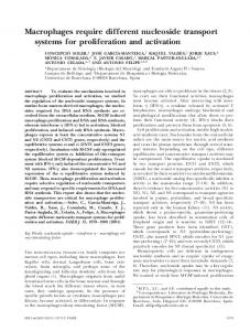
thesia. At 1 h postinactivation, all rats displayed behavioral sequelae characteristic of bilateral lesion of the vestibular apparatus. Two rats received bilateral transtympanic injections of 12.5 l of 0.9% sterile saline as a control for nonspecific effects of inactivation. During recovery from anesthesia, the isolation of two place cells was lost, probably due to the vigorous motor responses elicited during the first 30 min after injection, while under the influence of vestibular inactivation. The data shown below were collected from cells for which unit isolation was maintained over the entire experiment, from baseline through recovery of vestibular function. Vestibular inactivation disrupted the location-specific firing of hippocampal place cells. The place fields of all ten hippocampal cells (Fig. a–j) were severely degraded at 1 h postinjection, as compared to each respective baseline session. Place cell activity remained disrupted for a period of 36 –72 h postinjection. Despite disruption of baseline place fields (in particular, Fig. 1b– h), some hippocampal cells exhibited nonrandom firing patterns during vestibular inactivation. For example at 24 h postinjection, the cells in Figure 1a,i exhibited elevated firing in two locations, and cells in Fig. 1c,d,f,h,i had circular or crescent-shaped firing fields. Although many of the hippocampal neurons exhibited nonrandom patterns of firing after vestibular inactivation, these firing patterns were dramatically different from place fields in normal intact rats and could not be defined as place fields according to our working definition. Given the consistency and stability of cell waveforms (e.g., Fig. 1k), we are confident that electrical isolation of each cell was maintained over the course of inactivation. It is relevant to note the waveform traces of Figure 1k were acquired during the recording of the cell depicted in Figure 1c. In spite of its consistent waveforms over the course of the experiment, the place field of this cell exhibited a ⫺120° shift in position upon recovery, as compared to the baseline session (see below for recovery results). Complex-spike firing patterns, characteristic of hippocampal CA1 neurons, were present during vestibular inactivation (see Fig. 1l), suggesting that hippocampal network properties were preserved in the absence of vestibular input. Place cell activity was not disrupted by transtympanic injections of saline: spatial coherence, information content, and peak firing rate measures did not change appreciably following saline injections, and place fields remained stable in these animals for several days postinjection (data not shown). Vestibular inactivation significantly decreased place cell spatial coherence (Fig. 2a) (F(5,45) ⫽ 18.19, P ⬍ 0.0001), and spatial information content (F(5,45) ⫽ 4.89, P ⬍ 0.01), but did not affect place cell peak firing rate (F(5,45) ⫽ 1.21, n.s.) or average firing rate (F(5,45) ⫽ 0.90, n.s.). Although auditory function was disrupted by tympanic membrane perforation and blockade of auditory hair cell activity, hearing loss is unlikely to have influenced place fields, because location-specific firing remained intact in saline-injected control rats and in deaf rats (Hill and Best, 1981).
Place Cells Individual hippocampal CA1 neurons were recorded from rats moving freely about a cylindrical arena, foraging for food pellets. After recording baseline activity, rats received bilateral transtympanic injections of TTX (Beitz et al., 1995) under Brevital anes-
Locomotor Activity Vestibular inactivation did not disrupt rats’ ability to move about the cylinder and retrieve food pellets (Fig. 2b). All TTXinjected rats continued to forage for and consume food pellets
____________________________________
HIPPOCAMPAL SPATIAL FIRING AND VESTIBULAR INPUT
FIGURE 1. Vestibular inactivation disrupts location-specific firing in hippocampal place cells: examples of firing fields of all 10 cells recorded, before and after inactivation of the vestibular apparatus. a–j: For each map, increasing rates of discharge are coded from yellow, orange, red, green, blue, and purple, with yellow pixels depicting locations visited where no spikes were fired. Pixels that were never visited during the recording session are coded white. Each map was autoscaled such that the number of pixels in the next higher firing rate category was equal to 0.8 times the number of pixels in the lower firing rate category (Muller et al., 1987). For each example, Pre depicts activity recorded during the baseline session; postinjection activity is depicted in the remaining plots under the headings 1 hr, 24
295
hr, 48 hr, and Recovery. In each case, Recovery represents that activity acquired during the recording session at which vestibular function was judged as restored. Respective recovery time points for each cell were as follows: a, 60 h; b, 72 h; c, 60 h; d, 72 h; e, 48 h; f, 72 h; g, 60 h; h, 96 h; i, 72 h; and j, 72 h. k: Unit waveform traces acquired during recording the cell depicted in c, at each of the time points before and after vestibular inactivation (i–v: Pre; Post 1 hr; 24 hr; 48 hr; and Recovery). The calibration scale represents 50 V/200 s. l: Representative spike trace records depicting complex spike activity of the cell depicted in c, acquired at each of the time points before and after vestibular inactivation (i–v: Pre; Post 1 hr; 24 hr; 48 hr; and Recovery). Calibration scale represents 50 V/10 ms.
296
STACKMAN ET AL.
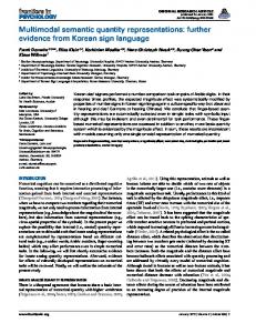
FIGURE 2. a: Vestibular inactivation disrupts spatial selectivity of firing of CA1 neurons: mean place field spatial coherence computed at each time point before and after vestibular inactivation for all 10 place cells. Asterisks indicate time points that were significantly different from Pre baseline, P < 0.05, Fisher’s PLSD test. b: Vestibular inactivation causes transient depression of locomotor activity: mean cumulative distance (in cm) traveled in the cylinder during each 16min recording session at each time point before and after vestibular inactivation. Measures of speed of locomotion reflected a similar de-
crease at 1 h postinjection and full recovery within 24 h. Asterisk indicates time point that was significantly different from Pre baseline, P < 0.05, Fisher’s PLSD test. c: Relative distribution of consequences to place field location upon recovery from vestibular inactivation, as compared to place field location during baseline recording sessions. Place fields were observed to: remain in a position consistent with baseline session (No Shift); undergo an angular shift in position (Angular Shift); undergo a radial shift in position (Radial Shift); or undergo both an angular and radial shift in position (Full Remapping).
throughout the entire postinjection period. However, at 1 h after TTX injection, we observed mild but significant ataxia. As shown in Figure 2b, TTX treatment caused a decrease in mean cumulative distance traveled, and in speed of movement (distance, (F(5,30) ⫽ 4.50, P ⬍ 0.05; speed, F(5,30) ⫽ 7.62, P ⬍ 0.05). This motor deficit was transient, as it was not observed at any other time point. Moreover, whatever motor deficits were apparent at 1 h postinjection had recovered well before the recovery of location-specific firing in place cells. During the first 12 h of vestibular inactivation, rats tended to display mild thigmotaxis, spending more time exploring the region of cylinder floor in near proximity to the wall. However, the place by firing rate maps (Fig. 1) clearly indicate that nearly all cylinder locations were still sampled by rats before and after TTX injection, including the 1-h postinjection time point. In short, rats under the influence of vestibular inactivation continued to locomote about the entire floor surface. Further, since all place fields but one were located adjacent to the cylinder wall during baseline sessions, mild thigmotaxis under vestibular inactivation did not disrupt visits to “former” place field locations. In sum, since hippocampal place coding did not recover until 48 –72 h postinactivation, it is unlikely that this brief alteration in locomotor behavior contributed significantly to the disruption of locationspecific firing.
defined recovery as the respective post-TTX recording session at which there was complete restoration of the contact-righting reflex and other vestibular-related behaviors (as defined above). As shown, many cells showed some recovery of spatially selective firing at 48 h postinactivation. Although we monitored place cell activity at 12-h intervals (activity is shown only at 24-h intervals in Fig. 1), it appeared that robust location-specific firing only returned once the rat showed a completely normal contact-righting reflex. However, monitoring place cell activity and contact-righting at intervals shorter than 12 h, along with applying quantitative measures to these parameters, would be necessary to firmly establish a tight correlation between the two parameters. Four different patterns of place field location were observed at recovery: 1) no shift; the place field remained in the same position as in the baseline session, 2) angular shift; the place field underwent an angular shift compared to the baseline session, 3) radial shift; the place field shifted radially compared to the baseline session, and 4) full remapping; the place field shifted both angularly and radially compared to the baseline session. Figure 2c depicts the distribution of these four observed consequences and shows that the most common response was a shift in place field location (n ⫽ 6). In 4 of these 6 cases, the place field remapped upon recovery (Fig. 1f– h,j). For the 2 other cases, upon recovery, one place field shifted angularly (Fig. 1c), while the other place field shifted radially (Fig. 1b). The cross-correlations between baseline and recovery conditions for cells that remapped were 0.36, 0.43, 0.45, and 0.64. For the two cells with shifted place fields upon recovery, the cross-correlations were 0.90 for the cell that exhibited a ⫺120° shift in its place field, and 0.59 for the cell that exhibited a radial shift in its place
Recovery Location-specific firing recovered approximately coincident with the restoration of vestibular function. However, place fields were often altered upon recovery (Fig. 1a–j). For each cell, we
____________________________________
HIPPOCAMPAL SPATIAL FIRING AND VESTIBULAR INPUT
field. In contrast, upon recovery, the remaining four cells had place fields that matched those observed during baseline (Fig. 1a,d,e,i), with a mean cross-correlation of 0.88 ⫾ 0.02 (range, 0.84 – 0.93). These cells exhibited shifts in their fields of 0°, 12°, ⫺18°, and ⫺6°, respectively, between baseline and recovery sessions. In two cases, two place cells were simultaneously recorded prior to, and over the course of, TTX-induced vestibular inactivation from the same rat. The place fields of the first pair (Fig. 1a,b) underwent a similar degree of TTX-induced disruption, and the place fields appeared to recover to within 12° of their baseline locations. The plane fields of the second pair of cells (Fig. 1c,g) were equally disrupted during the vestibular inactivation, and both fields exhibited a rotational shift upon recovery (cell c ⫽ 120° shift; cell g ⫽ 174° shift). These data suggest that rather than TTX inducing an individual cell to remap or angularly shift its place field, the vestibular inactivation produced a more generalized disruption of hippocampal spatial mapping. Two rats underwent a second vestibular inactivation after new place cells were isolated 1 month later. We did not find any commonality in the degree to which place fields were altered upon recovery of vestibular inactivation between the first and second vestibular inactivation. Specifically, for one animal, no shift in place field location was observed after the first inactivation, but a new place cell exhibited a radial shift after the second inactivation. For the other animal, an angular shift was observed after the first inactivation, while no shift in place field location was observed after the second inactivation. Therefore, it is unlikely that the manner is which place fields respond to vestibular inactivation (e.g., angular shift, remapping) is common to individual animals.
Theta Cells We also investigated the influence of vestibular inactivation on the activity of 14 hippocampal theta cells. Theta cells of the hippocampus 1) are putative interneurons located within the stratum oriens; 2) discharge at a characteristic 4 –12-Hz firing pattern; and 3) exhibit firing rates that are correlated with locomotor behaviors (Ranck, 1973) and minimally influenced by spatial location (Kubie et al., 1990). The observed mean spatial coherence of theta cells during baseline sessions, 0.22 ⫾ 0.03 (range, 0.03– 0.47), was considerably lower than that for place cells (0.62 ⫾ 0.05), although significantly different from zero (t(13) ⫽ 7.74, P ⬍ 0.001). Vestibular inactivation caused little change in the spatial properties of theta cell firing (Fig. 3a,c), and theta cell spatial coherence was not altered over the course of inactivation, (F(5,65) ⫽ 1.38, n.s.). Vestibular inactivation also did not influence theta cell peak firing rates (F(5,65) ⫽ 2.12, n.s.) or average firing rates (F(5,65) ⫽ 0.97, n.s.). Autocorrelograms (i.e., Fig. 3b,d), constructed to reveal the temporal characteristics of theta cell activity, indicated that rhythmic discharge was disrupted during inactivation for eight cells (Fig. 3d), and maintained for six cells (Fig. 3b). These data suggest the presence of two distinct populations of theta cells, one sensitive, and one resistant, to vestibular inactivation. In two cases, both types of theta cells were recorded simultaneously from the same animal. Hippocampal theta cells have been classified as theta On and theta Off, depending on their sensitivity or resistance to cho-
297
linergic drugs and medial septal inactivation (Smythe et al., 1991). It is not known whether the different sensitivities of theta cells to vestibular inactivation correspond to these two cell classes. Rhythmic firing properties were restored consummate with recovery of vestibular function. Representative EEG traces were acquired during each recording session for one theta cell (Fig. 3e). Baseline (Pre) traces revealed an increased rhythmic theta activity during episodes of walking as compared to episodes of immobility (Fig. 3e). This pattern of EEG activity is consistent with previous reports of hippocampal theta cells (Ranck, 1973; Kubie et al., 1990), and was preserved, for the most part, during (24 h) and following vestibular inactivation (Recovery; Fig. 3e). Theta rhythm was also evident in the EEG record of a second rat, at 12 and 24 h following vestibular inactivation. The preservation of movement-related hippocampal theta during vestibular inactivation is consistent with previous reports of intact movement-related hippocampal theta activity in vestibular-deficient rodents (Frederickson et al., 1982; Shoham et al., 1989).
Head Direction Cells Transtympanic TTX also disrupted the directional firing of three postsubicular cells. Head direction (HD) cell activity was disrupted 1 h after inactivation, and recovered with the restoration of vestibular function (48 –72 h postinjection). The preferred firing direction of two HD cells shifted upon recovery (30° and 96°; Fig. 4a– c), suggesting that vestibular inactivation caused some retuning among HD cells. The disruption of directional activity during vestibular inactivation is consistent with our previous report that permanent vestibular lesion abolished anterior thalamic HD cell activity (Stackman and Taube, 1997). We did not attempt to record both hippocampal place cells and HD cells simultaneously; therefore, we do not know whether place and HD cells undergo coincident remapping/reorientation.
DISCUSSION Many studies have demonstrated that the hippocampus plays a critical role in spatial memory (O’Keefe and Nadel, 1978; Jarrard, 1993; Whishaw and Maaswinkel, 1998; O’Keefe, 1999). Rodent and primate hippocampal neurons discharge with respect to spatial information (location and head direction; Rolls and O’Mara, 1995; Robertson et al., 1998), as well as nonspatial information (e.g., cue match/nonmatch; Colombo and Gross, 1994; Deadwyler et al., 1996; Wood et al., 1999). As outlined above, substantial research indicates that external cues or landmarks influence place cells, HD cells, and navigation. Recent data support the notion that in the absence of landmarks, internal (self-motion) cues also influence place cell and HD cell activity, suggesting a hierarchical involvement of external over internal cues. The precise involvement of self-motion cues in the firing of hippocampal place cells is less certain. Tight restraint of a rat within the place field of a hippocampal neuron causes a marked decline in location-specific firing, however if the rat is loosely restrained, location-specific
298
STACKMAN ET AL.
FIGURE 3
FIGURE 4
____________________________________
HIPPOCAMPAL SPATIAL FIRING AND VESTIBULAR INPUT
firing recovers (Foster et al., 1989). These data suggest that hippocampal spatial representations require a “preparedness for movement signal.” However, Gavrilov et al. (1998) recently showed that location-specific hippocampal neuronal firing is intact in rats that are gently restrained and passively transported about an enclosure. Taken together, these data indicate that multiple sources of movement-associated cues influence place cells, yet active motion may not be essential for the firing of hippocampal place cells. Importantly, the present data demonstrate that the capacity of the hippocampal formation to encode location-specific firing requires vestibular input. A consideration to note is that Hill and Best (1981) administered neomycin sulfate to rats daily for 40 – 60 days in order to produce deafness and reported that location-specific firing persisted in hippocampal place cells. Neomycin sulfate is known to be toxic to cochlear hair cells (Leake and Hradek, 1988; Lowenheim et al., 1999), and because of their similarity and close proximity to vestibular hair cells, the drug could also have been toxic to them. If there was a loss of vestibular hair cells following neomycin sulfate treatment, then to be consistent with the results reported here, one
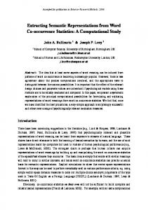
FIGURE 3. Representative examples of hippocampal theta cell activity recorded before and after vestibular inactivation. a, c: Place by firing rate plots reveal minimal alteration in location-specific firing over the course of vestibular inactivation. Spatial coherence values for these cells at baseline and recovery were as follows: a, 0.243 and 0.487; c, 0.200 and 0.205, respectively. b, d: Respective autocorrelation functions observed for the cells plotted in a and c. The autocorrelation function represents the measure of cell firing at each 1 msec interval (from 0 –350 msec), given a spike discharge at time 0. Autocorrelation functions were constructed by normalizing the spike count for each interval with respect to its peak value. The plot of the theta cell shown in b illustrates a preservation of rhythmic discharge over the course of inactivation, while the plot of the cell shown in d indicates a diminution of rhythmic discharge at 24 h and 48 h postinjection. e: Representative 1-s hippocampal EEG traces are depicted for the cell plotted in a, taken at each of the three time points indicated. Upper traces (Immobile) illustrate the EEG when the rat was stationary. In this case, voltage amplitude was decreased and theta rhythm was not evident. Lower traces (Walking) illustrate the EEG during episodes of locomotion when voltage amplitude was increased and theta rhythmicity was observed. Similar patterns of activity were reflected in EEG traces taken during and following vestibular inactivation; however, traces taken at 24 h suggest a slight irregularity in theta frequency over the course of the 1-sec trace. The calibration scale represents 2 mV/ 100 ms. FIGURE 4. Examples of firing rate by HD tuning curves of three postsubicular HD cells recorded from three different rats, before and after inactivation of the vestibular apparatus. For each cell, the baseline activity, or Pre, is represented by a heavy black line. The remaining lines illustrate the postinjection time course of inactivation: red, 1 h; green, 24 h; and blue, vestibular recovery, namely, a, 48 h; b, 72 h; and c, 72 h. For all three cells, directional discharge was abolished by 1 h postinjection. Upon recovery of vestibular function, two HD cells exhibited a marked shift in preferred firing direction as compared to the baseline session. Cross-correlation analyses of baseline vs. recovery session activity determined that the preferred firing direction of the cell in a shifted by 30°, the preferred firing direction of the cell in b shifted by 96°, and the preferred firing direction of the cell in c did not shift upon recovery.
299
would expect a disruption of location-specific firing following neomycin sulfate treatment. Unfortunately, Hill and Best (1981) did not assess vestibular function, although they reported that drugtreated rats performed well on a working memory task conducted on an elevated radial-arm maze. This finding suggests that vestibular functions may have been intact, since vestibular-lesioned rats exhibit difficulties traversing elevated radial mazes that have open (nonenclosed) arms (R.W. Stackman, unpublished results), and because other studies involving vestibular lesions reported impaired performance on a similar task (Ossenkopp and Hargreaves, 1993). In sum, without confirmation that vestibular function was compromised by neomycin sulfate treatment, the findings of Hill and Best (1981) can be considered consistent with our results. Vestibular inactivation disrupted place and HD cell activity despite the presence of the cylinder cue card, suggesting that vestibular input is a requirement for hippocampal spatial representations. Thus, in contrast to the notion that internal cues support location-specific firing only in the absence of external landmarks, our data suggest that hippocampal neurons may be continually monitoring vestibular input to maintain a current representation of spatial orientation. It is important to note that the principal locus of effect of vestibular inactivation is considerably distant from the hippocampus. Transtympanic TTX causes a neuronal blockade of peripheral vestibular activity. The consequence of inactivation of the vestibular end organs, and the resulting alteration in brain-stem vestibular nuclei activity, likely influence the tone of vestibular signals conveyed to the hippocampus. A consequence of the deprivation of vestibular input to the hippocampus may be impaired processing of spatial information. We previously reported that permanent lesions of the vestibular apparatus abolish HD cell activity in anterior thalamic nuclei (Stackman and Taube, 1997). It is possible that the loss of HD cell activity in postsubiculum contributes to the impaired locationspecific firing of place cells during vestibular inactivation. However, this possibility is unlikely, since bilateral lesions of either the anterior dorsal thalamus or postsubiculum do not abolish locationspecific firing of hippocampal place cells (Dudchenko et al., 1995; Archey et al., 1997). It is also unlikely that the loss of locationspecific firing is attributed to changes in locomotor behavior or in theta cell activity. First, normal motor behavior returned well before (at least 24 h) the recovery of location-specific firing. Second, although theta cell activity was disrupted in about half the theta cells recorded, other studies have shown that suppression of theta cell activity by either medial septal inactivation (Mizumori et al., 1989; Brazhnik et al., 1995), or intraventricular injection of a cholinergic antagonist (scopolamine; Brazhnik et al., 1994), does not abolish location-specific firing in place cells, although application of scopolamine will reduce the place cell’s in-field firing rate. It is possible that the inactivation of the vestibular apparatus causes dizziness and motion sickness in treated rats, which leads to disorientation and, in turn, disrupts the location-specific firing of hippocampal neurons. Manipulation of the vestibular system has been demonstrated to cause aversive symptoms in animals. For example, bilateral TTX-induced inactivation of brain-stem vestibular nuclei is an effective substitute unconditioned stimulus for taste aversion learning in rats (Ballesteros and Gallo, 2000). In
300
STACKMAN ET AL.
causing similar aversive distress and disorientation symptoms, transtympanic TTX may have prevented the normal expression of place fields in the familiar cylinder environment. Although we did not test whether our TTX-treated rats were disoriented by having them perform a spatial task, other studies have shown that vestibular-lesioned rats are impaired on two different spatial tasks (Potegal et al., 1977; Ossenkopp and Hargreaves, 1993). Of course, an animal can be impaired on a spatial task without being disoriented, so these behavioral data do not provide information on the extent to which these animals were disoriented. It is possible that a general illness experienced by rats after vestibular inactivation caused the rats to fail to attend to the familiar cue card, or experience it as an unstable landmark. If the rats perceived the cue card as unstable, one might have expected a deterioration of location-specific firing, or place fields that continually drifted around the cylinder. We examined the latter possibility by dividing 16-min recording sessions acquired during vestibular inactivation into four nonoverlapping 4-min sessions, and plotting firing rate by location maps for each. If a place field were present but perpetually drifting, we would expect to see evidence of the field in the 4-min session maps. We did not find any evidence for drifting fields using this procedure (data not shown). Further, all rats demonstrated that they used the cue card as a landmark prior to vestibular inactivation, because in cue card rotation experiments conducted prior to TTX-injections, we found that place fields shifted an amount similar to that of the rotation of the cue card. Thus, before inactivation, all rats demonstrated the use of the cue card as a polarizing stimulus for orientation. Clearly, this capacity was not intact during inactivation of the vestibular system. We cannot rule out, however, that the ill effects of vestibular inactivation either distracted the animals from using the cue as a landmark, or distorted their perception of the cue card so that it prevented them from using it as a landmark. Although illness remains a possible explanation for our data, we have no direct evidence that the rats were nauseous, distressed, or even disoriented because of the vestibular inactivation. Furthermore, TTX-treated rats did not exhibit appreciable loss of body weight during vestibular inactivation, suggesting that any consequence of the inactivation did not interrupt their normal feeding patterns. It is possible that with permanent loss of vestibular function, compensatory mechanisms would eventually enable the rats to perceive the cue as stable, thus allowing some recovery of place cell activity. However, we previously reported that permanent vestibular lesions caused a lasting disruption of HD cell firing (Stackman and Taube, 1997). The rats in that study had had considerable preand postlesion experience with the same cylinder and cue card; however, HD cell firing did not show any sign of recovery over the several months of postlesion evaluation. The possibility that compensatory mechanisms following longterm vestibular dysfunction might lead to a restoration of locationspecific firing cannot be ruled out in the present study. Humans with bilateral loss of vestibular function generally exhibit some degree of recovery of spatial function postlesion and do not report being disoriented under well-lit conditions in the presence of familiar landmarks. This recovery is often attributed to a compensatory reliance upon visual cues, as the deficits return if the subjects
are tested in the dark or in the absence of familiar landmarks (Beritoff, 1966). Ossenkopp and Hargreaves (1993) reported that spatial learning is impaired in rats tested 1 week after vestibular lesioning. We recently examined the long-term effects of vestibular lesions on spatial learning and memory. The results of our studies (Stackman and Herbert, 2002) demonstrated that lesions of the vestibular system did not impair acquisition of a spatial navigation task when a visible landmark was present. However, vestibularlesioned rats were markedly impaired during probe trials when the polarizing visual cue was removed. In contrast, the performance of control rats was spared during these no cue card probe trials. These data argue that despite the disorienting effects of the lesion, vestibular-deficient rats retain the ability to use a visual cue as a landmark to guide spatial behavior. It is interesting to note that impairment of navigation in the absence of a visual cue is consistent with clinical reports of humans with bilateral vestibular dysfunction (Beritoff, 1966; Pozzo et al., 1991; Brookes et al., 1993). Together, our data and those of others (Potegal et al., 1977) indicate that vestibular-lesioned rats exhibit persistent cognitive impairments on spatial navigational tasks, which are not a consequence of a lack of attention or of locomotor deficits.
Circular or Crescent-Shaped Firing Fields During Vestibular Inactivation It is noteworthy that several hippocampal cells exhibited a firing rate distribution that could be characterized as a circular firing pattern during vestibular inactivation (Fig. 1a,c,d,f,h,i). This pattern of firing during the 24-h and 48-h recording sessions corresponded with a propensity for thigmotaxic behavior, as determined from location by time plots generated for each cell (data not shown), however, the rats clearly moved about the entire floor surface, as indicated by the absence of significant nonsampled regions in the firing rate plots (Fig. 1, white pixels). Interestingly, at the 1-h time point, when none of the cells exhibited crescent- or circular-shaped fields, location by time plots revealed thigmotaxic behavior for 5 of the 10 cells. Therefore, it is not clear whether thigmotaxic behavior relates directly to the observed crescent- or circular-shaped firing fields, since not all place cells fired at the periphery during thigmotaxic behavior. One possibility is that this firing pattern reflects the disorienting consequence of a loss of vestibular function. It is interesting to consider that vestibular inactivation may have caused the animals to become disoriented, which in turn led to the disruption of hippocampal place fields. Alternatively, vestibular inactivation may have led to the disruption of place fields, which in turn caused the animals to become disoriented. Of course, neither issue can be resolved from our data; nor is the neural substrate for the sensation of disorientation known. Furthermore, we have no evidence that the rats were truly disoriented after vestibular inactivation. It is conceivable that the inactivation of the vestibular system impaired the rat’s ability to use the landmark cue to determine its location and bearing. Such a deficit might have caused decay in the precision of spatial firing to such a degree that the once-concise place fields became smeared into weakly firing, elongated crescent-shaped fields. Interestingly, McNaughton et al. (1995) theorized that hippocampal place cells
____________________________________
HIPPOCAMPAL SPATIAL FIRING AND VESTIBULAR INPUT
would adopt circular firing fields if directional input to the hippocampus were disrupted (McNaughton et al., 1995). Thus, one might conclude from our results that the disruption of place fields after vestibular inactivation is a secondary consequence of impaired HD cell activity. Again, as mentioned above, this conclusion is unlikely, since lesions to the postsubiculum or anterior dorsal thalamic nuclei (Dudchenko et al., 1995; Archey et al., 1997) fail to abolish hippocampal place cell activity.
Remapping Place cells are considered to have undergone remapping when 1) most of the recorded place fields undergo a substantial shift in both angular and radial dimensions, 2) new place fields appear in locations where they were previously absent, or 3) a previously active place cell becomes quiescent or silent (Bostock et al., 1991; Knierim et al., 1995; Muller, 1996). The finding that several place fields exhibited complete remapping, or an angular shift, upon recovery from vestibular inactivation was surprising, given that cell isolation remained consistent across each experiment (see Fig. 1k). In other words, the change in place field location from baseline to recovery cannot be explained as a loss of the original cell isolation. One possibility is that vestibular inactivation may have served as a cue for place cells to remap upon recovery. Alternatively, repeated exposure to the cylinder environment under the influence of vestibular inactivation may have encouraged place fields to remap. During vestibular inactivation, rats may have perceived the environment as distinct from that of the baseline session, which in turn may have influenced the degree of remapping, or place field shift observed upon recovery. In short, vestibular inactivation may have disrupted the rat’s use of familiar cylinder cues. When such conditions were experienced repeatedly during inactivation and as the animal recovered, a distinct set of stimuli may have driven hippocampal activity, causing the eventual emergence of a novel spatial representation. In fact, a similar explanation was offered for the complete remapping of hippocampal place cells when rats repeatedly experienced conflicting visuo-spatial cue information (Shapiro et al., 1997).
Vestibular-Hippocampal Interaction There is considerable neuroanatomical and neurophysiological support for vestibular-hippocampal interaction (reviewed in Smith, 1997). Our present data suggest that such an interaction plays a functionally significant role in hippocampal processing of spatial information. Vestibular input to the hippocampus may update spatial representations as the animal moves, so that the representations reflect current body position (McNaughton et al., 1996; Smith, 1997). Vestibular stimulation activates the hippocampal formation, parietal cortex, and retrosplenial cortex in humans (Vitte et al., 1996) and rats (Horii et al., 1994), and modulates primate hippocampal neuronal activity (O’Mara et al., 1994). Vestibular stimulation modulates rat hippocampal place cell activity (Sharp et al., 1995; Wiener et al., 1995; Bures et al., 1997), hippocampal theta rhythm (Gavrilov et al., 1995), and HD
301
cell activity (Knierim et al., 1995; Blair and Sharp, 1996). Recordings of place and HD cells following brief episodes of detectable and undetectable rotational stimulation indicate that vestibular information alone provides sufficient evidence to the animal that its position has changed. Thus, the hippocampus uses vestibular information (among other cues) to update representations of the animal’s spatial orientation. Neural network models of the place and HD cell systems include an angular velocity code that is necessary for the network to accurately track direction or location as the organism moves angularly or linearly. Several models propose that vestibular input is necessary for anterior thalamic HD cells to exhibit anticipatory firing, angular velocity modulation of HD cell firing, and the sustained firing of HD cells in the absence of visual input (Redish et al., 1996; Touretzky and Redish, 1996). A model of hippocampal spatial representations (Samsonovich and McNaughton, 1997) argues that the place cell (P) layer is influenced by a layer that integrates the output of the HD cell layer. Interestingly, their HD cell component, which is consistent with earlier models from the same laboratory (Skaggs et al., 1995; McNaughton et al., 1996), is directly influenced by rotation cells that integrate the output of angular velocity cells signals. Taken together, these neural models utilize a vestibular mechanism in order to enable the models to accurately represent an organism’s moment-to-moment spatial location and directional heading. Humans with bilateral vestibular deficits exhibit spatial impairments (Pozzo et al., 1991; Brookes et al., 1993; Grasso et al., 1996; Israel et al., 1997; Peruch et al., 1999) that may be a consequence of disturbed hippocampal coding of spatial information. Spatial navigation requires a perpetual integration of self-motion and landmark information in order to keep track of one’s current location and directional heading. The present data indicate that vestibular-hippocampal interaction provides an important influence on hippocampal spatial representations, and support the view that accurate navigation is a consequence of the monitoring of internal self-motion cues and landmarks (McNaughton et al., 1996; O’Keefe, 1999).
Acknowledgment The authors thank Ms. Siobhan Robinson for surgical assistance.
NOTE ADDED IN PROOF Saxon DW et al. (2001) recently reported that transtympanic tetrodotoxin produces a transient spontaneous nystagmus, decreases the vestibular-ocular reflex, and decreases brainstem vestibular neural activity. Taken together with the present findings, the transtympanic administration of tetrodotoxin appears to be a useful method for producing a temporary disruption of central vestibular function.
302
STACKMAN ET AL.
REFERENCES Archey WB, Stackman RW, Goodridge JP, Dudchenko PA, Taube JS. 1997. Place cells show directionality in an open field following lesions of the head direction cell system. Soc Neurosci Abstr 23:504. Ballesteros MA, Gallo M. 2000. Bilateral tetrodotoxin blockade of the rat vestibular nuclei substitutes the natural unconditioned stimulus in taste aversion learning. Neurosci Lett 279:161–164. Beitz AJ, Saxon DW, Anderson JH. 1995. Development of a novel, reversible labyrinthectomy model: behavioral and anatomical correlates. Soc Neurosci Abstr 21:919. Beritoff JS. 1966. Neural mechanisms of higher vertebrate behavior. Boston: Little, Brown and Co. Blair HT, Sharp PE. 1996. Visual and vestibular influences on headdirection cells in the anterior thalamus of the rat. Behav Neurosci 110:643– 660. Bostock E, Muller RU, Kubie JL. 1991. Experience-dependent modifications of hippocampal place cell firing. Hippocampus 1:193–206. Brazhnik ES, Fox SE, Muller RU. 1994. Either blockade or enhancement of cholinergic transmission affects the location-specific firing of hippocampal pyramidal cells. Soc Neurosci Abstr 20:343. Brazhnik ES, Muller RU, Fox SE. 1995. Temporary suppression of medial septal activity greatly reduces the activity of ca1 place cells. Soc Neurosci Abstr 21:1439. Brookes GB, Gresty MA, Nakamura T, Metcalfe T. 1993. Sensing and controlling rotational orientation in normal subjects and patients with loss of labyrinthine function. Am J Otol 14:349 –351. Bures J, Fenton AA, Kaminsky Y, Rossier J, Sacchetti B, Zinyuk L. 1997. Dissociation of exteroceptive and idiothetic orientation cues: effect on hippocampal place cells and place navigation. Philos Trans R Soc Lond [Biol] 352:1515–1524. Canady KS, Rubel EW. 1992. Rapid and reversible astrocytic reaction to afferent activity blockade in chick cochlear nucleus. J Neurosci 12: 1001–1009. Chen YC, Pellis SM, Sirkin DW, Potegal M, Teitelbaum P. 1986. Bandage backfall: labyrinthine and non-labyrinthine components. Physiol Behav 37:805– 814. Colombo M, Gross CG. 1994. Responses of inferior temporal cortex and hippocampal neurons during delayed matching to sample in monkeys (Macaca fascicularis). Behav Neurosci 108:443– 455. Deadwyler SA, Bunn T, Hampson RE. 1996. Hippocampal ensemble activity during spatial delayed-nonmatch-to-sample performance in rats. J Neurosci 16:354 –372. Dudchenko P, Goodridge JP, Taube JS. 1995. The effects of lesions of the postsubiculum on hippocampal place cell activity. Soc Neurosci Abstr 21:945. Dudchenko PA, Goodridge JG, Seiterle DA, Taube JS. 1997. Effects of repeated disorientation on the acquisition of two spatial reference memory tasks in rats: dissociation between the radial arm maze and the Morris water maze. J Exp Psychol [Anim Behav] 23:194 –210. Eichenbaum HB. 1999. The hippocampus and the mechanisms of declarative memory. Behav Brain Res 103:123–133. Etienne AS, Teroni E, Maurer R, Portenier V, Saucy F. 1985. Shortdistance homing in a small mammal: the role of exteroceptive cues and path integration. Experientia 41:122–125. Etienne AS, Maurer R, Se´guinot V. 1996. Path integration in mammals and its interaction with visual landmarks. J Exp Biol 199:201–209. Foster TC, Castro CA, McNaughton BL. 1989. Spatial selectivity of rat hippocampal neurons: dependence on preparedness for movement. Science 244:1580 –1582. Frederickson CJ, Frederickson MH, Lewis C, Howell GA, Smylie C, Wright CG. 1982. Hippocampal EEG in normal mice and in mice with congenital vestibular defects. Behav Neural Biol 34:121–131.
Gavrilov VV, Wiener SI, Berthoz A. 1995. Enhanced hippocampal theta EEG during whole body rotations in awake restrained rats. Neurosci Lett 197:239 –241. Gavrilov VV, Wiener SI, Berthoz A. 1998. Discharge correlates of hippocampal complex spike neurons in behaving rats passively displaced on a mobile robot. Hippocampus 8:475– 490. Goodridge JP, Taube JS. 1995. Preferential use of the landmark navigational system by head direction cells in rats. Behav Neurosci 109:49 – 61. Goodridge JP, Dudchenko PA, Worboys KA, Golob EJ, Taube JS. 1998. Cue control and head direction cells. Behav Neurosci 112:749 –761. Grasso R, Ivanenko Y, Israel I, Berthoz A. 1996. Vestibular spatial memory: perception of horizontal angular displacements in two-dimensional trajectories. J Vestib Res 6:16. Hill AJ, Best PJ. 1981. Effects of deafness and blindness on the spatial correlates of hippocampal unit activity in the rat. Exp Neurol 74:204 – 217. Horii A, Takeda N, Mochizuki T, Okakura-Mochizuki K, Yamamoto Y, Yamatodani A. 1994. Effects of vestibular stimulation on acetylcholine release from rat hippocampus: an in vivo microdialysis study. J Neurophysiol 72:605– 611. Horn KM, DeWitt JR, Nielson HC. 1981. Behavioral assessment of sodium arsanilate induced vestibular dysfunction in rats. Physiol Psychol 9:371–378. Hunt MA, Miller SW, Nielson HC, Horn KM. 1987. Intratympanic injection of sodium arsanilate (Atoxyl) solution results in postural changes consistent with changes described for labyrinthectomized rats. Behav Neurosci 101:427– 428. Israel I, Grasso R, Georges-Francois P, Tsuzuku T, Berthoz A. 1997. Spatial memory and path integration studied by self-driven passive linear displacement. I. Basic properties. J Neurophysiol 77:3180 – 3192. Jarrard LE. 1993. Review: on the role of the hippocampus in learning and memory in the rat. Behav Neural Biol 60:9 –26. Kaufman GD, Anderson JH, Beitz AJ. 1992. Fos-defined activity in rat brainstem following centripetal acceleration. J Neurosci 12:4489 – 4500. Knierim JJ, Kudrimoti HS, McNaughton BL. 1995. Place cells, head direction cells, and the learning of landmark stability. J Neurosci 15: 1648 –1659. Knierim JJ, Kudrimoti HS, McNaughton BL. 1998. Interactions between idiothetic cues and external landmarks in the control of place cells and head direction cells. J Neurophysiol 80:425– 446. Kubie JL, Muller RU, Bostock E. 1990. Spatial firing properties of hippocampal theta cells. J Neurosci 10:1110 –1123. Landeau B, Spelke E, Gleitman H. 1984. Spatial knowledge in a young blind child. Cognition 16:225–260. Leake PA, Hradek GT. 1988. Cochlear pathology of long term neomycin induced deafness in cats. Hear Res 33:11–34. Lowenheim J, Kil J, Gultig K, Zenner HP. 1999. Determination of hair cell degeneration and hair cell death in neomycin treated cultures of the neonatal rat cochlea. Hear Res 128:16 –26. Martin GM, Harley CW, Smith AR, Hoyles ES, Hynes CA. 1997. Spatial disorientation blocks reliable goal location on a plus maze but does not prevent goal location in the Morris maze. J Exp Psychol [Anim Behav] 23:183–193. McNaughton BL, Knierim JJ, Wilson MA. 1995. Vector encoding and the vestibular foundations of spatial cognition: Neurophysiological and computational mechanisms. In: Gazzaniga M, editor. The cognitive neurosciences. Cambridge, MA: MIT Press. p 585–595. McNaughton BL, Barnes CA, Gerrard JL, Gothard K, Jung M, Knierim JJ, Kudrimoti HS, Qin Y, Skaggs WE, Suster M, Weaver KL. 1996. Deciphering the hippocampal polyglot: the hippocampus as a path integration system. J Exp Biol 199:173–185. Miller S, Potegal M, Abraham L. 1983. Vestibular involvement in a passive transport and return task. Physiol Psychol 11:1–10.
____________________________________
HIPPOCAMPAL SPATIAL FIRING AND VESTIBULAR INPUT
Mittelstaedt ML, Mittelstaedt H. 1980. Homing by path integration in the mammal. Naturwissenschaften 67:566 –567. Mizumori SJ, Barnes CA, McNaughton BL. 1989. Reversible inactivation of the medial septum: selective effects on the spontaneous unit activity of different hippocampal cell types. Brain Res 500:99 –106. Morris RGM, Garrud P, Rawlins JNP, O’Keefe J. 1982. Place navigation impaired in rats with hippocampal lesions. Nature 297:681– 683. Muller RU. 1996. A quarter of a century of place cells. Neuron 17:813– 822. Muller RU, Kubie JL, Ranck JB. 1987. Spatial firing patterns of hippocampal complex-spike cells in a fixed environment. J Neurosci 7:1935–1950. O’Keefe J. 1999. Do hippocampal pyramidal cells signal nonspatial as well as spatial information? Hippocampus 9:352–364. O’Keefe J, Nadel L. 1978. The hippocampus as a cognitive map. Oxford: Clarendon Press. O’Mara SM, Rolls ET, Berthoz A, Kesner RP. 1994. Neurons responding to whole-body motion in the primate hippocampus. J Neurosci 14: 6511– 6523. Ossenkopp KP, Hargreaves EL. 1993. Spatial learning in an enclosed eight-arm radial maze in rats with sodium arsanilate-induced labyrinthectomies. Behav Neural Biol 59:253–257. Ossenkopp KP, Prkacin A, Hargreaves EL. 1990. Sodium arsanilate-induced vestibular dysfunction in rats: effects on open-field behavior and spontaneous activity in the automated Digiscan monitoring system. Pharmacol Biochem Behav 36:875– 881. Pasic TR, Rubel EW. 1989. Rapid changes in cochlear nucleus cell size following blockade of auditory nerve electrical activity in gerbils. J Comp Neurol 283:474 – 480. Paxinos G, Watson C. 1998. The rat brain in stereotaxic coordinates. 3rd ed. San Diego: Academic Press. Peruch P, Borel L, Gaunet F, Thinus-Blanc G, Magnan J, Lacour M. 1999. Spatial performance of unilateral vestibular defective patients in nonvisual versus visual navigation. J Vestib Res 9:37– 47. Potegal M, Day MJ, Abraham L. 1977. Maze orientation, visual and vestibular cues in two-maze spontaneous alternation of rats. Physiol Psychol 5:414 – 420. Pozzo T, Berthoz A, LeFort L, Vitte E. 1991. Head stabilization during various locomotor tasks in humans. II. Patients with bilateral peripheral vestibular deficits. Exp Brain Res 85:208 –217. Quirk GJ, Muller RU, Kubie JL. 1990. The firing of hippocampal place cells in the dark depends on the rat’s recent experience. J Neurosci 10:2008 –2017. Ranck JB Jr. 1973. Studies on single neurons in dorsal hippocampal formation and septum in unrestrained rats. I. Behavioral correlates and firing repertoires. Exp Neurol 41:461–531. Redish AD, Elga AN, Touretzky DS. 1996. A coupled attractor model of the rodent head direction system. Netw Comput Neural Syst 7:671– 685. Robertson RG, Rolls ET, Georges-Francois P. 1998. Spatial view cells in the primate hippocampus: effects of removal of view details. J Neurophysiol 79:1145–1156. Rolls ET, O’Mara SM. 1995. View-responsive neurons in the primate hippocampal complex. Hippocampus 5:409 – 424. Samsonovich A, McNaughton BL. 1997. Path integration and cognitive mapping in a continuous attractor neural network model. J Neurosci 17:5900 –5920. Saxon DW, Anderson JH, Beitz AJ. 2001. Transtympanic tetrodotoxin alters the VOR and Fos labeling in the vestibular complex. Neuroreport 12:3051–3055. Selye H. 1957. Lathyrism. Rev Can Biol 16:1– 82. Shapiro ML, Tanila H, Eichenbaum H. 1997. Cues that hippocampal place cells encode: dynamic and hierarchical representation of local and distal stimuli. Hippocampus 7:624 – 642.
303
Sharp PE, Blair HT, Tzanetos DB. 1995. Influences of vestibular and visual motion information on the spatial firing patterns of hippocampal place cells. J Neurosci 15:173–189. Shoham S, Chen YC, DeVietti TL, Teitelbaum P. 1989. Deafferentation of the vestibular organ: effects on atropine-resistant EEG in rats. Psychobiology 17:307–314. Skaggs WE, McNaughton BL, Gothard KM, Markus EJ. 1993. An information-theoretic approach to deciphering the hippocampal code. In: Hanson SJ, Cowan JD, Giles CL, editors. Advances in neural information processing systems. San Mateo, CA: Morgan Kaufmann. p 1030 –1037. Skaggs WE, Knierim JJ, Kudrimoti HS, McNaughton BL. 1995. A model of the neural basis of the rat’s sense of direction. In: Tesauro G, Touretzky DS, Leen T, editors. Advances in neural information processing systems. Cambridge, MA: MIT Press. p 173–180. Smith PF. 1997. Vestibular-hippocampal interactions. Hippocampus 7:465– 471. Smythe JW, Cristie BR, Colom LV, Lawson VH, Bland BH. 1991. Hippocampal theta field activity and theta-on/theta-off cell discharges are controlled by an ascending hypothalamo-septal pathway. J Neurosci 11:2241–2248. Stackman RW, Herbert AM. 2002. Rats with vestibular lesions require a visual landmark for spatial navigation. Behav Brain Res 128:27– 40. Stackman RW, Taube JS. 1997. Firing properties of head direction cells in the rat anterior thalamic neurons: dependence on vestibular input. J Neurosci 17:4349 – 4358. T’Ang Y, Wu C-F. 1936. The effects of unilateral labyrinthectomy in the albino rat. Chin J Physiol 10:571–598. T’Ang Y, Wu C-F. 1937. The effects of central compensation in labyrinthectomized rats. Chin J Physiol 12:117–124. Taube JS. 1998. Head direction cells and the neurophysiological basis for a sense of direction. Prog Neurobiol 55:225–256. Taube JS, Burton HL. 1995. Head direction cell activity monitored in a novel environment and during a cue conflict situation. J Neurophysiol 74:1953–1971. Taube JS, Muller RU, Ranck JB Jr. 1990. Head-direction cells recorded from the postsubiculum in freely moving rats. I. Description and quantitative analysis. J Neurosci 10:420 – 435. Telford L, Howard IP, Ohmi M. 1995. Heading judgments during active and passive self-motion. Exp Brain Res 104:502–510. Touretzky DS, Redish AD. 1996. Theory of rodent navigation based on interacting representations of space. Hippocampus 6:247–270. Vitte E, Derosier C, Caritu Y, Berthoz A, Hasboun D, Soulie D. 1996. Activation of the hippocampal formation by vestibular stimulation: a functional magnetic resonance imaging study. Exp Brain Res 112: 523–526. Whishaw IQ, Gorny B. 1999. Path integration absent in scent-tracking fimbria-fornix rats: evidence for hippocampal involvement in “sense of direction” and “sense of distance” using self-movement cues. J Neurosci 19:4662– 4673. Whishaw IQ, Maaswinkel H. 1998. Rats with fimbria-fornix lesions are impaired in path integration: a role for the hippocampus in “sense of direction.” J Neurosci 18:3050 –3058. Wiener SI, Korshunov VA, Garcia R, Berthoz A. 1995. Inertial, substratal and landmark cue control of hippocampal ca1 place cell activity. Eur J Neurosci 7:2206 –2219. Wood ER, Dudchenko PA, Eichenbaum H. 1999. The global record of memory in hippocampal neuronal activity. Nature 397:613– 616. Zola SM, Squire LR, Teng E, Stefanacci L, Buffalo EA, Clark RE. 2000. Impaired recognition memory in monkeys after damage limited to the hippocampal region. J Neurosci 20:451– 63.