RAPID COMMUNICATION
Histochemical Localization of Esterases in the Integument of the Female Boophilus microplus (Acari: Ixodidae) Tick M. A. VILLARINO,1 S. D. WAGHELA,
AND
G. G. WAGNER
Department of Veterinary Pathobiology, College of Veterinary Medicine, Texas A&M University, College Station, TX 77845Ð 4467
J. Med. Entomol. 38(6): 780Ð782 (2001)
ABSTRACT The cattle tick Boophilus microplus (Canestrini) is one of the most important ectoparasites affecting tropical cattle with worldwide distribution. Application of organophosphate compounds (OP) is extensively used as a tick control method. However, the appearance of ticks resistant to the OP decreases the therapeutic efÞcacy of such compounds. Esterases have been implicated as potential biochemical mechanisms for detoxiÞcation in B. microplus larvae. We found increased esterase activity in the inner layers of the integument of OP resistant adult female B. microplus ticks as compared with the OP susceptible ticks. We discuss the potential role of these enzymes during acaricide metabolism and propose future research. KEY WORDS Boophilus microplus, acaricide resistance, esterase activity, tick integument.
Boophilus microplus (CANESTRINI) ticks are economically important pests of cattle worldwide because of the direct damaged caused and the diseases causing organisms transmitted by them (Nun˜ ez et al. 1985). Just as in other arthropods, ticks have a chitinous exoskelton that serves as the primary protection against water loss and physical damage (Sonenshine 1991). Since the report of insecticide resistance in the houseßy, Musca domestica (L.), in 1946, an increased number of arthropods, including ticks, have been shown to be resistant to insecticides such as organophosphate (OP) compounds (Georghiou and Mellon 1983). Resistance to OP compounds frequently involves detoxiÞcation of the insecticide through increased esterase activity that can result from either qualitative or quantitative changes in the esterases of the resistant arthropods (Whyard et al. 1995). More than 22 esterases have been implicated in OP resistance in the Diptera (Healy et al. 1991). When an OP acaricide is applied, the active agent must reach the target molecule, acetylcholinesterase (Casida 1964). The acaricide can penetrate the tick, by penetrating the integument or by ingesting blood of the host feeding. Previously, increased esterase activity was detected in B. microplus larvae (Rosario-Cruz et al. 1997) and compared with other OP resistant insects (Villarino et al. 2000). Such increases in the esterase activity of OP resistant ticks implied that this group of enzymes was important in OP detoxiÞcation. In this article, we have determined that increased esterase activity occurs in the integument of engorged female B. microplus by histochemistry and light microscopy. 1
E-mail:
[email protected].
Materials and Methods Tick Strains. Engorged females of an OP-resistant strain (Mexico OP) (F14) and an OP susceptible (Gonzalez) (F9) strain were used during this experiment. Both strains were obtained from colonies maintained at the USDA-ARS, Mission, TX. The Mexico OP strain showed a LC90 ([AI], 95% CL) of 0.804 and the Gonzalez strain showed a LC90 ([AI], 95% CL) of 0.100 (Davey and George 1999) using the method described by FAO (Anonymous 1971). The ticks were collected before egg production began, frozen with dry ice and transported to the College of Veterinary Medicine, Texas A&M University, College Station, TX. Histochemical Analysis. Individual ticks were thawed and washed with 100 ml 0.01 M phosphate buffered saline (PBS) solution pH 7.2 that contained 2% Triton X-100 (Sigma, St. Louis, MO) for 15 min. A transverse section 1 by 3 mm was cut from the middle of the idiosoma of each tick. The sections obtained were carefully washed in PBS, and the internal contents removed. Each section was stored in PBS until further analysis. Histological Preparations. Sections of the integument of the ticks were Þxed in 4% formalin for 12 h, washed in distilled water for 15 min and embedded in warmed parafÞn. Serial sections of 3.5 m were cut with a microtome (Leitz 1512, Stuttgart Germany) Þtted with a metal knife. ParafÞn embedded sections were placed on a glass microscope slide, stained with hematoxylin and eosin (H&E) and Mallory stain (basic acid fuchsin, orange G and blue aniline) (Nun˜ ez et al. 1985), mounted in water base solution and covered with a glass cover slip. Cryostat Sections. Recently obtained integument sections of Þfteen ticks were covered with Tissue-Tek
0022-2585/01/0780Ð0782$02.00/0 䉷 2001 Entomological Society of America
November 2001
VILLARINO ET AL.: LOCALIZATION OF ESTERASES IN FEMALE B. microplus
781
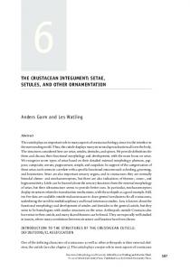
Fig. 1. Top left. Mexico OP engorged female integument. Mallory stained. 400⫻. The stain differentiates strata found in parafÞn embedded engorged female integument of Mexico OP strain. OC, outer cuticle; IC, inner cuticle. Top right: Mexico OP engorged female integument. H&E stain. 400⫻. Differences in tissue are evident: Muscles and connective tissue. PS, protein strata; BS, basophilic strata; M, muscle. Bottom right: Esterase B histochemistry, Mexico OP strain engorged female integument. Bottom left: Esterase B histochemistry, Gonzalez strain engorged female integument. Esterase activity is higher in Mexico OP strain, causing a darker dye precipitation in the IC strata of the integument.
O.C.T. compound (Sakura Laboratories, Torrence, CA) and frozen into chunks by immersion in liquid nitrogen. Sections were cut at 10 M with a Tissue-Tek cryostat (Sakura Laboratories), mounted on a microscope glass slide and air dried at 20⬚C for 15 min. Histochemistry. Mounted sections were Þxed in a methanol, acetic acid, water solution (3:1:6) for 1 h, then transferred to a ßat glass container and immersed for 12 h at room temperature in 100 ml of PBS that contained 10 mM -naphthyl acetate (diluted in 100 l of acetone), and 10 mM fast garnet (Sigma). The glass dish was covered to protect the slides from direct light. The slides were washed two times with 250 ml of PBS then air dried and mounted in a water base solution and covered with a glass cover slip. ParafÞn mounted and cryostat sections were stained at the same time under the same conditions. Integument samples were incubated without substrate and included for detection of nonspeciÞc reactions. Image analysis. All the samples were observed at 400⫻ magniÞcations using an Eclipse E-400 light microscope (Nikon, Tokyo, Japan). Images were recorded with a HV-C20 M digital camera (Hitachi, Tokyo, Japan) mounted on the microscope. The images were captured using Pax-it Version 3.0.4f (MIS, Houston, TX) for Windows (Microsoft, Roselle, IL), using a Dell Optißex personal computer (Dell, Austin, TX).
Results The Mallory stained parafÞn embedded tissues showed a red layer superimposed onto a blue layer (Fig. 1, top left). The sections stained with H&E showed a protein stratum and connective tissue layer beneath it. Also, a muscle stratum and a basophilic stratum under the lowest integument base formed by adipocite cells were observed (Fig. 1, top right). Because the chemicals used during Mallory and H&E staining process can affect esterase activity, frozen samples where taken and processed. After treating the cryostat tissues using -naphthyl acetate, a dark red precipitate of fast garnet was found between the inner and outer endocuticle. The tissue sections from Mexico OP engorged females processed under frozen circumstances showed darker brown color in the endocuticle part of the integument as a consequence of esterase activity, different than sections obtained from the Gonzalez females (Fig. 1, bottom left). This observation was found in 12 of the 15 OP-resistant ticks analyzed. No dark brown coloration was detected in any of the formalin Þxed tissues.
Discussion Most of the chemical agents used as pesticides for arthropod control are esters of substituted phospho-
782
JOURNAL OF MEDICAL ENTOMOLOGY
ric, carbamic or cyclopropane carboxylic acids, therefore subject to degradation by esterases (Devonshire 1991). Several authors have discussed the esterases as factors in the pesticide resistance phenomenon (Chounhury 1972). Naphthyl esters are routinely used as substrates to test esterase activity because the naphthol produced gives a strong color reaction with diazonium salts, and the insoluble resulting dye makes it ideal for histological studies (Devonshire and Moores 1982). Thus, the increased intensity of deposition of the dye in the Mexico OP strain of the resistant B. microplus is due to the increases esterase activity. These increased esterase activity suggests that in the adult female B. microplus, integument may be a part of a putative mechanism of resistance. Potentially, esterase detoxiÞcation of the OP compounds in the integument may reduce the amount of active OP compound reaching the nervous tissues as previously described (Nolan 1985). OP compounds are generally highly lipid soluble. Organic solvents and ionic detergents are often used as vehicles to facilitate their miscibility with water. This chemical characteristic can facilitate the penetration through the cuticle wax, process that could be affected if a detoxiÞcation mechanism is present in integument tissue. Previous metabolic analysis using a radio labeled OP compound showed signiÞcant greater concentration of hydrolytic products and a lower concentration of coumaphos and coroxon in treated adult ticks (Tuxtla strain) (Bull and Ahrens 1988). The Þnding that esterases are located in the integument strongly suggests that this may be the initial detoxifying mechanism in ticks that are resistant to OP. Strong conclusions about the role of the integument based just on the results as presented are incomplete; however, we believe our Þndings are important and further research on esterase characterization, synergy studies, and enzyme kinetic studies using commercial acaricides and their metabolites will be conducted and reported.
Acknowledgments We thank Ronald B. Davey (USDA-ARS, CFTRL, Mission, TX) for providing tick material and to the technical personnel of the Veterinary Histology Laboratory, College of Veterinary Medicine, Texas A&M University for their help in tick tissue preparation and staining.
Vol. 38, no. 6 References Cited
Anonymous. 1971. Recommended methods for detection and measurements of resistance of agricultural pests to pesticides: tentative method for larvae of cattle ticks, Boophilus spp. FAO method No. 7. FAO Plant Prot. Bull. 19: 15Ð18. Bull, O.D.L., and E. H. Ahrens. 1988. Metabolism of coumaphos in susceptible and resistant strains of Boophilus microplus (Acari: Ixodidae). J. Med. Entomol. 25: 94 Ð98. Casida, J. E. 1964. Esterase inhibitors as pesticide. Science 146: 1011Ð1017. Chounhury, S. R. 1972. The nature of nonspeciÞc esterases: a subunit concept. J. Histochem. Cytochem. 20: 507Ð512. Davey, R. B., and J. E. George. 1999. EfÞcacy of Coumaphos applied as a dip for control of an organophosphateresistant strain of Boophilus microplus (Acari: Ixodidae) on cattle. J. Econ. Entomol. 92: 1384 Ð1391. Devonshire, A. L., and G. D Moores. 1982. Pesticide. Biochem. Physiol. 18: 235Ð246. Devonshire, A. L. 1991. Role of esterases in resistance of insects to insecticides. Biochem. Soc. Trans. 19: 775Ð779. Georghiou, G. P., and R. B. Mellon. 1983. Pesticides resistance in time and space, pp. 1Ð 46. In G. P. Georghiou and T. Saito (eds.), Pest resistance to pesticides. Plenum, New York. Healy, M. J., M. M. Dumancic, and J. G. Oakeshott. 1991. Biochemical and physiological studies of soluble esterases from Drosophila melanogaster. Biochem. Genet. 29: 365Ð 388. Nolan, J. 1985. Mechanisms of resistance to chemicals in arthropods parasites of veterinary importance. Vet. Parasitol. 18: 155Ð166. Nun˜ ez, J. L., M. E. Mun˜ oz, and H. L. Moltedo. 1985. Boophilus microplus the common cattle tick, 2nd ed. Springer, Berlin. Rosario-Cruz, R., S. Miranda-Miranda, Z. Garcia-Vasquez, M. Ortiz-Estrada. 1997. Detection of esterase activity in susceptible and organophosphate resistant strains of the cattle tick Boophilus microplus (Acari: Ixodidae). Bull. Entomol. Rex. 53: 563Ð578. Sonenshine, D. E. 1991. Biology of ticks, vol. 1, 1st ed. Oxford University Press, New York. Villarino, M. A., S. D. Waghela, and G. G. Wagner. 2000. Deteccion de las enzimas detoxiÞcantes B Esterasas en Culex quinquefasciatus (Say) Boophilus microplus (Canestrini) Amblyomma cajennense (Fabricius) y Blatella germanica (L.) por medio de zimogramas. Rev. Vet. Mex. 31: 107Ð111. Whyard, S., E. R. Aylward, D. Walker, and V. Walker. 1995. Characterization of a Novel sterase conferring insecticide resistance in the mosquito Culex tarsalis. Arch. Insect. Biochem. Physiol. 29: 329 Ð342. Received for publication 19 April 2001; accepted 1 July 2001.