Aug 18, 2011 - The duration of the window period was established for HIV- ... immunochromatography principle for the specific detection and differentiation of ...
2 HIV-2: Testing Specificities Jean P. Ruelle
UCLouvain, AIDS Reference Laboratory Belgium 1. Introduction 1.1 Discovery of HIV-2: A story of antibody reactivity
Two years after the isolation of the first AIDS virus in 1983 (Barre-Sinoussi et al. 1983), a simian virus inducing a similar pathology was described in rhesus monkeys in captivity (Letvin et al. 1985). Apparently healthy individuals living in Senegal had antibodies that reacted better with the antigens of the simian virus than with those from the human virus (Barin et al. 1985). At the same period, two patients originating from West Africa were hospitalized in France and in Portugal with typical symptoms of AIDS, whereas their serology was negative for the human virus now called HIV-1. The second virus causing AIDS was isolated in 1986 (Clavel et al. 1986). It was then described in various countries of West Africa, and was later called Human immunodeficiency virus type 2 (HIV-2) (BrunVezinet et al. 1987; Clavel et al. 1987). It is classified in the Retroviridae family within the lentivirus genus. Among the lentiviruses, the two HIV are phylogenetically closer to simian lentiviruses than those infecting other animal species, explaining the antibody crossreactivity observed at first (Chakrabarti et al. 1987). The HIV-2 genome is closest to SIVsm infecting sooty mangabeys (Hirsch et al. 1989), whereas HIV-1 is closest to SIVcpz infecting chimpanzees (Huet et al. 1990). 1.2 Clinical outcome HIV-2 differs from HIV-1 in its lower rate of disease progression and infectivity (Jaffar et al. 2004). The routes of transmission are identical to those described for HIV-1, but with lower rates for both horizontal and vertical transmissions, and correlate with the mean lower viral load in HIV-2 infected patients (O'Donovan et al. 2000). The majority of them are long-term non-progressors (LTNP), meaning that they don’t develop symptoms and that the infection does not significantly affect their survival (Rowland-Jones & Whittle 2007). Nevertheless, patients experiencing disease progression and AIDS share the same likelihood of morbidity and mortality as seen in HIV-1 infection (Schim van der Loeff et al. 2002), and will be eligible for antiretroviral therapy. AIDS-defining events are comparable in both types of infections (Martinez-Steele et al. 2007). The distinction between LTNP, patients who remain asymptomatic for at least 8 years while CD4 counts are above 500 cells/ul, and controllers, who control HIV replication without therapy, was recently analysed in the French ANRS cohort; they represented respectively 6.1% and 9.1% (Thiébaut et al. 2011). Although those low percentages contrast with previous publications, it remains clear that LTNP in HIV-2
14
HIV Testing
are far most frequent compared to HIV-1 cohorts, and that low viral load is the main feature of non-progression. Cellular immunity and the maintenance of early-differentiated CD8+ Tcells contribute to low immune activation and low viral replication (Leligdowicz et al. 2010). Understanding the mechanisms underlying HIV-2 biology and the immune response, as a model for attenuated-HIV disease, are important in the concept of HIV vaccines (Leligdowicz & Rowland-Jones 2008). 1.3 Sensitivity to ARV drugs HIV-2 is naturally resistant to non-nucleosidic reverse transcriptase inhibitors, including those of second generation, and to the fusion inhibitor enfuvirtide (Witvrouw et al. 2004; Andries et al. 2004; Poveda et al. 2004). The sensitivity to some protease inhibitors is reduced: amprenavir and its prodrug are not active, and contradictory results were published for atazanavir and tipranavir (Desbois et al. 2008; Brower et al. 2008). Moreover, the genetic barrier to resistance is reduced for several NRTIs and PIs (Smith et al. 2009). In the context of that reduced therapeutic arsenal, recent drug classes represent welcome options. Theoretically, CCR-5 antagonists can be used to treat HIV-2 infection, but two main issues need to be solved: no tropism assay is currently available for clinical routine, and the possible impact of the broader co-receptor usage compared to HIV-1 is not known (Calado et al. 2010). Nevertheless some experimental case studies showed treatment success (Armstrong-James et al. 2010). Integrase inhibitors are active on HIV-2; several in vitro studies on patient isolates and case series showed good activity (Roquebert et al. 2008; Damond et al. 2008a): raltegravir is therefore an option in case of failure or intolerability to other drugs (Francisci et al. 2011), but long-term data are lacking at this point (Gottlieb et al. 2011). No randomized clinical trials were performed to investigate response to treatment in HIV-2 patients (Gottlieb et al. 2008a). Data on treatment efficacy are obtained through cohort analysis, case series or collaborative networking between cohorts: a double-NRTI backbone combined to a boostedPI is the preferred first-line regimen (Benard et al. 2011). Therapy limitations underline the importance of an accurate diagnosis of HIV-2 infection, particularly in countries where drug classes availability is limited (Peterson et al. 2011). 1.4 Epidemiology The evaluation of the total number of case varies between 1 and 2 million infected people worldwide, the majority living in West African countries (Gottlieb et al. 2008a). The highest prevalence was noticed in Guinea-Bissau around 1990, where 17% of blood donors were positive (Poulsen et al. 1993; Naucler et al. 1989). Since then, the HIV-2 prevalence has declined, and is nowadays lower than 1% in the global population in most countries, with the exceptions of the Gambia, Guinea-Bissau and Côte d’Ivoire (da Silva et al. 2008; Sangare et al. 1998). The prevalence of HIV-1 infection has increased during the last two decades in West Africa, and has now exceeds that of type 2 (van Tienen et al. 2010). Although declining, HIV-2 remains of concern particularly in urban areas. The prevalence is more important in older groups, above 45 years of age (van Tienen et al. 2010). Outside West Africa, the virus is present in European countries, essentially in Portugal and France who share colonial histories with endemic regions (Valadas et al. 2009; Barin et al. 2007). Sporadic
HIV-2: Testing Specificities
15
cases are found in other African and European countries, in Brazil, in the Middle East, in Japan, Korea and in India. The majority of patients in these countries were born in or had a possible transmission link with West Africa (Campbell-Yesufu & Gandhi 2011). The virus is very rare in North America (Torian et al. 2010; Centers for Disease Control 2011). 1.5 Diversity of HIV-2: Origins and impact on laboratory assays Eight groups of HIV-2 have been described, named A to H (Damond et al. 2004). Each of those phylogenetically distinct groups corresponds to a cross-species transmission from monkey to man (Sharp et al. 2001). Only groups A and B spread in the human population while other groups seem epidemic dead-ends possibly because of weak adaptation to the human organism (Gao et al. 1994). A/B recombinant forms appeared (Yamaguchi et al. 2008) and disseminated outside West Africa (Ibe et al. 2010). Based on HIV-2 sequences with available sampling dates, the date of the most recent common ancestor was inferred by phylogeny: group A and B strains appeared respectively around 1940 and 1945 (Lemey et al. 2003). Molecular clock analysis favours the hypothesis of a zoonotic transmission during the first half of the 20th century, followed by an epidemic dissemination during the 1960s. The independence war of Guinea-Bissau between 1963 and 1974 offered the circumstances favouring human transmissions (Poulsen et al. 2000; Gomes et al. 2003). Genetic variability between HIV-2 groups, and between strains of the same group is important. The most conserved genomic regions are the LTRs, followed by the gag and pol genes (Kanki 1991). Several examples in the next paragraphs will illustrate the impact of viral diversity on laboratory assays: the use of conserved epitopes can lead to cross-reactivity between HIV types in serology. On the opposite, if an assay targets particularly variable regions, the probability of a false negative result or an underestimated quantification will rise. Serological screening and confirmatory assays, as well as nucleic acid tests (NAT) used for diagnosis or clinical follow-up, must thus take into account HIV-2 diversity from design through field validation.
2. Diagnosis of HIV-2 infection 2.1 Screening assays The diagnosis of HIV-1 or -2 infection is based on serology, mostly using enzyme immunoassays (EIA). As antibodies appear in the serum several weeks post-infection, antibody detection was progressively improved in order to reduce the window period between infection and test positivity. Sensitive assays detecting IgM and IgG are positive a mean of 3 weeks after transmission. Fourth generation assays sensitive to both HIV-antigen and anti-HIV antibodies, also called “combo-tests”, are able to detect an acute infection as early as 2 weeks after the transmission event, when the viral replication level is extremely high (review in Branson 2010). The duration of the window period was established for HIV1 infections and is largely unknown for HIV-2. A study on recent seroconverters showed that viral loads are on average 28-fold times lower compared to HIV-1 (Andersson et al. 2000). As replication capacity is lower and viral turnover slower, there exists the possibility that the time between HIV-2 infection and seropositivity is longer. Initially designed for the detection of HIV-1 group M, most commercial tests are nowadays reactive to anti-HIV-2 and anti-HIV-1 group O antibodies, but not all of them. As disease
16
HIV Testing
progression is on the mean slower in HIV-2 and that the majority of patients are asymptomatic, we can expect to underdiagnose HIV-2 infection in countries or regions where HIV screening assays do not recognize anti-HIV-2 antibodies. It is therefore recommended to opt for tests sensitive to anti-HIV-1 and -2. For antigen detection, fourth generation assays reacting with HIV-1 p24 Ag have no specific component to detect HIV-2 antigens. However, because of conserved regions in the Gag capsid protein and its epitopes, p24-sensitive antigen assays can detect the HIV-2 related antigen, the p26. We cannot exclude a difference of sensitivity for HIV-2 antigen detection in HIV combo assays. That difference has probably a negligible impact in clinical practice, because the probability to test a patient in early acute HIV-2 infection is very low. Nevertheless, as for HIV-1 infection, in the presence of an HIV negative test and clinical signs or seroconversion illness, the patient should be retested 2 to 4 weeks later (Poljak et al. 2009). If a risky behaviour is suspected in an HIV-2 prevalent area, a period of 3 months is recommended to ascertain the negativity. Several rapid tests using immunoprecipitation techniques for antibody detection are approved for clinical use, using serum, whole blood or saliva. As the result can be obtained in 20 minutes, it may be appropriate for the screening of individuals who may not return for the results in conventional settings. They require a minimum of reagents and infrastructure: their use is widespread in resource-constrained countries, in point-of-care facilities, or in centres for voluntary testing. Rapid assays of four generation are currently developed, such as Alere Determine HIV1/2 combo assay (Inverness Medical, UK). Although its performance for antibody detection is comparable to reference EIA, the assay’s antigen sensitivity is weaker than references and is unable to detect HIV-2 antigens (Beelaert & Fransen 2010). Antibody screening assays relying on other methods than EIA or immunoprecipitation are currently in development, with the goal to enhance specificity and to have a higher throughput (Talha et al. 2011). Present day screening assays have an excellent sensitivity close to 100%, and a specificity higher than 99%. Their negative predictive value is very high, but the positive predictive value is low, depending on the local prevalence. False positive results are thus common in practice, and a second independent test should be used to confirm the initial result. As the first-line assays give a positive signal in screening whether due to anti-HIV-1 or anti-HIV-2 antibodies, confirmatory tests used in clinical settings should be able to discriminate the type of virus and arrive to the final diagnosis of HIV-2 infection. 2.2 Discrimination of anti-HIV-2 antibodies Three types of assays are widespread in clinical laboratories for the discrimination of HIV type by serology: Western Blots, line immunoassays, and rapid tests. Commercial Western blot registered for diagnostics such as New LAV Blot II (Bio-Rad, CA) or HIV Blot 1.2 WB (Genelabs, CA) use immobilised HIV antigens to detect specific IgG antibodies to different viral proteins. A result is interpreted as positive if bands reveal a reaction with two or more of the following HIV antigens: p24, gp41, or gp120/160. The result is considered as indeterminate if bands are present, but fewer than two reacting with the antigens cited above. Western blot has several pitfalls. It can overestimate HIV-2 infection, particularly in
HIV-2: Testing Specificities
17
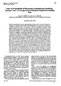
regions with very low prevalence, because of frequent cross-reactions (Qiu et al. 2009; McKellar et al. 2008). Moreover, the turnaround time is higher compared to line immunoassay or rapid tests, it needs more laboratory material, and generates more waste. The use of a commercial line peptide assay such as Inno-Lia (InnoGenetics, Belgium) engineered with synthetic peptides, allows more standardised results and gives less significant cross reactivity for the discrimination between type 1 and 2 infections (Amor et al. 2009). The rapid test Genie HIV-1/HIV-2 (Bio-Rad, CA) is a test based on the immunochromatography principle for the specific detection and differentiation of HIV-1 and HIV-2 antibodies. In several West African countries, national algorithms are based on rapid tests including Genie I/II (Kania et al. 2010). Double reactive samples as defined in Burkina Faso were retested with line peptide assay: only 59% of dually reactive serums had concordant results. Among dual positive samples, 15 % had a detectable HIV-2 plasma viral load while 60% of HIV-2 seropositive patients had a detectable viraemia (Ruelle et al. 2007). Double HIV-1/HIV-2 infections are thus largely overestimated. Double-reactive samples in serology tested with specific qualitative PCRs confirmed only 32% of double infections (Ciccaglione et al. 2010). The group of HIV-2 may also influence serological cross-reactivity: a strong reaction was described between group B and HIV-1 env antigen, mostly in the gp transmembrane glycoprotein (Damond et al. 2001a). 2.3 Nucleic acid tests (NAT) Molecular tests are available for the quantitative detection of HIV-1 plasma RNA, beside commercial plasma viral load assays. No commercial qualitative or quantitative test is available for HIV-2. Several in-house methods have been published and validated (Schutten et al. 2000; Ruelle et al. 2004; Damond et al. 2001b; Ferns & Garson 2006), but are restricted to specialised laboratory. As many HIV-2 patients has very low plasma viral load, without any ARV therapy, RNA counts will often fall under the PCR limit of detection. As a consequence, the viraemia can often not be considered as a diagnostic marker. However, proviral DNA can be theoretically detected in all HIV-2 seropositive patients, at least if the PCR is sensitive enough, and if the primers target a sufficiently conserved region. The amount of DNA present in PBMC was thought to be similar to that in HIV-1 infection. It seems to be the case in patients experiencing low CD4 counts, but can be lower for controllers (Gueudin et al. 2008; Gottlieb, Hawes et al. 2008). Globally the ratio plasma RNA/proviral DNA is lower in HIV-2 infection, due to putative differences in the replicative cycle. It is estimated that HIV proviral DNA is present in 1 cell out of 10,000 PBMC. Laboratory NAT protocols should therefore use a sufficient number of cells to avoid false negative PCR results: to detect 10 HIV DNA copies, we should introduce in the PCR reaction a DNA volume corresponding to the extraction of a minimum of 100,000 cells. 2.4 Proposed HIV-2 testing algorithm A decisional algorithm is shown in Figure 1; it avoids the shortcomings of Western Blot detailed above, allows differentiating HIV-2 from HIV-1 upon confirmation, and reduces the window period in case of acute infection. When a confirmatory test gives an HIV untypable
18
HIV Testing
* Alternatively one single test if high HIV prevalence ** Alternatively a rapid test discriminant for HIV type *** If not available, retest a new sample 2 to 4 weeks later. Not informative about HIV type in case of positivity
Fig. 1. Algorithm for HIV-2 confirmation result, we should consider a NAT amplifying specifically HIV-2 proviral DNA. A single or dual-infection result will strongly influence the therapeutic management, as recommended first-line therapies are based on NNRTI in most countries. Although no study is available, the proviral DNA approach in case of dual-reactivity should be cost-effective by avoiding therapeutic failures.
HIV-2: Testing Specificities
19
2.5 Children born to HIV-2 seropositive mothers Vertical transmission occurs rarely, reported rates vary between 0.1 and 2% (Burgard et al. 2010; Padua et al. 2009). Undetectable viral load during the third trimester of pregnancy and at delivery is a good predictor of prevention success. Due to passive transfer of maternal antibodies, the diagnosis of a mother-to-child HIV transmission is based on viral genome amplification. In addition to the issues of low DNA amount and primer mismatches discussed above for provirus detection, viral turnover is lower in HIV-2 infection and we have virtually no data on what occurs after the child’s infection. Plasma RNA should be amplified in parallel with proviral DNA, or alternatively different sets of primers should be used to amplify the same DNA sample. As viral genetic variability can be responsible of PCR failure (Padua et al. 2009), some authors recommend amplifying one sample from the mother, with the same sets of primers, as a positive control. Ideally, the follow-up of the baby should include the search for proviral DNA: if viral genome is not detected at the age of 3 months, vertical transmission did not occur. In countries where NATs are not available, prevention with antiretroviral therapy including PIs will considerably reduce the chances of transmission.
3. Plasma viral load 3.1 Available assays and standardisation Plasma viral load correspond to the number of RNA genomes present in the patient’s blood, expressed in copies per millilitre (ml) of plasma or in log copies/ml. It is a predictor of disease progression: viral replication will enhance CD4 count drop (Gottlieb et al. 2002). RNA quantification also allows the identification of HIV-2 controllers. No commercial assay was approved for HIV-2 viral load monitoring in clinical settings. Some companies developed PCR primers sets, other offer a biochemical assay based on reverse transcriptase activity, but no major HIV-1 viral load platform has a version dedicated to HIV-2. Some studies described the use of the Nuclisens HIV-1 assay (Biomérieux, France), an isothermal RNA amplifiction method, applied to HIV-2 group A (Rodes et al. 2007). When compared to in-house RT-PCR assays, sensitivity was not optimal (Damond et al. 2008b). Plasma viral load assays are mainly restricted to some reference laboratories across Europe, and in some African centres. Real-time PCR is by far the most common technology used by those laboratories. If the coverage is sufficient for the HIV-2 cohorts in Europe, most West African countries lack reliable assays to monitor their patients. RNA quantification is ensured by an external standard curve in PCR. Several methods were published using an electron microscopy counted reference strain. The main advantages of such standard is that the process of extraction and amplification is exactly the same as a sample, avoiding extraction yield discrepancies, and that one quantified stock conserved in aliquots at -80°C can be used for a long period of time. As a drawback, the viral load is expressed in RNA genomes per ml and not in viral particles: some non-infectious virions present in a viral stock do not contain nucleic acid and can therefore bias the result. As an example, if 1000 viral particles contain less than the 2000 supposed genome copies, the standard will be too high and the samples tested will be under-quantified. Another option
20
HIV Testing
is the use of nucleic acids quantified by spectrophotometry as standards. The count of RNA standards is more close to the real number of genome copies than counted particles, but RNAs are less stable and can easily be degraded by environmental RNAse or by too long conservation periods. Moreover, a supplementary control on the extraction step must be added to the assay, and traces of parental DNA serving as template for RNA synthesis can also bias the RNA quantification. The quality and reproducibility of HIV-2 viral load assays was evaluated across Europe and in the Gambia through the AcHIeV2e collaborative network (Damond et al. 2008b). A first round showed that most of the assays gave reproducible results, but important discrepancies were seen for absolute quantifications. As those were possibly linked to the use of different standards, a second round of controls was sent around together with one aliquot of counted particles. Inter-laboratory homogeneity was then better, and primer mismatches were suspected as the origin of result variability (Damond et al. 2011). A good correlation of results was obtained for HIV-2 group A samples, but quantification of group B strains is extremely variable between laboratories. The latter evaluations underscore the need of a common standard if multi-centric assessments using viral load as a parameter are foreseen, and the need for an enhanced quality for HIV-2 group B quantifications. Two reference standards are now available, made respectively from strains ROD and CAM, belonging to group A. Their titres are expressed in international units (IU), which raises the unsolved issue about the use of IU vs. RNA copies in HIV clinical practice. 3.2 Indications and interpretation Plasma viral load is indicated to monitor the absence or degree of viral replication during antiretroviral therapy. If we hypothesise that HIV-2 patients in need of therapy have the same likelihood to progress as compared to HIV-1, we can assume that recommendations for the follow-up are the same: viral load measurement at therapy initiation, one month after and then every 3 months, with the goal of achieving durable suppression. In resourceconstraint settings, clinical monitoring alone should be used to expand antiretroviral therapy, although it is not the optimal solution (Laurent et al. 2011). Given the mean slower disease progression and the high proportion of non-progressors, the monitoring of HIV-2 viral load could be spaced for untreated patients from 6 months to 1 year, but no study support clearly that recommendation until now. In case CD4 counts drop or if disease progresses despite an undetectable viral load, the plasma should be retested with an alternative assay to avoid a possible problem of genetic variability (Gilleece et al. 2010).
4. Resistance testing 4.1 Genotypic assays To determine the sensitivity of an HIV-1 isolate to antiretroviral drugs, genotypic assays are the most widespread in clinical laboratories by sequencing the viral gene coding for the drug target (protease, reverse transcriptase, integrase and envelope glycoproteins). The translated amino acid sequence is compared to that of a reference strain to establish a list of mutations. From that list, sensitivity to each drug is inferred by interpretation rules. Although some phenotypic assays can be used in the clinic, their use is restricted by much
HIV-2: Testing Specificities
21
higher costs, the need for a biosafety level 3 laboratory, and higher turnaround times; they are usually developed for research purposes. Alternatively, virtual HIV-1 phenotypes are available, translating genotypic data into IC50 fold changes and activity cut-offs (review in Schutten 2006). Clinically relevant cut-offs were inferred using clinical trials and cohort data (Winters et al. 2008). None of the commercial assays available for HIV-1 resistance testing can be used for HIV-2: resistance assays are restricted to some reference laboratories, mostly in Europe, and rely on home-brew protocols. Classically Sanger sequencing is performed after RT-PCR amplification from plasma RNA: sensitive PCR protocols are needed, as HIV-2 viral loads are low. Next-generation sequencing allowing detection of more variants in quasi-species would help to understand HIV-2 specific viral dynamics and resistance pathways. 4.2 Interpretation rules Even if the two types of HIV share some major resistance mutations, the genetic barrier, the pathways leading to resistance and the frequency of mutations differ (Ntemgwa et al. 2009; Smith et al. 2009). Therefore the interpretation rules developed to determine HIV-1 resistance do not apply to HIV-2. Some natural polymorphisms in the HIV-2 pol gene correspond to resistance mutations in HIV-1 algorithms (Bercoff et al. 2010; Rodes et al. 2006; Colson et al. 2005). Compared to HIV-1, the RT multi-drug resistance mutation Q151M is far more frequent after therapy failure, as the K65R mutation (Descamps et al. 2004). Two sets of HIV-2 specific interpretation rules have been published so far: the ANRS (Agence Nationale de Recherche sur le SIDA et les Hépatites, Paris, France) and the Rega (Rega Institute, KULeuven, Belgium) rules. The latest versions of those algorithms are available on line (ANRS-AC11 2011; Gomes et al. 2009). The first one is more specific, as the list includes only mutations for which the impact was clearly demonstrated in several publications: K65R, Q151M, M184V and S215 changes in the RT (Damond et al. 2005), as well as mutations at positions 143, 148 and 155 in the integrase (Charpentier et al. 2011). The second algorithm is probably more sensitive as the list includes other minor mutations for which an impact has been described, as well as primary protease mutations absent from the ANRS list. Nevertheless, in the absence of large clinical studies on HIV-2 treatment failures, the evidence related to some mutations is based on small case series. Moreover the lists of mutations refer to the strain ROD from HIV-2 group A: it may skew the interpretation for group B, as some mutations are natural polymorphisms with no known effect on drug sensitivity in B strains. Besides the analysis of clinical samples related to virological failures, more studies depicting the phenotypic impact of mutations on isolates in vitro are warranted. 4.3 Indications HIV-2 resistance tests are indicated in case of virological failure, i.e. presence of viral replication (detectable plasma viral load) under therapy. European guidelines for the clinical use of resistance tests recommend genotypic assays (Vandamme et al. 2011). As genetic barrier to resistance is low and therapeutic options limited, a resistance test should not be deferred once viral replication is detected.
22
HIV Testing
In antiretroviral-naïve patients, HIV-2 resistance tests are not indicated in clinical practice. Even though some studies demonstrated the transmission of drug-resistant strains in West Africa and in Europe (Ruelle et al. 2007; Ruelle et al. 2008; Jallow et al. 2009), no data support the cost-effectiveness of such indication for HIV-2. 4.4 Tropism testing No HIV-2 tropism assay suitable for clinical use has been currently developed. Besides the lack of phenotypic or genotypic assay, the guidelines on the clinical management of HIV-1 tropism (Vandekerckhove et al. 2011) do not apply to HIV-2 for the following reasons: the clinical outcome of CCR-5 antagonists containing regimen is unknown, few studies correlating the gp120 coding sequences and the phenotype are published and thus no genotypic interpretation rules exist (Dimonte et al. 2011), and the viral tropism extends to broader chemokine receptors for which the clinical relevance is controversial (Calado et al. 2010; Blaak et al. 2005).
5. Conclusions Several challenges related to the diagnosis and the follow-up of HIV-2 infections need to be addressed:
Although most HIV screening assays now detect antibodies directed against type 1 and 2 viruses, fourth generation tests have poorer sensitivity to HIV-2 antigens. The algorithms defining the number and which tests to use for HIV-2 diagnosis differ widely. This is related to varying prevalences between countries or continents, but is also related to the availability of tests, particularly nucleic acid tests. Those decision trees should be harmonised to ensure an accurate diagnosis: misidentification of HIV type has hazardous consequences for the clinical management. Reference standards for plasma viral load quantification will facilitate multi-centric collaborations, as a lack of consistency between assays has been observed. Although standards are now available for group A, quantification of other groups remains problematic. No commercial HIV-1 viral load platform is up to now applicable for HIV2 in clinical settings. No clinically validated tropism assay is available, preventing the use of CCR-5 antagonists for HIV-2 treatment. The apparent reduced genetic barrier to antiretroviral resistance imposes a careful choice of drugs and a fine-tuning of genotypic interpretation rules. The majority of patients are long-term non-progressors or controllers; prediction of evolution and applying different follow-up patterns to controllers or progressors would rationalise the use of resources.
We have to keep in mind that the majority of HIV-2 patients live in countries where the diagnostics tools discussed here are not all available. More field evaluations in endemic regions, monitoring the impact of new laboratory tools, defining the best antiretroviral regimen, evaluating the prevalence of resistance and understanding better the pathways leading to treatment failure would supplement expert opinion rules with evidence-based data.
HIV-2: Testing Specificities
23
6. References Amor, A., A. Simon, M. Salgado, B. Rodes, V. Soriano, and C. Toro. 2009. Lack of significant cross-reactivity for HIV-2 immunoblots in HIV-1-infected patients. J Acquir Immune Defic Syndr 50 (3):339-40. Andersson, S., H. Norrgren, Z. da Silva, A. Biague, S. Bamba, S. Kwok, C. Christopherson, G. Biberfeld, and J. Albert. 2000. Plasma viral load in HIV-1 and HIV-2 singly and dually infected individuals in Guinea-Bissau, West Africa: significantly lower plasma virus set point in HIV-2 infection than in HIV-1 infection. Arch Intern Med 160 (21):3286-93. Andries, K., H. Azijn, T. Thielemans, D. Ludovici, M. Kukla, J. Heeres, P. Janssen, B. De Corte, J. Vingerhoets, R. Pauwels, and M. P. de Bethune. 2004. TMC125, a novel next-generation nonnucleoside reverse transcriptase inhibitor active against nonnucleoside reverse transcriptase inhibitor-resistant human immunodeficiency virus type 1. Antimicrob Agents Chemother 48 (12):4680-6. ANRS-AC11. Genotype interpretation for HIV-2, www.hivfrenchresistance.org 2011. Consulted 2011 Aug 18. Armstrong-James, D., J. Stebbing, A. Scourfield, E. Smit, B. Ferns, D. Pillay, and M. Nelson. 2010. Clinical outcome in resistant HIV-2 infection treated with raltegravir and maraviroc. Antiviral Res 86 (2):224-6. Barin, F., S. M'Boup, F. Denis, P. Kanki, J. S. Allan, T. H. Lee, and M. Essex. 1985. Serological evidence for virus related to simian T-lymphotropic retrovirus III in residents of west Africa. Lancet 2 (8469-70):1387-9. Barin, F., F. Cazein, F. Lot, J. Pillonel, S. Brunet, D. Thierry, F. Damond, F. Brun-Vezinet, J. C. Desenclos, and C. Semaille. 2007. Prevalence of HIV-2 and HIV-1 group O infections among new HIV diagnoses in France: 2003-2006. AIDS 21 (17):2351-3. Barre-Sinoussi, F., J. C. Chermann, F. Rey, M. T. Nugeyre, S. Chamaret, J. Gruest, C. Dauguet, C. Axler-Blin, F. Vezinet-Brun, C. Rouzioux, W. Rozenbaum, and L. Montagnier. 1983. Isolation of a T-lymphotropic retrovirus from a patient at risk for acquired immune deficiency syndrome (AIDS). Science 220 (4599):868-71. Beelaert, G., and K. Fransen. 2010. Evaluation of a rapid and simple fourth-generation HIV screening assay for qualitative detection of HIV p24 antigen and/or antibodies to HIV-1 and HIV-2. J Virol Methods 168 (1-2):218-22. Benard, A., A. van Sighem, A. Taieb, E. Valadas, J. Ruelle, V. Soriano, A. Calmy, C. Balotta, F. Damond, F. Brun-Vezinet, G. Chene, and S. Matheron. 2011. Immunovirological response to triple nucleotide reverse-transcriptase inhibitors and ritonavir-boosted protease inhibitors in treatment-naive HIV-2-infected patients: The ACHIEV2E Collaboration Study Group. Clin Infect Dis 52 (10):1257-66. Bercoff, D. P., P. Triqueneaux, C. Lambert, A. A. Oumar, A. M. Ternes, S. Dao, P. Goubau, J. C. Schmit, and J. Ruelle. 2010. Polymorphisms of HIV-2 integrase and selection of resistance to raltegravir. Retrovirology 7:98. Blaak, H., P. H. Boers, R. A. Gruters, H. Schuitemaker, M. E. van der Ende, and A. D. Osterhaus. 2005. CCR5, GPR15, and CXCR6 are major coreceptors of human immunodeficiency virus type 2 variants isolated from individuals with and without plasma viremia. J Virol 79 (3):1686-700. Branson, B. M. 2010. The future of HIV testing. J Acquir Immune Defic Syndr 55 Suppl 2:S1025.
24
HIV Testing
Brower, E. T., U. M. Bacha, Y. Kawasaki, and E. Freire. 2008. Inhibition of HIV-2 protease by HIV-1 protease inhibitors in clinical use. Chem Biol Drug Des 71 (4):298-305. Brun-Vezinet, F., M. A. Rey, C. Katlama, P. M. Girard, D. Roulot, P. Yeni, L. Lenoble, F. Clavel, M. Alizon, S. Gadelle, and et al. 1987. Lymphadenopathy-associated virus type 2 in AIDS and AIDS-related complex. Clinical and virological features in four patients. Lancet 1 (8525):128-32. Burgard, M., C. Jasseron, S. Matheron, F. Damond, K. Hamrene, S. Blanche, A. Faye, C. Rouzioux, J. Warszawski, and L. Mandelbro. 2010. Mother-to-child transmission of HIV-2 infection from 1986 to 2007 in the ANRS French Perinatal Cohort EPF-CO1. Clin Infect Dis 51 (7):833-43. Calado, M., P. Matoso, Q. Santos-Costa, M. Espirito-Santo, J. Machado, L. Rosado, F. Antunes, K. Mansinho, M. M. Lopes, F. Maltez, M. O. Santos-Ferreira, and J. M. Azevedo-Pereira. 2010. Coreceptor usage by HIV-1 and HIV-2 primary isolates: the relevance of CCR8 chemokine receptor as an alternative coreceptor. Virology 408 (2):174-82. Campbell-Yesufu, O. T., and R. T. Gandhi. 2011. Update on human immunodeficiency virus (HIV)-2 infection. Clin Infect Dis 52 (6):780-7. Centers for Disease Control CDC 2011. HIV-2 infection surveillance - United States, 19872009. Morbidity and mortality weekly report (MMWR) 60 (29):977-1008. Chakrabarti, L., M. Guyader, M. Alizon, M. D. Daniel, R. C. Desrosiers, P. Tiollais, and P. Sonigo. 1987. Sequence of simian immunodeficiency virus from macaque and its relationship to other human and simian retroviruses. Nature 328 (6130):543-7. Charpentier, C., B. Roquebert, O. Delelis, L. Larrouy, S. Matheron, R. Tubiana, M. Karmochkine, X. Duval, G. Chene, A. Storto, G. Collin, A. Benard, F. Damond, J. F. Mouscadet, F. Brun-Vezinet, and D. Descamps. 2011. Hot spots of integrase genotypic changes leading to HIV-2 resistance to raltegravir. Antimicrob Agents Chemother 55 (3):1293-5. Ciccaglione, A. R., M. Miceli, G. Pisani, R. Bruni, P. Iudicone, A. Costantino, M. Equestre, E. Tritarelli, C. Marcantonio, P. Tataseo, M. C. Marazzi, S. Ceffa, G. Paturzo, A. M. Altan, M. M. San Lio, S. Mancinelli, M. Ciccozzi, A. Lo Presti, G. Rezza, and L. Palombi. 2010. Improving HIV-2 detection by a combination of serological and nucleic acid amplification test assays. J Clin Microbiol 48 (8):2902-8. Clavel, F., D. Guetard, F. Brun-Vezinet, S. Chamaret, M. A. Rey, M. O. Santos-Ferreira, A. G. Laurent, C. Dauguet, C. Katlama, C. Rouzioux, and et al. 1986. Isolation of a new human retrovirus from West African patients with AIDS. Science 233 (4761):343-6. Clavel, F., K. Mansinho, S. Chamaret, D. Guetard, V. Favier, J. Nina, M. O. Santos-Ferreira, J. L. Champalimaud, and L. Montagnier. 1987. Human immunodeficiency virus type 2 infection associated with AIDS in West Africa. N Engl J Med 316 (19):1180-5. Colson, P., M. Henry, N. Tivoli, H. Gallais, J. A. Gastaut, J. Moreau, and C. Tamalet. 2005. Polymorphism and drug-selected mutations in the reverse transcriptase gene of HIV-2 from patients living in southeastern France. J Med Virol 75 (3):381-90. da Silva, Z. J., I. Oliveira, A. Andersen, F. Dias, A. Rodrigues, B. Holmgren, S. Andersson, and P. Aaby. 2008. Changes in prevalence and incidence of HIV-1, HIV-2 and dual infections in urban areas of Bissau, Guinea-Bissau: is HIV-2 disappearing? AIDS 22 (10):1195-202.
HIV-2: Testing Specificities
25
Damond, F., C. Apetrei, D. L. Robertson, S. Souquiere, A. Lepretre, S. Matheron, J. C. Plantier, F. Brun-Vezinet, and F. Simon. 2001. Variability of human immunodeficiency virus type 2 (hiv-2) infecting patients living in france. Virology 280 (1):19-30. Damond, F., D. Descamps, I. Farfara, J. N. Telles, S. Puyeo, P. Campa, A. Lepretre, S. Matheron, F. Brun-Vezinet, and F. Simon. 2001. Quantification of proviral load of human immunodeficiency virus type 2 subtypes A and B using real-time PCR. J Clin Microbiol 39 (12):4264-8. Damond, F., M. Worobey, P. Campa, I. Farfara, G. Colin, S. Matheron, F. Brun-Vezinet, D. L. Robertson, and F. Simon. 2004. Identification of a highly divergent HIV type 2 and proposal for a change in HIV type 2 classification. AIDS Res Hum Retroviruses 20 (6):666-72. Damond, F., G. Collin, S. Matheron, G. Peytavin, P. Campa, S. Delarue, A. Taieb, A. Benard, G. Chene, F. Brun-Vezinet, and D. Descamps. 2005. Letter. In vitro phenotypic susceptibility to nucleoside reverse transcriptase inhibitors of HIV-2 isolates with the Q151M mutation in the reverse transcriptase gene. Antivir Ther 10 (7):861-5. Damond, F., S. Lariven, B. Roquebert, S. Males, G. Peytavin, G. Morau, D. Toledano, D. Descamps, F. Brun-Vezinet, and S. Matheron. 2008. Virological and immunological response to HAART regimen containing integrase inhibitors in HIV-2-infected patients. AIDS 22 (5):665-6. Damond, F., A. Benard, J. Ruelle, A. Alabi, B. Kupfer, P. Gomes, B. Rodes, J. Albert, J. Boni, J. Garson, B. Ferns, S. Matheron, G. Chene, and F. Brun-Vezinet. 2008. Quality control assessment of human immunodeficiency virus type 2 (HIV-2) viral load quantification assays: results from an international collaboration on HIV-2 infection in 2006. J Clin Microbiol 46 (6):2088-91. Damond, F., A. Benard, C. Balotta, J. Boni, M. Cotten, V. Duque, B. Ferns, J. Garson, P. Gomes, F. Goncalves, G. Gottlieb, B. Kupfer, J. Ruelle, B. Rodes, V. Soriano, M. Wainberg, A. Taieb, S. Matheron, G. Chene, and F. Brun-Vezinet. 2011. An international collaboration to standardize HIV-2 viral load assays: Results from the 2009 ACHIEV2E quality control study. J Clin Microbiol. Desbois, D., B. Roquebert, G. Peytavin, F. Damond, G. Collin, A. Benard, P. Campa, S. Matheron, G. Chene, F. Brun-Vezinet, and D. Descamps. 2008. In vitro phenotypic susceptibility of human immunodeficiency virus type 2 clinical isolates to protease inhibitors. Antimicrob Agents Chemother 52 (4):1545-8. Descamps, D., F. Damond, S. Matheron, G. Collin, P. Campa, S. Delarue, S. Pueyo, G. Chene, and F. Brun-Vezinet. 2004. High frequency of selection of K65R and Q151M mutations in HIV-2 infected patients receiving nucleoside reverse transcriptase inhibitors containing regimen. J Med Virol 74 (2):197-201. Dimonte, S., V. Svicher, R. Salpini, F. Ceccherini-Silberstein, C. F. Perno, and M. BabakirMina. 2011. HIV-2 A-subtype gp125(C2-V3-C3) mutations and their association with CCR5 and CXCR4 tropism. Arch Virol. Ferns, R. B., and J. A. Garson. 2006. Development and evaluation of a real-time RT-PCR assay for quantification of cell-free human immunodeficiency virus type 2 using a Brome Mosaic Virus internal control. J Virol Methods 135 (1):102-8.
26
HIV Testing
Francisci, D., L. Martinelli, L. E. Weimer, M. Zazzi, M. Floridia, G. Masini, and F. Baldelli. 2011. HIV-2 infection, end-stage renal disease and protease inhibitor intolerance: which salvage regimen? Clin Drug Investig 31 (5):345-9. Gao, F., L. Yue, D. L. Robertson, S. C. Hill, H. Hui, R. J. Biggar, A. E. Neequaye, T. M. Whelan, D. D. Ho, G. M. Shaw, and et al. 1994. Genetic diversity of human immunodeficiency virus type 2: evidence for distinct sequence subtypes with differences in virus biology. J Virol 68 (11):7433-47. Gilleece, Y., D. R. Chadwick, J. Breuer, D. Hawkins, E. Smit, L. X. McCrae, D. Pillay, N. Smith, and J. Anderson. 2010. British HIV Association guidelines for antiretroviral treatment of HIV-2-positive individuals 2010. HIV Med 11 (10):611-9. Gomes, P., A. Abecasis, M. Almeida, R. Camacho, and K. Mansinho. 2003. Transmission of HIV-2. Lancet Infect Dis 3 (11):683-4. Gomes, P. , K. Van Laethem, A-M. Geretti, R. Camacho, and A-M. Vandamme. Algorithm for the interpretation of genotypic HIV-2 resistance data, www.kuleuven.ac.be/rega/cew/links 2009. Consulted 2011 Aug 18. Gottlieb, G. S., P. S. Sow, S. E. Hawes, I. Ndoye, M. Redman, A. M. Coll-Seck, M. A. FayeNiang, A. Diop, J. M. Kuypers, C. W. Critchlow, R. Respess, J. I. Mullins, and N. B. Kiviat. 2002. Equal plasma viral loads predict a similar rate of CD4+ T cell decline in human immunodeficiency virus (HIV) type 1- and HIV-2-infected individuals from Senegal, West Africa. J Infect Dis 185 (7):905-14. Gottlieb, G. S., S. P. Eholie, J. N. Nkengasong, S. Jallow, S. Rowland-Jones, H. C. Whittle, and P. S. Sow. 2008. A call for randomized controlled trials of antiretroviral therapy for HIV-2 infection in West Africa. AIDS 22 (16):2069-72; discussion 2073-4. Gottlieb, G. S., S. E. Hawes, N. B. Kiviat, and P. S. Sow. 2008. Differences in proviral DNA load between HIV-1-infected and HIV-2-infected patients. AIDS 22 (11):1379-80. Gottlieb, G. S., R. A. Smith, N. M. Dia Badiane, S. Ba, S. E. Hawes, M. Toure, A. K. Starling, F. Traore, F. Sall, S. L. Cherne, J. Stern, K. G. Wong, P. Lu, M. Kim, D. N. Raugi, A. Lam, J. I. Mullins, and N. B. Kiviat. 2011. HIV-2 Integrase Variation in Integrase Inhibitor-Naive Adults in Senegal, West Africa. PLoS One 6 (7):e22204. Gueudin, M., F. Damond, J. Braun, A. Taieb, V. Lemee, J. C. Plantier, G. Chene, S. Matheron, F. Brun-Vezinet, and F. Simon. 2008. Differences in proviral DNA load between HIV-1- and HIV-2-infected patients. AIDS 22 (2):211-5. Hirsch, V. M., R. A. Olmsted, M. Murphey-Corb, R. H. Purcell, and P. R. Johnson. 1989. An African primate lentivirus (SIVsm) closely related to HIV-2. Nature 339 (6223):38992. Huet, T., R. Cheynier, A. Meyerhans, G. Roelants, and S. Wain-Hobson. 1990. Genetic organization of a chimpanzee lentivirus related to HIV-1. Nature 345 (6273):356-9. Ibe, S., Y. Yokomaku, T. Shiino, R. Tanaka, J. Hattori, S. Fujisaki, Y. Iwatani, N. Mamiya, M. Utsumi, S. Kato, M. Hamaguchi, and W. Sugiura. 2010. HIV-2 CRF01_AB: first circulating recombinant form of HIV-2. J Acquir Immune Defic Syndr 54 (3):241-7. Jaffar, S., A. D. Grant, J. Whitworth, P. G. Smith, and H. Whittle. 2004. The natural history of HIV-1 and HIV-2 infections in adults in Africa: a literature review. Bull World Health Organ 82 (6):462-9. Jallow, S., T. Vincent, A. Leligdowicz, T. De Silva, C. Van Tienen, A. Alabi, R. Sarge-Njie, P. Aaby, T. Corrah, H. Whittle, A. Jaye, G. Vanham, S. Rowland-Jones, and W. Janssens. 2009. Presence of a multidrug-resistance mutation in an HIV-2 variant
HIV-2: Testing Specificities
27
infecting a treatment-naive individual in Caio, Guinea Bissau. Clin Infect Dis 48 (12):1790-3. Kania, D., P. Fao, D. Valea, C. Gouem, T. Kagone, H. Hien, P. Somda, P. Ouedraogo, A. Drabo, S. Gampini, N. Meda, S. Diagbouga, P. Van de Perre, and F. Rouet. 2010. Low prevalence rate of indeterminate serological human immunodeficiency virus results among pregnant women from Burkina Faso, West Africa. J Clin Microbiol 48 (4):1333-6. Kanki, P. J. 1991. Biologic features of HIV-2. An update. AIDS Clin Rev:17-38. Laurent, C., C. Kouanfack, G. Laborde-Balen, A. F. Aghokeng, J. B. Mbougua, S. Boyer, M. P. Carrieri, J. M. Mben, M. Dontsop, S. Kaze, N. Molinari, A. Bourgeois, E. MpoudiNgole, B. Spire, S. Koulla-Shiro, and E. Delaporte. 2011. Monitoring of HIV viral loads, CD4 cell counts, and clinical assessments versus clinical monitoring alone for antiretroviral therapy in rural district hospitals in Cameroon (Stratall ANRS 12110/ESTHER): a randomised non-inferiority trial. Lancet Infect Dis doi:10.1016/S1473-3099(11)70168-2 Leligdowicz, A., and S. Rowland-Jones. 2008. Tenets of protection from progression to AIDS: lessons from the immune responses to HIV-2 infection. Expert Rev Vaccines 7 (3):319-31. Leligdowicz, A., C. Onyango, L. M. Yindom, Y. Peng, M. Cotten, A. Jaye, A. McMichael, H. Whittle, T. Dong, and S. Rowland-Jones. 2010. Highly avid, oligoclonal, earlydifferentiated antigen-specific CD8+ T cells in chronic HIV-2 infection. Eur J Immunol 40 (7):1963-72. Lemey, P., O. G. Pybus, B. Wang, N. K. Saksena, M. Salemi, and A. M. Vandamme. 2003. Tracing the origin and history of the HIV-2 epidemic. Proc Natl Acad Sci U S A 100 (11):6588-92. Letvin, N. L., M. D. Daniel, P. K. Sehgal, R. C. Desrosiers, R. D. Hunt, L. M. Waldron, J. J. MacKey, D. K. Schmidt, L. V. Chalifoux, and N. W. King. 1985. Induction of AIDSlike disease in macaque monkeys with T-cell tropic retrovirus STLV-III. Science 230 (4721):71-3. Martinez-Steele, E., A. A. Awasana, T. Corrah, S. Sabally, M. van der Sande, A. Jaye, T. Togun, R. Sarge-Njie, S. J. McConkey, H. Whittle, and M. F. Schim van der Loeff. 2007. Is HIV-2- induced AIDS different from HIV-1-associated AIDS? Data from a West African clinic. AIDS 21 (3):317-24. McKellar, M. S., P. Jongthavorn, and H. Khanlou. 2008. False-positivity of HIV-2 immunoblots in a cohort of elite suppressors infected with HIV-1. J Acquir Immune Defic Syndr 47 (5):644. Naucler, A., P. A. Andreasson, C. M. Costa, R. Thorstensson, and G. Biberfeld. 1989. HIV-2associated AIDS and HIV-2 seroprevalence in Bissau, Guinea-Bissau. J Acquir Immune Defic Syndr 2 (1):88-93. Ntemgwa, M. L., T. d'Aquin Toni, B. G. Brenner, R. J. Camacho, and M. A. Wainberg. 2009. Antiretroviral drug resistance in human immunodeficiency virus type 2. Antimicrob Agents Chemother 53 (9):3611-9. O'Donovan, D., K. Ariyoshi, P. Milligan, M. Ota, L. Yamuah, R. Sarge-Njie, and H. Whittle. 2000. Maternal plasma viral RNA levels determine marked differences in motherto-child transmission rates of HIV-1 and HIV-2 in The Gambia. MRC/Gambia
28
HIV Testing
Government/University College London Medical School working group on mother-child transmission of HIV. AIDS 14 (4):441-8. Padua, E., C. Almeida, B. Nunes, H. Cortes Martins, J. Castela, C. Neves, and M. T. Paixao. 2009. Assessment of mother-to-child HIV-1 and HIV-2 transmission: an AIDS reference laboratory collaborative study. HIV Med 10 (3):182-90. Peterson, K., S. Jallow, S. L. Rowland-Jones, and T. I. de Silva. 2011. Antiretroviral Therapy for HIV-2 Infection: Recommendations for Management in Low-Resource Settings. AIDS Res Treat 2011:463704. Poljak, M., E. Smit, and J. Ross. 2009. 2008 European Guideline on HIV testing. Int J STD AIDS 20 (2):77-83. Poulsen, A. G., P. Aaby, A. Gottschau, B. B. Kvinesdal, F. Dias, K. Molbak, and E. Lauritzen. 1993. HIV-2 infection in Bissau, West Africa, 1987-1989: incidence, prevalences, and routes of transmission. J Acquir Immune Defic Syndr 6 (8):941-8. Poulsen, A. G., P. Aaby, H. Jensen, and F. Dias. 2000. Risk factors for HIV-2 seropositivity among older people in Guinea-Bissau. A search for the early history of HIV-2 infection. Scand J Infect Dis 32 (2):169-75. Poveda, E., B. Rodes, C. Toro, and V. Soriano. 2004. Are fusion inhibitors active against all HIV variants? AIDS Res Hum Retroviruses 20 (3):347-8. Qiu, M., X. Liu, Y. Jiang, J. N. Nkengasong, W. Xing, L. Pei, and B. S. Parekh. 2009. Current HIV-2 diagnostic strategy overestimates HIV-2 prevalence in China. J Med Virol 81 (5):790-7. Rodes, B., J. Sheldon, C. Toro, L. Cuevas, E. Perez-Pastrana, I. Herrera, and V. Soriano. 2007. Quantitative detection of plasma human immunodeficiency virus type 2 subtype A RNA by the Nuclisens EasyQ Assay (version 1.1). J Clin Microbiol 45 (1):88-92. Rodes, B., J. Sheldon, C. Toro, V. Jimenez, M. A. Alvarez, and V. Soriano. 2006. Susceptibility to protease inhibitors in HIV-2 primary isolates from patients failing antiretroviral therapy. J Antimicrob Chemother 57 (4):709-13. Roquebert, B., F. Damond, G. Collin, S. Matheron, G. Peytavin, A. Benard, P. Campa, G. Chene, F. Brun-Vezinet, and D. Descamps. 2008. HIV-2 integrase gene polymorphism and phenotypic susceptibility of HIV-2 clinical isolates to the integrase inhibitors raltegravir and elvitegravir in vitro. J Antimicrob Chemother 62 (5):914-20. Rowland-Jones, S. L., and H. C. Whittle. 2007. Out of Africa: what can we learn from HIV-2 about protective immunity to HIV-1? Nat Immunol 8 (4):329-31. Ruelle, J., B. K. Mukadi, M. Schutten, and P. Goubau. 2004. Quantitative real-time PCR on Lightcycler for the detection of human immunodeficiency virus type 2 (HIV-2). J Virol Methods 117 (1):67-74. Ruelle, J., M. Sanou, H. F. Liu, A. T. Vandenbroucke, A. Duquenne, and P. Goubau. 2007. Genetic polymorphisms and resistance mutations of HIV type 2 in antiretroviralnaive patients in Burkina Faso. AIDS Res Hum Retroviruses 23 (8):955-64. Ruelle, J., F. Roman, A. T. Vandenbroucke, C. Lambert, K. Fransen, F. Echahidi, D. Pierard, C. Verhofstede, K. Van Laethem, M. L. Delforge, D. Vaira, J. C. Schmit, and P. Goubau. 2008. Transmitted drug resistance, selection of resistance mutations and moderate antiretroviral efficacy in HIV-2: analysis of the HIV-2 Belgium and Luxembourg database. BMC Infect Dis 8:21.
HIV-2: Testing Specificities
29
Sangare, K. A., I. M. Coulibaly, and A. Ehouman. 1998. [Seroprevalence of HIV among pregnant women in the ten regions of the Ivory Coast]. Sante 8 (3):193-8. Schim van der Loeff, M. F., S. Jaffar, A. A. Aveika, S. Sabally, T. Corrah, E. Harding, A. Alabi, A. Bayang, K. Ariyoshi, and H. C. Whittle. 2002. Mortality of HIV-1, HIV-2 and HIV-1/HIV-2 dually infected patients in a clinic-based cohort in The Gambia. AIDS 16 (13):1775-83. Schutten, M., B. van den Hoogen, M. E. van der Ende, R. A. Gruters, A. D. Osterhaus, and H. G. Niesters. 2000. Development of a real-time quantitative RT-PCR for the detection of HIV-2 RNA in plasma. J Virol Methods 88 (1):81-7. Schutten, M. 2006. Resistance assays. In Antiretroviral resistance in clinical practice, edited by A.-M. Geretti. London: Mediscript. Sharp, P. M., E. Bailes, R. R. Chaudhuri, C. M. Rodenburg, M. O. Santiago, and B. H. Hahn. 2001. The origins of acquired immune deficiency syndrome viruses: where and when? Philos Trans R Soc Lond B Biol Sci 356 (1410):867-76. Smith, R. A., D. J. Anderson, C. L. Pyrak, B. D. Preston, and G. S. Gottlieb. 2009. Antiretroviral drug resistance in HIV-2: three amino acid changes are sufficient for classwide nucleoside analogue resistance. J Infect Dis 199 (9):1323-6. Talha, S. M., T. Salminen, S. Swaminathan, T. Soukka, K. Pettersson, and N. Khanna. 2011. A highly sensitive and specific time resolved fluorometric bridge assay for antibodies to HIV-1 and -2. J Virol Methods 173 (1):24-30. Thiébaut, R., S. Matheron, A. Taieb, F. Brun-Vezinet, G. Chene, and B. Autran. 2011. Longterm nonprogressors and elite controllers in the ANRS CO5 HIV-2 cohort. AIDS 25 (6):865-7. Torian, L. V., J. J. Eavey, A. P. Punsalang, R. E. Pirillo, L. A. Forgione, S. A. Kent, and W. R. Oleszko. 2010. HIV type 2 in New York City, 2000-2008. Clin Infect Dis 51 (11):133442. Valadas, E., L. Franca, S. Sousa, and F. Antunes. 2009. 20 years of HIV-2 infection in Portugal: trends and changes in epidemiology. Clin Infect Dis 48 (8):1166-7. van Tienen, C., M. S. van der Loeff, S. M. Zaman, T. Vincent, R. Sarge-Njie, I. Peterson, A. Leligdowicz, A. Jaye, S. Rowland-Jones, P. Aaby, and H. Whittle. 2010. Two distinct epidemics: the rise of HIV-1 and decline of HIV-2 infection between 1990 and 2007 in rural Guinea-Bissau. J Acquir Immune Defic Syndr 53 (5):640-7. Vandamme, A. M., R. J. Camacho, F. Ceccherini-Silberstein, A. de Luca, L. Palmisano, D. Paraskevis, R. Paredes, M. Poljak, J. C. Schmit, V. Soriano, H. Walter, and A. Sonnerborg. 2011. European recommendations for the clinical use of HIV drug resistance testing: 2011 update. AIDS Rev 13 (2):77-108. Vandekerckhove, L. P., A. M. Wensing, R. Kaiser, F. Brun-Vezinet, B. Clotet, A. De Luca, S. Dressler, F. Garcia, A. M. Geretti, T. Klimkait, K. Korn, B. Masquelier, C. F. Perno, J. M. Schapiro, V. Soriano, A. Sonnerborg, A. M. Vandamme, C. Verhofstede, H. Walter, M. Zazzi, and C. A. Boucher. 2011. European guidelines on the clinical management of HIV-1 tropism testing. Lancet Infect Dis 11 (5):394-407. Winters, B., J. Montaner, P. R. Harrigan, B. Gazzard, A. Pozniak, M. D. Miller, S. Emery, F. van Leth, P. Robinson, J. D. Baxter, M. Perez-Elias, D. Castor, S. Hammer, A. Rinehart, H. Vermeiren, E. Van Craenenbroeck, and L. Bacheler. 2008. Determination of clinically relevant cutoffs for HIV-1 phenotypic resistance
30
HIV Testing
estimates through a combined analysis of clinical trial and cohort data. J Acquir Immune Defic Syndr 48 (1):26-34. Witvrouw, M., C. Pannecouque, W. M. Switzer, T. M. Folks, E. De Clercq, and W. Heneine. 2004. Susceptibility of HIV-2, SIV and SHIV to various anti-HIV-1 compounds: implications for treatment and postexposure prophylaxis. Antivir Ther 9 (1):57-65. Yamaguchi, J., A. Vallari, N. Ndembi, R. Coffey, C. Ngansop, D. Mbanya, L. Kaptue, L. G. Gurtler, S. G. Devare, and C. A. Brennan. 2008. HIV type 2 intergroup recombinant identified in Cameroon. AIDS Res Hum Retroviruses 24 (1):86-91.