tribution of allelic polymorphism in the interaction between CD4 and MHC class II molecules. Using mouse DAP-3-transfected cells expressing different isotypes ...
H L A - D R Polymorphism Affects the Interaction with CD4 By S. Fleury,*~J. Thibodeau,* G. Croteau,* N. Labrecque,*r H.-E. Aronson, II C. Cantin,* E. O. Long,g and K.-P. S6kaly*r From the "Laboratoire d'Immunologie, Institut de Recherches Cliniques de Montrdal, and r de Microbiologie et lmmunologie, FacuM de Mddecine, Universit~ de Montrdal, Montttal, Qudbe6 Canada H2W IR7; SLaboratory of lmmunogenetics, National Institute of Allergy and Infectious Diseases, Rockville, Maryland 20852; and IlDepartment of Biochemistry and Molecular Biophysics, College of Physicians and Surgeons, Columbia University, New York 10032
Stlmmal'y
Major histocompatibility complex (MHC) class II molecules are highly polymorphic and bind peptides for presentation to CD4 + T cells. Functional and adhesion assays have shown that CD4 interacts with MHC class II molecules, leading to enhanced responses of CD4 + T cells after the activation of the CD4-associated tyrosine kinase p56 uk. We have addressed the possible contribution of allelic polymorphism in the interaction between CD4 and MHC class II molecules. Using mouse DAP-3-transfected cells expressing different isotypes and allelic forms of the HLADR molecule, we have shown in a functional assay that a hierarchy exists in the ability of class II molecules to interact with CD4. Also, the study of DR4 subtypes minimized the potential contribution of polymorphic residues of the peptide-binding groove in the interaction with CD4. Chimeras between the DR4 or DR1 molecules, which interact efficientlywith CD4, and DRw53, which interacts poorly, allowed the mapping of polymorphic residues between positions 3180 and 189 that can exert a dramatic influence on the interaction with CD4.
ature T lymphocytes are divided into two major subsets. CD8 + T cells recognize nominal antigens in M the context of MHC class I molecules, while CD4 + T cells recognize antigen bound to the MHC dass II molecules (1-3). Experimental evidence obtained from a variety of functional systems has shown that the interaction between CD8 and MHC class I molecules or between CD4 and MHC class II molecules leads to significant enhancement of T cell activation, probably by recruiting CD4- and CD8-associated tyrosine kinase p56 ~ to the vicinity of the TCR complex (4, 5). Adhesion assays have further confirmed the physical association of CD8 to MHC class I (6, 7) and CD4 to class II molecules (8). These assays have also been used to determine the molecular features of the interactions between these molecules. Polymorphism in the c~3 domain of class I leads to severe perturbations of class I-restricted T cell responses (9), including positive and negative selection in the thymus (10). Mutagenesis analysis of class I further demonstrated that CD8 molecules interact with a highly conserved determinant in the a3 domain of MHC class I molecules (11). Similarly, CD4 interacts with a highly conserved region in the 32 domain of MHC class II molecules (12). Interestingly, this domain in class II bears significant homology with the abovementioned domain in class I. However, very little is known 733
about the effect of isotypic and allelic diversity of MHC class II molecules on their interaction with CD4. MHC class II molecules are highly polymorphic. The presence of several dass II isotypes (DR, DP, and DQ) further increases their diversity. As previously mentioned, polymorphism of MHC class I molecules affects their interaction with CD8. To evaluate the effect of MHC class II polymorphisms on the interaction with CD4, we have concentrated our efforts on HLA-DR molecules. These MHC class II molecules are all composed of the same monomorphic ot chain, but their 3 chains can be encoded by four different isotypic genes, namely, the B1, B3, B4, and B5 genes (13). The DKB1 gene is the most polymorphic, while the products of the DRB3 and DRB4 can be coexpressed with several DKB1 alleles. The DRB5 product is only coexpressedin individuals bearing the DR2 haplotype. While most of these DR molecules present Ag to CD4 + T cells, the majority of class II-restricted T cell responses has been shown to be restricted by products of the BI gene (14). However, peptides presented in the context of DR2 haplotype are predominantly restricted by the product of the B5 gene (15, 16). Such differences could be attributed to variationsin the affinityof CD4 for these different MHC class II molecules. Indeed, MHC class I alleles that fail to interact with CD8 are inefficient in stimulating allo-
The Journal of Experimental Medicine 9 Volume 182 September 1995 733-741
geneic and antigen-specific responses (9, 11). The possibility of such a hierarchy in the capacity of different HLA-DK molecules to interact with CD4 was assessed in an assay that specifically isolates the CD4-dass II component in the interaction between effector T ceUs and APC. This assay was previously used to identify residues on CD4 that are involved in the interaction with MHC dass II molecules (17-19). Our results clearly indicate that aUdic polymorphism and isotypic diversity of HLA-DK molecules lead to variations in their capacity to interact with CD4.
Materials and Methods Cells and Transfectants. The generation and characterization of the 3DT52.5.8 murine T cell hybridomaand of its variant expressing the human CD4 molecule(I1B-3) havebeen described(19-22). The I1B-3 T cell hybridoma is maintained in culture medium consisting of KPMI 1640 supplemented with 10% FCS, 10 ~M 2-ME, 2 mM r-glutamine, and 500 #g/ml of G-418 (GIBCO BILL, Gaithersburg, MD). The B4.2.3 T cell hybridoma is H-2D d restricted and was generatedby the fusion of BW1100 thymoma with lymph node cells of BALB/c mice immunized with p18 in complete Freund's adjuvant (23). The p18 peptide corresponds to residues 315-329 of the gp160 protein of the HIV-1 strain IIIB (24). The B4.2.3 T cell hybridoma is maintained in DMEM medium supplemented with 10% FCS, 10 #M 2-ME, and 2 mM of L-glutamine.The HLADK o~chain is encoded by a full length eDNA (25). The HLA-DR B chain cDNAs correspond to the different alleles of HLA-DK previouslydescribed (26-32). The routine fibroblasticclass II-negative DAP-3 cell line was transfected using calcium phosphate as previously described (33). Briefly, DAP-3 cells (3 x 10³) were transfectedwith 10 #g of ILSV.5or KSV.3 plasmids (34) encoding the HLA-DR o~ and one of the different allelic or isotypic forms of the B chain, together with 5 #g of the plasmid containing the H-2D d gene. Neomycin (G-418) or mycophenolicacid (10/~g/ml) and xanthine (100 #g/ml) sdection were applied 48 h after transfection. Aseptic cell sorting using a FACStar Plus| (Becton Dickinson & Co., Cockeysville,MD) was used to obtain homogeneous populations of cells expressing comparable levels of D a and the different HLA-DK alleles. FACS* Analysis. T cell hybridomas (I1B-3) were stained with either OKT4 (anti-human CD4) and with KJ12-98 (anti-murine TCK idiotype), followed by fluorescein-coupledgoat anti-mouse Igs (Becton Dickinson & Co.). DAP-3 cell lines were stained with either 34.5.8 (anti-mouse class I H-2D a) or L-243 (mouse anti-human class II HLA-DR) antibody. Cells were analyzed on a FACScan| flow cytometer (Becton Dickinson & Co). Mean fluorescence values (M.F.V.)1 are expressed in arbitrary units. For each fluorescencehistogram, 10,000 live cells were analyzed, using a four-decade logarithmic scale. Dead ceUs were excluded by propidium iodide (0.5 mg/ml) gating. As a control, the cells were stained only with the FITC-goat anti-mouse Igs. HI.A-DR Nomer~lature. DPd-Dwl = DRB1 0101 (27); DK2BDw2 = DRB1 1501(28); DR2A-Dw2 = DRB5 0101 (28); DKw6b III= DILB30201 (29); DKw53 ~ DILB40101 (30); DK4-Dw4 = DILB1 0401 (31); DK4-Dwl0 = DRB1 0402 01); DK4-Dw14 = DILB1 0404 (31); DR4-Dw15 = DKB1 0405 (31); DKw11.1 = DP.~I 1101 (32).
I1B3 Stimulation Assays. A fixed number of T cells (75 x 103) expressingthe human wild-type CD4 moleculewas coculturedwith different DAP-3 target cells (75 x 103)expressing H-2D d and various HLA-DK molecules. The CD4- 3DT52.5.8 T cell hybridoma was cocultured under the same conditions. The assaywas performed in 200 #1 of complete medium for 18 h at 37~ 5% COs, in 96-well flat-bottom culture plates (Flow Laboratories, Inc., McLean, VA). Supernatants from the coculture were tested for the presence of IL-2by their ability to support the proliferation of the Ib2-dependent cell line CTLL.2 using the hcxosaminidase colorimetric assay (18, 19). A calibration curve was performed in parallel to determine the IL-2 concentration (U/ml). In previous experiments, we had demonstrated that an anti-CD4 antibody (OKT4B or b68) could abrogate Ib2 production (19). Growth Inhibition Assay with B4.2.3. DAP-3 cell transfectants (104) expressing H-2Dd alone, DKw53 alone, H-2Dd and DKw53 or H-2D a, and DR4-Dw4 were used as APCs. DAP cells were pretreated with 50 #g/ml of mitomycin (Sigma Chemical Co., St. Louis, MO) for 45 min and then washed fivetimes with PBS. These APC were pulsed for 4 h with 0.004, 0.02, 0.1, and 0.5 #g/ml of the p18 peptide diluted in complete DMEM without FCS and then washed three times with PBS. B4.2.3. T cell hybridomas (104) were then cocultured with transfectedDAP cells in complete DMEM containing 10% FCS for 18 h at 37~ 5% COs in 96well culture plates. This T cell concentration does not allow antigen presentationbetween themselves.After 20 h, cells were pulsed with [3H]thymidinefor 6 h and were then collected and counted.
1 Abbreviation used in this paper: M.F.V.,mean fluorescencevalue. 734
CD4-MHCClass II Interactions
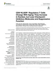
Results and Discussion Polymorphism in M H C Class II Molecules Affects the Interaction with CD4. A T call hybridoma specific for the murine MHC class I molecule H-2D a and dependent on the CD4class II interaction for high levels of IL-2 production was used in these experiments. As previously demonstrated, expression of class II molecules in H-2D a+ APCs leads to a clear enhancement of IL-2 production by T cell hybridomas, when cffector cells also express CD4 molecule (18, 19, 22, 35). Cocultures were performed between the CD4 + murine T cell hybridoma (I1B-3) and transfected murine fibroblastic ceUs (DAP-3) expressing comparable levels of various aUdes and isotypes of DK molecules together with the TCK ligand D d (Fig. 1). DK molecules encoded by different DILB genes interact with CD4; the DKw6bIII, DKw53, and DP,2A-Dw2, which are products of the DtLB3, B4, and B5 genes, respectively, can trigger enhanced Ib2 production levels, as compared with DAP-3 cells expressing only H-2D a molecules (Fig. 2 A). Increase in IL-2 production ranged between 3- and 17-fold (Fig. 2), when compared with control DAP-3 that express only H-2D d. Results from Fig. 2 A indicate that DK4-Dw4 and DKw6B III molecules are more efficient than DKw53 and DR.2A in stimulating IL-2 production by the I1B-3 T cell hybridoma. Among DRBI alleles (Fig. 2 B), DK4-Dw4 and DIL5 are capable of stimulating high levds of IL-2 production by the T cells (14-15-fold), as compared with cells expressing D a alone. However, coculture of CD4 § T cells with DAP-3 expressing DK2B-Dw2 or DK1 constantly yidded lower levels of II,2 (20-fold diff~ence). Similar differences were also obtained when cocultures were carried
;1B'3
DdDR2.B.Ow2
- - 0 K T 4 I~'~ --KJI2 18
- - L - 2 4 3 152 D
-t
~
ii i i
i{ /',.,
)~R2A-Dw2
M.FI DdORw6bm9
34.5.8 187 L-243 47
9
m
}
:
/-
L'243
20
A
M.F.V OdOR4.DwlO
34.5.8 i l l L-243 43
M,EV D~iDR4.Dw15 34.58 i21 L-243 20
103
10o
i0 t
i02
103
10o
101
101
~t..=V 34..~.8 122 L-243 5 3
-!
j i, tO 2
:,
M.~V: DdDRwltt 34~.8 181 L-243 30
A
~0~
tj_l