Injury, Int. J. Care Injured 45 (2014) 1359–1364
Contents lists available at ScienceDirect
Injury journal homepage: www.elsevier.com/locate/injury
How early should VATS be performed for retained haemothorax in blunt chest trauma? Hsing-Lin Lin b,e,g, Wen-Yen Huang c, Chyan Yang c, Shih-Min Chou f, Hsin-I. Chiang d, Liang-Chi Kuo b,e,g, Tsung-Ying Lin b,e,g, Yi-Pin Chou a,h,* a
Division of Trauma, Department of Emergency, Veterans General Hospital-Kaohsiung, Kaohsiung, Taiwan Department of Emergency Medicine, Kaohsiung Medical University Hospital, Kaohsiung Medical University, Kaohsiung, Taiwan Institute of Business and Management, National Chiao Tung University-Taipei, Taipei, Taiwan d Shih Chien University-Kaohsiung, Kaohsiung, Taiwan e Division of Trauma, Department of Surgery, Kaohsiung Medical University Hospital, Kaohsiung Medical University, Kaohsiung, Taiwan f Department of Administration Center, Kaohsiung Medical University Hospital, Kaohsiung Medical University, Kaohsiung, Taiwan g Department of Emergency Medicine, Faculty of Medicine, College of Medicine, Kaohsiung Medical University, Kaohsiung, Taiwan h Research Center for Industry of Human Ecology, Chang Gung University of Science and Technology, Kweishan, Taoyuan, Taiwan b c
A R T I C L E I N F O
A B S T R A C T
Article history: Accepted 24 May 2014
Background: Blunt chest injury is not uncommon in trauma patients. Haemothorax and pneumothorax may occur in these patients, and some of them will develop retained pleural collections. Video-assisted thoracoscopic surgery (VATS) has become an appropriate method for treating these complications, but the optimal timing for performing the surgery and its effects on outcome are not clearly understood. Materials and methods: In this study, a total of 136 patients who received VATS for the management of retained haemothorax from January 2003 to December 2011 were retrospectively enrolled. All patients had blunt chest injuries and 90% had associated injuries in more than two sites. The time from trauma to operation was recorded and the patients were divided into three groups: 2–3 days (Group 1), 4–6 days (Group 2), and 7 or more days (Group 3). Clinical outcomes such as the length of stay (LOS) at the hospital and intensive care unit (ICU), and duration of ventilator and chest tube use were all recorded and compared between groups. Results: The mean duration from trauma to operation was 5.9 days. All demographic characteristics showed no statistical differences between groups. Compared with other groups, Group 3 had higher rates of positive microbial cultures in pleural collections and sputum, longer duration of chest tube insertion and ventilator use. Lengths of hospital and ICU stay in Groups 1 and 2 showed no statistical difference, but were longer in Group 3. The frequency of repeated VATS was lower in Group 1 but without statistically significant difference. Discussion: This study indicated that an early VATS intervention would decrease chest infection. It also reduced the duration of ventilator dependency. The clinical outcomes were significantly better for patients receiving VATS within 3 days under intensive care. In this study, we suggested that VATS might be delayed by associated injuries, but should not exceed 6 days after trauma. ß 2014 The Authors. Published by Elsevier Ltd. This is an open access article under the CC BY-NC-ND license (http://creativecommons.org/licenses/by-nc-nd/3.0/).
Keywords: Video-assisted thoracoscopic surgery Haemothorax Blunt injury Length of stay Infection Ventilator Thoracic injury
Introduction Blunt injury is the leading cause of chest trauma. Pneumothorax and haemothorax often occur after severe chest trauma. Approximately 85% of patients with pneumothorax and haemothorax can
* Corresponding author at: Division of Trauma, Department of Emergency, Veterans General Hospital-Kaohsiung, 386 Ta-Chung 1st Rd., Kaohsiung 81362, Taiwan. Tel.: +886 73125895x7553; fax: +886 73208255. E-mail address:
[email protected] (Y.-P. Chou).
be successfully treated with pain control or simple tube thoracostomies [1–3]. The remaining patients with retained pleural collections should be managed with further surgical interventions to prevent complications such as empyema in the early phase or fibrothorax in the late phase. These complications will increase morbidity and mortality [4–6]. Video-assisted thoracoscopic surgery (VATS) has become a common and acceptable method for diagnosis of intra-thoracic lesions from the 1990s because of advanced developments in this surgical technique [7–9]. It is also widely used in treating retained pleural collections because it is only slightly more invasive
http://dx.doi.org/10.1016/j.injury.2014.05.036 0020–1383/ß 2014 The Authors. Published by Elsevier Ltd. This is an open access article under the CC BY-NC-ND license (http://creativecommons.org/licenses/by-nc-nd/ 3.0/).
1360
H.-L. Lin et al. / Injury, Int. J. Care Injured 45 (2014) 1359–1364
compared with chest tube thoracostomy, and has been shown as a simple technical alternative to open thoracotomy [10–12]. Although VATS offers many benefits, the optimal timing for performing the surgery and its effects on patient’s outcomes are not clearly understood. Numerous recent studies describe that the earlier the interventions of injured chest are performed, the better the prognosis of patients [13–15]. However, there exist wide variations in the exact recommended time for operation after trauma in these studies, especially when complicated with associated injuries [7–9,14–16]. The main reasons for prolonging the hospitalisation of trauma patients are infections and respiratory failure [1,17,18]. We hypothesised that early VATS may play a role in preventing post-traumatic infections and decrease the duration of acute respiratory failure, which in turn can shorten the whole treatment course. The purpose of this study was to identify the most appropriate time for implementing VATS to treat pleural collection and its effects on respiratory failure in patients with multiple traumas. Materials and methods Setting and patients This retrospective study was conducted in a level-1 trauma medical centre located in southern Taiwan, which has 1300 beds with approximately 1200 emergent traumatic visits per month. All patients with blunt thoracic injuries that were admitted to the emergency department (ED) during the study period were included. Data of patients that developed residual haemothorax and received video-assisted thoracoscopy (VATS) were prospectively collected. Patient data including demographics, mechanism of injury, numbers of rib fractures (confirmed by chest CT), associated injuries, injury severity score (ISS), pulmonary contusion score, postoperative complications, respiratory failure, number of ventilator days, length of stay (LOS) in intensive care unit (ICU), and LOS in hospital were all collected. This study was approved by the ethics committee of the hospital in which this study was conducted. Patients older than 14 years of age that were admitted to the trauma unit in our hospital with blunt chest trauma alone or with other associated injuries in different anatomic regions were included in this study. All patients with haemothorax needing to be drained were treated with a 36Fr (This size of chest tube is chosen due to Asian people having smaller body size) straight thoracostomy tube at the time of initial evaluation. These procedures were performed by trauma surgeons with first-line service. Patients had minimal pleural collections initially without thoracostomy tubes inserted but admitted for further observation were not included. In this study, patients that were hemodynamically unstable with more than 1500 mL of blood drainage from the initial tube thoracostomy or had ongoing blood losses of more than 250 mL/h lasting for at least 4 h and received emergency thoracotomy were excluded. Furthermore, patients with disruptions of mediastinal structures (including heart, great vessels and tracheobronchial trees) were excluded. Patients with severe medical diseases like liver cirrhosis, chronic obstructive pulmonary disease, chronic renal disease under hemodialysis, and chronic heart failure that would increase complications after trauma were also excluded. All patients received chest-computed tomography (CT) in the emergency department. After completing both primary and secondary surveys at trauma bay, all patients were admitted into trauma ICU for further care because nearly all patients had multiple injured sites. Chest roentgenogram was performed routinely after admission. If the density of the following chest
X-ray showed increasing density, secondary chest CT was performed to diagnose and estimate the retained pleural collections. There are two indications for VATS to be performed. One is the retained volumes estimated to exceed 300 mL. The other is formation of para-pneumonic effusions which appeared in CT as separate lobulated pleural collections. Often, retained pleural collections do not happen immediately; hence, all secondary CT and VATS are performed at least 48 h after trauma. The associated injuries were recorded with abbreviated injury score (AIS) following the 2005 edition. Patients with AIS exceeding grade 3 were also excluded. All patients received VATS performed by thoracic surgeons according to the same standard procedures. The surgery was performed in the operating room under general anaesthesia with the patients in lateral position on the unaffected side. All patients received prophylactic antibiotics before surgical intervention. A double-lumen endobronchial tube was used for one lung procedure and provided a better view for the assessment of the chest and its contents. Two thoracostomies were performed for inserting thoracoscopes and endoscopic instruments, respectively. All patients had collections evacuated, clots removed, the pleura decorticated, and were irrigated with normal saline solutions. Thereafter, the lung was re-expanded. Finally, two 36Fr drainage tubes were fixed to the pleural cavity and the patient was transferred to the trauma ICU for further postoperative care. Routine chest roentgenograms were performed after operations. In case of recurrence of retained pleural collections, secondary VATS would be performed for adequate drainages. Following either procedure, the thoracostomy tubes were removed at the discretion of the thoracic surgeon when the drainage was less than 100 mL/ 24 h and no air leak was present. Post-trauma complications included the development of infections and prolonged ventilations. Postoperative outcomes indicated hospital mortality, number of ventilator days, ICU, and hospital LOS. Empyema was defined as an accumulation of pus in the pleural space (confirmed by bacteria culture). Pneumonia was defined as an infective condition with bacteria cultured from the sputum after trauma. Sepsis was defined as a systemic response to lung infection with bacteremia. Statistical analysis Patients were divided into three groups according to when VATS was performed, namely Group 1, within 2–3 days; Group 2, within 4–6 days; and Group 3, after more than 6 days. Group 1 was formed on the basis of Meyer’s prospective study that showed very early VATS within 3 days provided better clinical outcomes [8]. Group 3 was based initially on intervals that were greater than the average, with exploratory analysis confirming that this gave better discrimination of outcome than a later cut-off point. An initial descriptive analysis was performed for every variable, and the frequencies and averages in the groups were determined. Numerical variables were presented as mean and standard deviation (SD) or median and quartiles. The chi-square or Fisher’s test was employed to evaluate the categorical or proportional variables between groups. The continuous variables were compared between groups using tests of analysis of variance. A P < 0.05 was considered statistically significant. All data were analysed using the SPSS 16.0 statistical software. Results A total of 1663 patients with chest injuries were admitted to our hospital from January 1, 2003 to December 31, 2011. A total of 145 thoracoscopic drainages performed for clotted post-traumatic haemothorax were selected. A 2-year-old patient and 8 adult
H.-L. Lin et al. / Injury, Int. J. Care Injured 45 (2014) 1359–1364
1361
Table 1 Demographic analysis of patients treated with VATS for blunt trauma (n = 136). Mean age (yr) (SD) Males (%) Mechanisms of injury Motorcyclist Vesicle driver or passenger Fall accident Cyclist or pedestrian Anatomic injury score (AIS) of thoracic injury (mean, SD) Associated injuries Head injury (%) Abdominal injury (%) Extremity injury (%) Multiple trauma ISS (mean, SD) Positive microbial cultures after trauma From sputum From pleural effusion From blood Time from trauma to perform VATS (days) (mean, SD) Secondary VATS Time of ventilator use (days) (mean, SD) Time of chest tube use (days) (mean, SD) ICU length of stay (days) (mean, SD) In-hospital length of stay (days) (mean, SD) Mortality (none related to VATS)
51.9 (16.2) 107 (78.7%) 87 14 21 14 3.3 (0.5) 55 (40.4%) 39 (28.7%) 100 (73.5%) 120 (88.2%) 18.1 (6.0) 54 (39.7%) 29 (21.3%) 16 (11.8%) 5.9 (4.6) 14 (10.3%) 7.8 (11.4) 12.7 (9.1) 8.7 (7.1) 24.1 (18.4) 3 (2.2%)
patients with penetrating injuries were excluded. The characteristics of each group are shown in Table 1. There were 107 men and 29 women aged from 16 to 82 years (average 51.9, SD = 16.2). The mechanism of injury was motorcycle-related (motorcyclist or passenger) injury for 87 patients, vehicle-related (driver or passenger) injury for 14 patients, falling injury for 21 patients, and cyclist or pedestrian for 14 patients. Median ISS for all patients was 18 (quartile, 13, 22) and their mean ISS was 18.1 (SD, 6.1). The severity of chest injury is recorded by anatomic abbreviated injury score (AIS) of thoracic injuries. Four patients were recorded as grade 2, eighty-four patients were grade 3 and forty-eight patients were grade 4. Mean AIS of chest in all patients was 3.3 (SD, 0.5) and median was 3 (quartiles, 3, 4). All patients had tube thoracostomy at the trauma bay of emergency department before VATS. Mean time from trauma to residual pleural collections diagnosed from secondary CT is 52.8 h (SD, 9.1). The average time period between the trauma and performance of VATS was 5.9 days (SD, 4.6). No patients had their VATS procedures converted to thoracotomies during surgical interventions. However, 14 patients received secondary VATS because of persistent pleural collections after operation. The mean LOS of ICU and hospital were 8.7 (SD, 7.1) and 24.2 (SD, 18.4) days. Rate of post-trauma infections cultured from
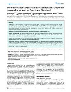
Fig. 1. Relation between LOS in hospital and timing of VATS.
sputum, pleural effusion and blood were 40.5%, 21.3% and 11.8% respectively. Overall, the mortality of these patients was 2.2% (3/ 136). Fig. 1 shows that shorter hospital lengths of stay were most common in those with shorter intervals from trauma to VATS. Patients with positive microbial culture had longer mean time periods from trauma to VATS (9.1 days vs. 5.0 days, p = 0.004), as did those with positive microbial culture of sputum (7.8 days vs. 4.6 days, p = 0.001). When patients were divided into three groups according to the time from trauma to performance of the VATS (2– 3 days, average 2.6, SD, 0.5; 4–6 days, average 4.6, SD, 0.8; >6 days after trauma, average 11.9, SD, 5.2), they were compared for demographic and preoperative status (Table 2). Group 3 has higher head AIS than the others but the Glasgow coma scale at emergency department, AIS of other regions and the overall ISS showed no statistical significant differences between the three groups. Other basic demographics, such as age, gender, mechanism of injury, and number of rib fractures, rate of flail chest, pulmonary contusion scores, rate of acute respiratory failure within 4 h after trauma, retained pleural collections diagnosed time showed no statistical differences between these groups; thus, the demographic and preoperative status of patients in the three groups were similar. The outcomes of the three groups are shown in Table 3. As can be seen, Group 3 had longer ICU LOS and in-hospital LOS than the
Table 2 Comparison of basic demographics between patient groups.
Age (mean, SD) Gender (male) Mechanism of injury Motorcycle Vesicle Fall Pedestrian Numbers of fractured rib (mean, SD) Flail chest Pulmonary contusion scores (mean, SD) Acute respiratory failure within 4 h after trauma Head AIS (mean, SD) Thoracic AIS (mean, SD) Abdomen AIS (mean, SD) Extremity AIS (mean, SD) ISS (mean, SD) GCS at ED (mean, SD)
Group 1 (n = 43)
Group 2 (n = 57)
Group 3 (n = 36)
p
50.1 (14.2) 32 (74%)
50.1 (16.1) 44 (77%)
56.9 (17.9) 31 (86%)
0.099 0.422 0.407
26 7 5 5 5.0 (2.5) 14 (33%) 6.9 (2.3) 18 (42%) 0.5 (1.0) 3.3 (0.5) 0.5 (1.0) 1.6 (1.0) 16.7 (5.7) 14.0 (1.2)
40 5 7 5 5.8 (3.0) 24 (42%) 7.1 (2.4) 30 (53%) 1.1 (1.3) 3.3 (0.5) 0.6 (1.0) 1.5 (1.0) 18.7 (6.4) 14.1 (1.1)
21 2 9 4 6.1 (0.2) 17 (47%) 8.0 (2.7) 18 (50%) 1.2 (1.3) 3.4 (0.6) 0.7 (1.1) 1.3 (1.1) 19.0 (5.7) 13.8 (1.3)
0.243 0.394 0.109 0.554 0.017 0.532 0.789 0.295 0.149 0.465
1362
H.-L. Lin et al. / Injury, Int. J. Care Injured 45 (2014) 1359–1364
Table 3 Comparison of clinical outcomes between patient groups.
ICU LOS (mean, SD) Hospital LOS (mean, SD) Number of total ventilator days (mean, SD) Number of post-VATS ventilator days (mean, SD) Duration of total tube drainage (mean, SD) Duration of post-VATS tube drainage (mean, SD) Secondary VATS (mean, SD) Post trauma infections Sputum culture positive Pleural effusion culture positive Blood culture positive Mortality
Group 1 (n = 43)
Group 2 (n = 57)
Group 3 (n = 36)
p
5.0 (2.3) 16.2 (9.6) 2.6 (1.9) 0.9 (1.4) 8.2 (3.7) 6.9 (3.0) 1 (2%)
7.7 (5.0) 21.3 (13.0) 6.8 (8.0) 5.2 (9.1) 11.6 (6.3) 8.3 (5.6) 8 (14%)
14.6 (9.6) 38.2 (25.0) 15.5 (17.2) 12.1 (17.2) 19.8 (12.7) 9.5 (9.3) 5 (14%)