AFM AND NANOINDENTATION STUDIES OF A WET-CURED EPOXY ADHESIVE B.B. Johnsen, B.R.K. Blackman, A.J. Kinloch, W.S. Teo Department of Mechanical Engineering, Imperial College London, London SW7 2AZ, UK Email:
[email protected] Introduction The presence of water in the substrate or in the adhesive at the time of manufacture of an adhesive joint can have a very deleterious effect on the performance of the joint. Previous research has shown that moisture being present in a composite laminate prior to bonding significantly decreased the fracture energy of the bonded joints [1,2], even though the fracture was always cohesive in the adhesive layer. The mode I fracture energy, GIc, was reduced from 960 J/m2 for fully dried laminates to 100 J/m2 for laminates containing 0.6% w/w water. For comparison, substrates which had picked up moisture in the laboratory gave a GIc value of 200 J/m2. This loss in GIc was caused by water diffusing from the laminate and being absorbed by the adhesive during the high-temperature curing process. The decrease in fracture energy was accompanied by a decrease in the glass transition temperature, Tg, of the adhesive and, at the highest levels of absorbed moisture, by the formation of steam cavities in the adhesive bondline. The present study was undertaken to investigate what effect the presence of water during curing has on the microstructure and the mechanical properties of the different phases in the adhesive. The same adhesive that was used to bond the composite joints was studied. Cured bulk adhesive samples were prepared by mixing water into the adhesive prior to curing. Tapping mode atomic force microscopy (AFM) was then used to investigate the microstructure of the dry- and wet-cured adhesives, while the mechanical properties of the epoxy matrix were investigated using nanoindentation techniques.
Experimental A rubber-toughened epoxy-paste adhesive was investigated. Pre-bond moisture was introduced by blending water with the paste adhesive, followed by thorough mixing using a high speed mixer. Plates of the conditioned adhesive were prepared by placing the mixture in a metal mould and curing it at 150oC for 45 minutes. Differential scanning calorimetry (DSC) was used to determine the Tg of the cured adhesive plates. The Tg of the neat epoxy polymer (with no rubber) was found to be 125oC, while the Tg of the CTBN rubber is -46oC. AFM studies were undertaken using a MultiMode scanning probe microscope from Veeco, Santa Barbara,
CA, USA, equipped with a ‘J-scanner’ and a ‘NanoScope IV controller’. A smooth surface was first prepared by cutting samples on a cryo-ultramicrotome at temperatures down to -100°C. The AFM scans were performed in tapping mode using silicon probes with a nominal spring constant of 40 N/m and probe tip radius < 10 nm. Both height and phase images were recorded, typically at a scan rate of 1 Hz. The AFM micrographs were analysed with the help of image analysis software. Nanoindentation was performed using a ‘DNISP’ indentation probe from Veeco. This probe has a diamond indenter with a Berkovich triangular pyramid geometry. The nominal spring constant of the cantilever arm was 150 N/m and the probe tip radius was 40 nm. The sample surface was first imaged in tapping mode AFM using the indentation probe to locate an area of interest. The AFM software was then switched to indent mode and the indentation was performed, the maximum load used in these experiments was 5.8 µN. This was followed by subsequent imaging of the plastic impression that was left in the surface. The size of the projected contact area and the depth of the plastic impression could then be measured.
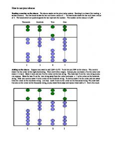
Results 1-Tapping Mode AFM The cured epoxy adhesive with no added water was found to contain phase-separated rubber particles with an average diameter of about 0.25 μm. It can be seen clearly by AFM that these rubber particles contain phase-inverted epoxy (Fig. 1A). The adhesive also contains two types of inorganic particles, namely (i) fumed silica nanoparticles which are added to adjust the thixotropic properties of the adhesive, these are mainly present as agglomerates in the epoxy matrix, and (ii) large particles of an aluminium filler with widely varying sizes up to a maximum of tens of microns. The addition of water to the adhesive prior to curing altered the microstructure of the resulting cured adhesive. The most obvious change, which was also evident using optical microscopy, was the formation of cavities of different sizes in the wet-cured adhesive at the higher water concentrations. These were steam cavities that also have been observed previously [2]. However, it is not believed that these cavities can alone explain the loss in fracture energy of the bonded laminates as a significant reduction in GIc was even observed for relatively low levels of water content, before voiding became apparent [2]. Interestingly, several micro-structural changes could 1
be observed by AFM in the wet-cured samples. Firstly, both the mean diameter and the volume fraction of the rubber particles were increased, see Table 1. Secondly, it was increasingly difficult to detect phase-inverted epoxy in the rubber particles in the wet-cured samples (Fig. 1B).
A
ably caused by steam diffusing through the hot-curing adhesive and in the process driving the hydrophobic fumed silica particles away from that particular region. Still, the degree of silica agglomeration appeared to be the same for all samples. Also, no effect of water on the aluminium filler was observed.
Results 2-Nanoindentation Load-displacement curves were recorded during indentation in to the epoxy matrix of the dry- and the wetcured adhesive samples. The epoxy adhesive displays both viscoelastic and plastic behaviour. This can be clearly seen by the shape of the loading and unloading curves in Fig. 2, and the fact that a plastic impression is left in the surface, see Fig. 3. The maximum displacement for all the indentations in the epoxy matrix, for both dry- and wet-cured samples, was about 80 nm. The probe had then been indented 80 nm into the samples. The final depth of penetration when the indenter was fully unloaded and no longer in contact with the material was about 35 nm. The elastic modulus can be deduced from the slope of the upper portion of the unloading curve during the initial stages of unloading [3]. However, the slopes of the different samples were all very similar. Thus, it appeared that any changes in the elastic modulus were very small and were beyond the resolution limit of the apparatus used. The Berkovich hardness of the epoxy matrix was calculated by dividing the maximum applied load by the projected contact area, H = Pmax/A, of the plastic impression. The samples all had a hardness of ~450 MPa which was independent of water content. This value is typical for epoxies [4]. This would also indicate that the yield stress of the epoxy matrix was independent of water content.
2 μm
B
2 μm Figure 1. AFM phase images: (A) dry-cured epoxy adhesive and (B) example of wet-cured epoxy adhesive. Table 1. Glass transition temperature, mean rubber particle size and rubber volume fraction in the adhesive. (± Standard deviation in brackets.) % w/w water in adhesive Tg (oC) Diameter (μm) Volume fraction
0
0.3
0.6
1.0
107 0.23 (0.12) 0.043 (0.006)
98 0.34 (0.14) 0.063 (0.022)
94 0.45 (0.18) 0.077 (0.019)
89 0.37 (0.19) 0.057 (0.013)
Thirdly, there were areas with no or very little fumed silica, particularly close to the steam cavities. This was prob-
Loading Unloading
Figure 2. Typical indentation load-displacement data for the epoxy matrix in the epoxy adhesive. The final plastic impressions were all imaged within 5-10 minutes after the indentation was performed. An interesting observation was that the depth of the plastic impression left in the surface was a function of the amount of water present during curing, see Fig. 4. From the unloading data, it was found that the depth of penetration imme2
diately on unloading was 35 nm for all samples. However, the depths of the final plastic impressions in the surfaces were different, i.e. 9 and 27 nm for the 0 and 1.0% w/w water samples, respectively. This indicates that a timedependent visco-elastic recovery of the material was taking place after unloading, and that there was less recovery in the wet samples. One uncertainty with these experiments is that the outer sample surface might dry out, the results may therefore not be representative of the joints employed in the fracture tests.
250 nm Figure 3. AFM height image showing four plastic impressions in the epoxy matrix in the dry-cured adhesive.
had been measured. Also, a decrease in Tg with increasing water content was observed. This might indicate that more rubber was dispersed in the epoxy matrix and that less rubber phase-separates. However, as mentioned above, the observations made by AFM indicate that more rubber was phase-separating into the formation of particles in the ‘wet’ samples; the rubber volume fraction was increased (vf=7.7+1.9% at 0.6% water content) and certainly less phase-inverted epoxy in the particles was detected (Fig 1). These apparently conflicting observations can be explained by the fact that water, which has a very low Tg of -137oC [5], is present in the system during the hot-curing of the epoxy adhesive. One possible effect of water is that it could induce subtle chemical changes to the epoxy matrix, which would result in a different crosslink density of the polymer. This would again alter the Tg of the polymer. Physically absorbed water in the epoxy polymer would also decrease the Tg. The combined effect of less rubber in solution and increased water content could therefore explain the loss in Tg from 107 to 89oC for the 0 and 1.0% w/w bulk adhesive samples, respectively. Although an intriguing observation, it is considered unlikely that the observed changes to the rubber morphology in the wet-cured adhesive could explain the substantial reduction in GIc for the bonded joints. It is more likely that the explanation lies in subtle chemical changes to the epoxy matrix induced by water. The difference in viscoelastic recovery of the epoxy matrix after nanoindentation would suggest that there is such a difference in chemistry. This could lead to a change in the mechanical properties of the epoxy matrix. These changes might suppress the toughening mechanisms (plastic void growth and shear yield) that are acting in the ‘dry’ epoxy matrix and cause the loss of toughness that is observed in the ‘wet’ samples. More AFM and nanoindentation work is currently being undertaken to investigate these effects in more detail.
Figure 4. Depth of the plastic impressions in the epoxy matrix 5-10 minutes after indentation.
Discussion & Conclusions The presence of phase-separated rubber particles in the cured epoxy adhesive is a major contributor to toughness in the dry bonded adhesive joints. In the present work the epoxy adhesive contains 8.5 wt% rubber, and based on the measured Tg values, see Table 1, the Fox-Flory equation was used to estimate that 5 wt% rubber has phaseseparated upon curing while 3.5 wt% remained in solution in the epoxy matrix of the ‘dry’ samples. This is in general agreement with the measured rubber volume fraction, as shown in Table 1 (vf rubber =4.3+0.6% for the dry adhesive). Scanning electron microscopy of the fracture surfaces of the bonded joints showed that the toughening mechanisms acting in the adhesive in the absence of water were (i) rubber particle cavitation followed by plastic void growth and (ii) shear yielding in the epoxy matrix [2]. However, very little evidence of these mechanisms could be found in the wet samples when very low values of GIc
Acknowledgements The authors would like to thank Dr Terry Gordon (BondMaster UK) and Dr John Bishopp (Star Adhesion) for useful discussion. We would also like to thank the EPSRC and A*STAR (Singapore) for financial support.
References 1. 2.
3. 4.
5.
B.R.K. Blackman, A.J. Kinloch and M. Paraschi, J. Mater. Sci. Lett., 2001, 20, pp 265-267. B.R.K. Blackman, A.J. Kinloch and W.S. Teo, 26th Annual Meeting of the Adhesion Society, 2003, Myrtle Beach, SC, USA. W.C. Oliver and G.M. Pharr, J. Mater. Res., 2004, 19, pp 3-20. X. Li, H. Gao, W.A. Scrivens, D. Fei, X. Xu, M.A. Sutton, A.P. Reynolds and M.L. Myrick, Nanotechnology, 2004, 15, pp 1416-1423. R.S. Smith and B.D. Kay, Nature, 1999, 398, pp 788791. 3