5367
Development 127, 5367-5378 (2000) Printed in Great Britain © The Company of Biologists Limited 2000 DEV2638
Ectopic Hoxa2 induction after neural crest migration results in homeosis of jaw elements in Xenopus Massimo Pasqualetti1,*, Michela Ori1,2,*, Irma Nardi2 and Filippo M. Rijli1,‡ 1Institut
de Génétique et de Biologie Moléculaire et Cellulaire, CNRS/INSERM/ULP, Collège de France, BP 163 – 67404 Illkirch Cedex, C.U. de Strasbourg, France 2Laboratori di Biologia Cellulare e dello Sviluppo, Universita’ di Pisa, 56010 Ghezzano, Pisa, Italy *These authors contributed equally to this work ‡Author for correspondence (e-mail:
[email protected])
Accepted 29 September; published on WWW 14 November 2000
SUMMARY Hox genes are required to pattern neural crest (NC) derived craniofacial and visceral skeletal structures. However, the temporal requirement of Hox patterning activity is not known. Here, we use an inducible system to establish Hoxa2 activity at distinct NC migratory stages in Xenopus embryos. We uncover stage-specific effects of Hoxa2 gainof-function suggesting a multistep patterning process for hindbrain NC. Most interestingly, we show that Hoxa2 induction at postmigratory stages results in mirror image homeotic transformation of a subset of jaw elements, normally devoid of Hox expression, towards hyoid
morphology. This is the reverse phenotype to that observed in the Hoxa2 knockout. These data demonstrate that the skeletal pattern of rhombomeric mandibular crest is not committed before migration and further implicate Hoxa2 as a true selector of hyoid fate. Moreover, the demonstration that the expression of Hoxa2 alone is sufficient to transform the upper jaw and its joint selectively may have implications for the evolution of jaws.
INTRODUCTION
presumptive mandibular crest, already predetermined before migration. In contrast, more recent experiments from a number of laboratories have shown that, in addition to prepatterning influences, the cranial NC displays a degree of plasticity after leaving the neuroepithelium (e.g. Couly et al., 1996, 1998; Hunt et al., 1998; Saldivar et al., 1996, 1997; Trainor and Krumlauf, 2000). However, little is known about the temporal patterning of the NC contributing to jaw elements. In Xenopus embryos, NC streams are initially contiguous and become separated only at later migratory stages (Sadaghiani and Thiebaud, 1987). A high level of integration is maintained between the rhombomeres, their associated NC and the pharyngeal arches throughout development, resulting in precise spatial relationships between skeletal, muscle and neuronal elements of the same axial origin (Kontges and Lumsden, 1996). A remarkable conservation of these anatomical relationships is present in vertebrates, indicating that this is a key mechanism in the construction of the vertebrate head, prompting the question of its genetic control. A Hox code was suggested for the patterning of the branchial region of the head (Hunt et al., 1991). The knockout of mouse Hoxa2 provided support to this hypothesis and suggested a tight causal link between Hox code and final morphology of the pharyngeal arches (Gendron-Maguire et al., 1993; Rijli et al., 1993). Hoxa2 is expressed up to the r1/r2 border and in the NC of the 2nd and more posterior arches, though not in the NC of the 1st pharyngeal arch which is devoid of Hox expression
The early organisation of the vertebrate head depends on three key events. First, the generation of the neural crest (NC), which gives rise to most of the skull bones as well as to the facial and visceral skeleton (Couly et al., 1993). Second, the segmentation of the hindbrain into a series of lineage-restricted compartments, the rhombomeres (r), which provide a framework for the segmental migration of the NC (Fraser et al., 1990). Third, the generation of the pharyngeal arches, which are patterned for later differentiation into skeletal elements according to the segmental origin of their populating NC (Couly et al., 1993; Kontges and Lumsden, 1996; Lumsden et al., 1991; Sechrist et al., 1993). A working hypothesis for many years has been that the spatial pattern of skeletogenesis of facial structures is fixed in the NC before the onset of migration from the neural tube (Noden, 1983). When premigratory 2nd arch NC was replaced with rostral rhombencephalic (r1+r2) and mesencephalic 1st arch NC (normally contributing to mandibular arch structures) in chick embryos, the crest cells generated from the graft migrated into the 2nd arch, according to their new location, but gave instead rise to a second set of 1st arch-like mandibular structures, appropriate for their original position. Such experiments appeared to demonstrate that, while the 2nd arch environment is permissive for migration of 1st arch NC, the spatial pattern of skeletogenesis is an intrinsic property of
Key words: Hox, Hindbrain, Cranial neural crest, Craniofacial development, Jaw evolution, Inducible gain-of-function, Xenopus
5368 M. Pasqualetti and others (Maconochie et al., 1999; Prince and Lumsden, 1994; Prince et al., 1998; this study). In Hoxa2 mutants, 2nd arch NC derivatives have been replaced by a mirror-image duplication of a subset of 1st arch-like structures. Based on fate-mapping studies in chick, and homologies between birds and mammals, it was suggested that the 2nd arch derivatives in Hoxa2 mutant mice were homeotically transformed in those normally derived from r1+r2 crest (Kontges and Lumsden, 1996). These findings also suggested that part of the mandibular and hyoid arch shared a common default developmental ‘ground pattern’ corresponding to the r1+r2 crest programme (Rijli et al., 1993). However, conclusive evidence for such a proposal is awaited, as Hoxa2 has not yet been shown to be sufficient to induce a reverse homeotic transformation of mandibular (r1+r2 crest) towards hyoid (r4 crest) morphology. Moreover, the knockout experiment did not provide information about the temporal requirement of Hoxa2 activity. Here, we have performed gain-of-function experiments injecting mouse, zebrafish and frog Hoxa2 orthologues into Xenopus embryos. We have also used an inducible system to establish Hoxa2 activity at distinct NC migratory stages. We uncover stage-specific effects of Hoxa2 gain-of-function, indicating a multistep patterning process for hindbrain NC. Most interestingly, induction of Hoxa2 after NC migration selectively results in mirror-image homeotic transformation of a subset of 1st arch structures towards hyoid morphology, opposite to the Hoxa2 knockout phenotype. The implications of these results both on developmental and evolutionary grounds are discussed. MATERIALS AND METHODS Cloning of frog, zebrafish and mouse full-length Hoxa2 A 799 bp fragment of mouse Hoxa2 genomic DNA corresponding to the second exon and 3′-UTR was amplified using PCR (Tan et al., 1992) and used to probe a stage 28 Xenopus embryo cDNA library (kindly provided by R. Harland). In brief, after plaque lifts, filters were incubated in hybridisation solution (5× SSC, 5× Denhardt’s solution, 0.1% SDS, 0.1mg/ml of denatured salmon sperm DNA) for 4 hours at 65°C. The PCR fragment was labelled with 32P-dCTP and added to the hybridisation solution. Incubation of the filters continued for a further 18 hours at 60°C. Highest stringency washing condition was 2× SSC, 0.1% SDS, 60°C. Seventeen positive clones were identified from which two distinct sets of cDNAs encoding Hoxa2related sequences were isolated. Two full-length clones from each set were fully sequenced on both strands. One 1530 bp cDNA encoded XHoxa2, and the other 1553 bp cDNA encoded XHoxa2b. Sequence analysis was carried out with the University of Wisconsin GCG computer package. The same probe and identical conditions were used to screen an oligo-dT-primed zebrafish embryo cDNA library (18-40 hours) prepared in λZapII. Sequence analysis of 26 positive clones revealed them to be all derived from the same cDNA sequence. The longest clone contained a 1797 bp insert, encoding the full-length zebrafish Hoxa2 cDNA, as assessed by comparison with the reported partial cDNA. Three nucleotide (nt) changes (nt979, nt1530 and nt1548) were found when compared with (Prince et al., 1998). The change at position 1530 results in a P to H change at position 360 of the predicted protein sequence. The mouse Hoxa2 full-length cDNA was screened from a 10.0 days postcoitus (dpc) mouse embryo cDNA library at high stringency. Nucleotide sequence was identical to that reported by Tan et al. (1992).
In situ hybridisation Digoxigenin (DIG)-labelled antisense RNA probes were generated for XHoxa2, XSlug (Mayor et al., 1995), XAP-2 (Winning et al., 1991), Xbap and Xgsc (Newman et al., 1997). Whole-mount in situ hybridisation was performed on embryos fixed in MEMFA as described in Harland (1991), except that BM purple (Roche) was used as a substrate for the alkaline phosphatase. After colour development, embryos were postfixed and bleached (Mayor et al., 1995). In situ hybridisation on 10 µm cryostat embryo sections were as described in Pasqualetti et al. (1999). Constructs To generate pCS2.XHoxa2, nucleotides 174 to 1280 of the XHoxa2 cDNA were PCR amplified using primers ZE216 AATAGAATTCATGAATTACGAATTTGAGCGAGAG and ZE217 AATAGAATTCGTAGTTCAAATGCTGCAAGTCTAT (restriction enzyme sites are underlined), digested with EcoRI, and cloned into the EcoRI site of pCS2+. For pCS2.XHoxa2:GR, primers ZE218 ATAACAGTCGACGAACCTCTGAAAATCCTGGTAACAAAA and ZE219 ATAACAGTCGACCTTTTGATGAAACAGAAGTTTTTTGA were used to PCR amplify the glucocorticoid ligand binding domain cDNA from the pSP64T.MyoD:GR vector (Kolm and Sive, 1995). The amplified fragment was digested with SalI and then subcloned into the XhoI site of the pCS2.XHoxa2 vector. To generate the pCS2.mHoxa2 construct, the StyI/HindIII Klenow-filled fragment containing the mouse Hoxa2 whole coding region (Tan et al., 1992) was subcloned into the StuI site of pCS2+. The zebrafish Hoxa2 full-length cDNA (nucleotides 452-1540) was PCR amplified using the primers XE171 CATAACATGTACTCGAGATGAATTACGAATTCGAGCG and XE172 CATCTTGAAGTTCTAGAGTTAGTAGCTCAAGTGTTGC. The amplified fragment was digested and subcloned in the XhoI/XbaI site of pCS2+ and of the Myc-tagged version pCS2+MT to generate the pCS2.zHoxa2 and pCS2MT.zHoxa2 constructs, respectively. The control construct lacking the homeobox, pCS2+.z∆Hoxa2, was made using primers XE171 and XI244 CATCTTGAAGTTCTAGAATCAGAAATCTCGGGCGAGCC to amplify nucleotides 452-832 of the zebrafish Hoxa2 cDNA, and cloned in the XhoI/XbaI site of pCS2+. All constructs were confirmed by sequencing. The pCMTEGFP (a gift from D. Gilmour) contains the EGFP reporter inserted into pCS2+MT. pAdMLARE reporter construct and pSGpbx1 were from Di Rocco et al. (1997). Transfections Transfection assays were performed in COS-1 cells by calcium phosphate precipitation, as reported by Di Rocco et al. (1997). A CMV-β-galactosidase plasmid was cotransfected as an internal measure of transfection efficiency. Dexamethasone (DEX, Sigma) was added to a final concentration of 1 µM to the culture medium 24 hours after transfection. Cells were harvested 48 hours after transfection, and the relative luciferase activity calculated. Embryo injections and treatments Xenopus laevis embryos obtained by in vitro fertilisation were allowed to develop in 0.1× Marc’s modified Ringer (MMR) and staged according to Nieuwkoop and Faber (1967). Capped RNAs were synthesised in vitro from pCS2.XHoxa2, pCS2.mHoxa2, pCS2.zHoxa2, pCS2MT.zHoxa2, pCS2+.z∆Hoxa2, pCS2.XHoxa2:GR, and pCMTEGFP template DNAs using the SP6 Message Machine kit (Ambion). RNAs were injected into one blastomere at the two-cell stage in 4% Ficoll in 0.1× MMR and incubated overnight at 14°C. The following day, embryos were transferred to 0.1× MMR and incubated at 22°C until fixation. Glucocorticoid receptor (GR) fusion proteins were activated by adding DEX 10 µM into the culture medium (Kolm and Sive, 1995). Culture medium plus DEX was prepared fresh and changed daily. Specimens were visualised with a Leica MS FL-III stereo dissecting
Hoxa2 patterns postmigratory cranial neural crest 5369 microscope equipped with epifluorescence optics, and digital images were generated using a Photometrics COOLSNAP CCD camera. Skeletal and muscle staining Stage 49 embryos were fixed in 4% paraformaldehyde, rinsed in PBT (PBS, 0.1% Tween 20) and stained for 6 hours in 0.05% Alcian Blue in 80% ethanol/20% acetic acid. After destaining overnight in acidic alcohol, specimens were transferred into 1% KOH/3% H2O2 for 3 hours. The tissue was cleared in 0.05% trypsin in saturated sodium tetraborate for another 2 hours. Embryos were washed in PBT and then stored in PBT/0.1% NaN3. For double staining, the musclespecific 12/101 monoclonal antibody (Developmental Studies Hybridoma Bank) diluted 1:10 was first used following a standard immunohistochemistry procedure. Skeletal staining of tadpoles was subsequently performed as above. The skin was dissected out and the ethmoidal plate cut along the midline and specimens flat-mounted on slides. Sections from stage 44 embryos were stained for cartilage with Alcian Blue and counterstained with Sefranine O.
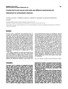
RESULTS Cloning and expression pattern of Hoxa2 in Xenopus embryos From the screening of a Xenopus embryo cDNA library using a mouse Hoxa2 probe (Tan et al., 1992), two classes of clones were isolated bearing highly similar, although not identical, Hoxa2-related sequences (Fig. 1A,B). Full-length clones representative of the two classes were named XHoxa2 and XHoxa2b, respectively. Comparison of their deduced amino acid sequences with those of other vertebrate Hoxa2 proteins indicates high homology with mouse (79%) (Tan et al., 1992), chick (78%) (Prince and Lumsden, 1994) and zebrafish (69%) (Prince et al., 1998) Hoxa2 (Fig. 1B) as compared with only 49% with human and zebrafish Hoxb2 (data not shown). Moreover, XHoxa2 and XHoxa2b display identical developmental expression patterns, including diagnostic r2 expression (Fig. 2B; data not shown). Altogether, these data indicate the presence of two Hoxa2 genes in Xenopus laevis. Although, it cannot be ruled out that these two Hoxa2-related sequences represent polymorphic alleles of the same gene, two distinct genetic loci instead of one present in other vertebrates have been reported for several genes in Xenopus laevis, including Hox genes (Fritz et al., 1989). XHoxa2 expression pattern was analysed by in situ hybridisation and RT-PCR analysis (Fig. 2A-H; data not shown). Transcripts were detectable from stage 12 on (not shown). XHoxa2 rostralmost expression domains were found in r2 and the NC of the 2nd pharyngeal arch, as in other vertebrates. By stage 26 (Nieuwkoop and Faber, 1967), XHoxa2-expressing NC has penetrated into the 2nd and more posterior arches (Fig. 2A). By stage 28, XHoxa2-expressing hyoid NC segregates from the mesodermal mass located in the centre of the arch (Sadaghiani and Thiebaud, 1987) (Fig. 2C,D). Strong XHoxa2 expression persisted in 2nd arch NC up to late developmental stages. At stage 41 XHoxa2 transcripts could still be readily detected and identify the procartilaginous ceratohyal condensation (arrow, Fig. 2E,G). By stage 44, the ceratohyal (C) cartilage is clearly identified by Alcian Blue staining (Fig. 2F,H), whereas XHoxa2 expression is no longer detectable. Injection of vertebrate Hoxa2 orthologues Full-length constructs derived from XHoxa2 as well as from
mouse and zebrafish Hoxa2 cDNAs were generated for comparative gain-of-function studies. The different Hoxa2 RNAs were injected into single blastomeres of two-cell stage Xenopus embryos. Specific alterations of the NC-derived skeleton of the pharyngeal arches were observed. Similar phenotypes were obtained with mouse, zebrafish or Xenopus Hoxa2 RNAs (Fig. 3F-L; data not shown), demonstrating functional conservation of vertebrate Hoxa2 proteins in vivo (see also the analysis of their transcriptional activities in vitro; Fig. 6A). Overall, more than 4000 embryos were injected. Embryos injected with zebrafish Hoxa2 RNA lacking the homeobox or with β-galactosidase (lacZ) RNA as control never displayed abnormalities (not shown). The optimal concentration range was found between 50 and 100 pg of injected wild-type Hoxa2 RNAs. Above 100 pg, gastrulation defects were induced, affecting survival (not shown). Below 50 pg no developmental abnormalities were observed. Within the optimal range, the survival rates of injected embryos with either full-length or control RNA were similar and close to 100% at 24 hours (stage 18). Increasing amounts of injected Hoxa2 (between 50 and 100 pg) resulted in more frequent and penetrant skeletal phenotypes in stage 49 tadpoles, demonstrating a dose-specific effect of Hoxa2 overexpression (not shown). The spatiotemporal distribution of exogenous Hoxa2 was analysed by immunostaining analysis up to stage 30 in embryos injected with 50 pg of a Myc-tagged zebrafish Hoxa2 RNA (not shown). Expressing cells showed random mosaic distribution varying from one embryo to another (not shown). To assess the correlation between spatial restriction of injected Hoxa2 and skeletal phenotype, co-injection experiments were carried out with green fluorescent protein (GFP) RNA, as a vital lineage tracer (Fig. 3A-C). Fig. 3A summarises the distributions of normal and abnormal skeletal phenotypes in tadpoles coinjected with 50 pg of XHoxa2 and GFP RNAs (n=443). Control embryos injected only with GFP had normal skeletal phenotypes (not shown). In contrast, XHoxa2 coinjected embryos selected at stage 49 (i.e. just before skeletal analysis) for widespread GFP expression (GFP+; n=336; Fig. 3A,C) displayed a high frequency of skeletal abnormalities (68%), which, however, affected only a subset of the pharyngeal arch skeletal derivatives (see below). On the contrary, almost 90% of the embryos with no detectable anterior GFP expression (GFP–; n=107; Fig. 3A,B) displayed normal skeletons. These results indicate a strong correlation between the presence of exogenous XHoxa2 and the observed phenotype. Analysis of the craniofacial and visceral skeleton Fig. 3D,E shows the normal pattern of NC derivatives in the head and visceral skeleton of the Xenopus tadpole (Sadaghiani and Thiebaud, 1987). Based on fate-mapping studies (Kontges and Lumsden, 1996; Sadaghiani and Thiebaud, 1987), the 1st arch skeleton has a composite origin. The upper jaw (palatoquadrate; Q) and the proximal part of lower jaw (Meckel’s; M) cartilages are mostly r1+r2 derived (Met MCS), whereas the distal portion of the Meckel’s and the ethmoid (Et) plate are exclusively midbrain derived (Mes MCS). The 2nd (hyoid) arch crest (HCS) gives origin to the ceratohyal (C) cartilage, whereas the 3rd (aBCS) and 4th (pBCS) arch NC contribute to the gill cartilages (G).
5370 M. Pasqualetti and others
A agtttaatagtagcacagtccccatacggctgtaatcagtgaattagaaaaaaaaaaacacaccggcaaaggatttttat gatttttttttttttttaccccctcgtcgttacggggcctttaaacgtaaaagtaaggggctaagttgtttaaagggctt ggaggagagggccATGAATTACGAATTTGAGCGAGAGATTGGTTTTATCAATAGTCAGCCGTCGCTTGCTGAGTGCCTGA M N Y E F E R E I G F I N S Q P S L A E C L
80 160 240 22
CATCCTTTCCCCCTGTCGGTGATACATTTCAAAGTTCATCAATCAAGAGCTCGGCGCTTTCACACTCGACACTCATTCCT T S F P P V G D T F Q S S S I K S S A L S H S T L I P
320 49
CCTCCTTTTGAGCAGACAATCCCCAGCTTGAATCCAGCCAGCCACCCTCGCCACAGCCGGCCCAAACACAGCCCAAATGG P P F E Q T I P S L N P A S H P R H S R P K H S P N G
400 76
CCACGGAGACAGCCCGGTGCCAGCAGGATCCCTGCCTCCCGAGTACCCCTGGATGAAAGAGAAAAAGGCTTCCAAGAAAG H G D S P V P A G S L P P E Y P W M K E K K A S K K
480 102
CCCCTATAGTCACAGCAACAGCAGCCGCCAACGTTGCCCCGGCCACCACCGCCTGCCTCAGCCACAAAGAAATTATTGAG A P I V T A T A A A N V A P A T T A C L S H K E I I E
560 129
ATCCAGGATAACAGTGGTGGGGGATCGAGACGATTAAGGACAGCTTATACCAACACTCAGCTCTTAGAACTGGAAAAAGA I Q D N S G G G S R R L R T A Y T N T Q L L E L E K E
640 156
ATTTCATTTCAACAAGTATCTGTGCAGACCGAGGAGGGTGGAAATTGCAGCCCTGTTGGATCTGACCGAAAGGCAGGTCA F H F N K Y L C R P R R V E I A A L L D L T E R Q V
720 182
AAGTCTGGTTCCAGAACAGGAGAATGAAGCACAAGAGGCAAACCCAGTGCAAGGAGAACCAGAATGGAGATGGGGGCAAA K V W F Q N R R M K H K R Q T Q C K E N Q N G D G G K
800 209
TTCAAGCATCTGGACGATTGTGAGAAGGGAGAGGAGGAGACGGAGAAATCCCTGTTTGAGCAAGCCTTGAATAGTGTCTC F K H L D D C E K G E E E T E K S L F E Q A L N S V S
880 236
TGGGGCTTTGCTGGAGAGAGACAGCTATAGTTTCCAGCAGAATGCCTTAGCCCAGCAGCAGTCTCACAACTCACACAATG G A L L E R D S Y S F Q Q N A L A Q Q Q S H N S H N
960 262
GAGATTCCCAGAGTTTCCCTGTATCGCCTTTATCTAACAGTGAGAAAAATCTCAGACATGTCCAGCAACAGTCGCCCAAC G D S Q S F P V S P L S N S E K N L R H V Q Q Q S P N
1040 289
TGCTTATCAACAATTGCCCTGGACTGTGCAGCTGGACTCAACAATGATAGTCCCGAGGCCTTAGATGTGGCTTCACTCTC C L S T I A L D C A A G L N N D S P E A L D V A S L S
1120 316
GGACTTCAATGTCTTCTCCTCGGATTCCTGCCTTCAGCTCTCAGACACAGTCTCCCCCAGCCTGCCTGGCTCTTTGGACA D F N V F S S D S C L Q L S D T V S P S L P G S L D
1200 342
GTCCTGTAGACATTTCAGCTGACAGTTTCGATTTTTTCACAGATACATTTACCACAATAGACTTGCACCATTTGAACTAC S P V D I S A D S F D F F T D T F T T I D L H H L N Y
1280 369
tgaagcaacccacattattacatagtgaatttatttttattttttaagtaaaaattctcccccttagatccagtttcagt tttaacatccagttcagttgagttaatccaacaccattgaaagcaataaccgaggcctctatgaaagtgacttttgtctg aacgtatctgcactttcttttatttttctattgcattaaaatgcgctgtcatagaagtatttttggtagaataaatgagt atttgccatg
1360 1440 1520 1530
B XHoxa2 XHoxa2b mouse Hoxa2 chick Hoxa2 fish Hoxa2
MNYEFEREIGFINSQPSLAECLTSFPPVGDTFQSSSIKSSALSHSTLIPPPFEQTIPSLNPASHPRHSR-----PKHSPNGHGDSPVPAGSL-PPEYPWM 94 MNYEFEREIGFINSQPSLAECLTSFPPVGDTFQSSSIKSSALSHSTLIPPPFEQTIPSLNPASHPRHSR-----PKHSPNGHGDSPVPAGSL-PPEYPWM 94 MNYEFEREIGFINSQPSLAECLTSFPPVADTFQSSSIKTSTLSHSTLIPPPFEQTIPSLNPGSHPRHGAGVGGRPKSSPAGSRGSPVPAGALQPPEYPWM 100 MNFEFEREIGFINSQPSLAECLTSFPPVGDTFQSSSIKNSTLSHSTLIPPPFEQTIPSLNPGGHPRHS-G-GGRPKASPRARSGSPGPAGAPPPPEYPWM 98 MNYEFERETGFINSQPSLAECLTSFPPVGDAFQSSSIKSSTLSHSTLIPPPFEQTIPSLNPGSHPRHSR-----PKQNPNG--SCPLPAASL-PPEYPWM 92
XHoxa2 XHoxa2b mouse Hoxa2 chick Hoxa2 fish Hoxa2
KEKKASKKAPIVTATAAANVAPAT---TACLSHKEIIEIQDNSGGGSRRLRTAYTNTQLLELEKEFHFNKYLCRPRRVEIAALLDLTERQVKVWFQNRRM KEKKASKKTPIAAATAAAAAASASPVTTACLSHKEIIEIQDNSGGGSRRLRTAYTNTQLLELEKEFHFNKYLCRPRRVEIAALLDLTERQVKVWFQNRRM KEKKAAKKTALPPAAAST-----G---PACLGHKESLEIADGSGGGSRRLRTAYTNTQLLELEKEFHFNKYLCRPRRVEIAALLDLTERQVKVWFQNRRM KEKKASKRSSLPPASASASAA--G---PACLSHKDPLEIPDSGSGGSRRLRTAYTNTQLLELEKEFHFNKYLCRPRRVEIAALLDLTERQVKVWFQNRRM KEKKASKKNQTTSTAATTDPG------PLYFSPQGSPEISDGGSGATRRLRTAYTNTQLLELEKEFHFNKYLCRPRRVEIAALLDLTERQVKVWFQNRRM
191 194 192 193 186
XHoxa2 XHoxa2b mouse Hoxa2 chick Hoxa2 fish Hoxa2
KHKRQTQCKENQNGDGGKFKHLDDCEKGEEE--TEKSLFEQALNSVSGALLERDSYSFQQNALAQQQSHNSHNGDSQSFPVSPLSNSEKNLRHVQQQS-KHKRQTQCKENQNGDGGKFKHLDDSEKGEEE--KEKSLFEQALNSVSGALLERDSYSFQQNALAQQQSHNSHNGDSQSFPVSPLSNSEKNLRHFQQQSPT KHKRQTQCKENQNSEG-KFKNLEDSDKVEED-EEEKSLFEQAL-SVSGALLEREGYTFQQNALSQQQAPNGHNGDSQTFPVSPLTSNEKNLKHFQHQSPT KHKRQTQCKENQNGEG-KFKGSEDPEKAAEDDEEEKALFEQALGTVSGALLEREGYAFQQNALSQQQAQNAHNGESQSFPVSPLTSNEKNLKHFQHQSPT KHKRQTQCKENHHGDG-KPPSLEEAGGRGDG----KSFFEQVANNVSGALLEREGYPFQQNTLTSQQSQNGHNSDSQSATVSPLGSNDKHLKHFPNPSPT
287 292 289 292 281
XHoxa2 XHoxa2b mouse Hoxa2 chick Hoxa2 fish Hoxa2
-PNCLSTIALDCAAGLNNDSPEALDVASLSDFNVFSSDSCLQLSDTVSPSLPGSLDSPVDISADSFDFFTDTFTTIDLHHLNY APNCLSTIAQDCAAGLDNDSPEALDVASLSDFNVFSSDSCLQLSDTVSPSLPGSLDSPVDISADSFDFFTDTFTTIDLQHLNY VPNCLSTMGQNCGAGLNNDSPEAIEVPSLQDFNVFSTDSCLQLSDALSPSLPGSLDSPVDISADSFDFFTDTLTTIDLQHLNY VQNCLSTMAQNCAAGLNNDSPEALEVPSLQDFNVFSTDSCLQLSDAVSPSLPGSLDSPVDISADSFDFFTDTLTTIDLQHLNY VPICTTTMAPDCASAQDNGSPSALDVS-LQDFNVFSNDSCLHLSDAVSPSLSESVDSPIGLTTEAFDFFSETLTTIDLQHLSY
369 375 372 375 363
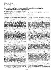
Hoxa2 patterns postmigratory cranial neural crest 5371 Fig. 1. Frog Hoxa2 (XHoxa2) cDNA, deduced protein sequence and alignment with vertebrate Hoxa2 proteins. (A) cDNA and deduced protein sequences. Stop codon is in bold. The underlined nucleotide sequence is a putative polyadenylation signal. The hexapeptide and homeobox domains are underlined and double underlined, respectively. (B) Alignment of vertebrate Hoxa2 protein sequences was performed using the ClustalW program. XHoxa2, XHoxa2b and fish Hoxa2 sequences have been submitted to GenBank (accession numbers AF307008, AF307009 and AF307010, respectively).
Two main classes of morphological alterations were found in the pharyngeal arch skeleton of injected tadpoles (n=443; Fig. 3F-L): (1) a ‘segmentation’ phenotype (n=161), involving mandibular-hyoid or 3rd (first gill)-hyoid arch fusions (Fig. 3F,G,K), and (2) a ‘homeotic’ phenotype (n=60), involving morphological changes of mandibular and/or 3rd arch elements towards hyoid morphology (Fig. 3H-J,L). Mandibular-hyoid arch fusions selectively involved the palatoquadrate, the Meckel’s and ceratohyal cartilages with various degree of penetrance, while not affecting the ethmoid or infrarostral (I) cartilages (Fig. 3F; compare with the control, uninjected side). In some specimens, mandibular arch structures were not readily identifiable (although they may be partially included in the fused elements) and the ceratohyal cartilage appeared much thicker (Fig. 3G). Third-hyoid arch fusions were also observed in some embryos, resulting in posterior thickening and partially split ceratohyal cartilage with gill-supporting branchial rays appearing on its posterior surface (arrow, Fig. 3K). In the homeotic class of phenotypes, 1st and 3rd arch elements were replaced by ectopic ceratohyal cartilages (Fig. 3H,L). Most strikingly, in several embryos the palatoquadrate and the proximal portion of the Meckel’s appeared as a single element resembling a partial mirror-image duplication of the ceratohyal cartilage (n=17, out of 443). Fig. 3I,J shows similar mirror-image duplications obtained by injecting mouse and frog Hoxa2 RNAs, respectively. Altogether, these data indicate that subsets of 1st and 3rd arch NC develop with an hyoid morphology, as a result of Hoxa2 overexpression. Patterns of skeletomuscular connectivity in Hoxa2injected embryos A double staining procedure was set up to visualise the pattern of skeletomuscular connectivity in experimental embryos (Fig. 4). As NC of the same rhombomeric origin forms both the connective tissue of individual branchial muscles and the skeletal domain on which they attach (Kontges and Lumsden, 1996), fate changes of 1st and 3rd arch NC should be expected to alter the muscle pattern selectively as well. In wild-type tadpoles (Fig. 4A), the 2nd arch-specific muscles include the interhyoideus (Ih) and the jaw-opening muscles, such as the orbitohyoideus (Oh), which inserts on both the ceratohyal and palatoquadrate cartilages, the hyoangularis (Ha) and quadratoangularis (Qa), originating from the ceratohyal and palatoquadrate, respectively, and inserting on the ventral side of the posterior third of the Meckel’s (arrow) (Hanken et al., 1997). Among the 1st arch-specific muscles, the complex of the levator mandibulae (Lm) inserts on both the ethmoid and the middle portion of the Meckel’s (Fig. 4A). The intermandibular muscle (Im) originates just rostral to the attachment sites of the Ha and Qa on the Meckel’s (arrow, Fig. 4A; see also Fig. 4B), and inserts on the medial aspect of the
ceratohyal. For the purpose of this work, two other muscles are relevant. The genohyoideus (Gh) muscle, which inserts on the infrarostral (I) and on the 3rd arch cartilages. This muscle has a likely multisegmental origin, as it spans three arches and receives innervation from different cranial nerves (Hanken et al., 1997). The other muscle is the subarcuales rectus (Sr) connecting the posterior edge of the ceratohyal to the anterior portion of the 3rd arch cartilage. In XHoxa2-injected tadpoles, selective alterations of the muscle pattern were observed (Fig. 4B-D). Fig. 4B,C shows the muscle phenotype of XHoxa2-injected tadpoles displaying
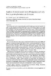
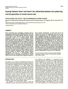
Fig. 2. XHoxa2 developmental expression pattern. (A,B) Lateral and dorsal view of a stage 26 embryo showing XHoxa2 expression in branchial arches and hindbrain up to the r1/r2 boundary (arrow). (C,D) Lateral view and horizontal section, respectively (broken line in C represents level of section in D), of stage 28 embryos showing higher XHoxa2 transcript levels in PA2 (arrows) than more posterior arches (C), as well as a specific distribution in NC cells (D). (E,G) Coronal and horizontal sections, respectively, of stage 41 embryos showing XHoxa2 expression in the procartilaginous condensation of the ceratohyal (arrow), which is identified at stage 44 by Alcian Blue/Sefranine O staining (F,H). BH, basihyal; C, ceratohyal; CG, cement gland; E, eye; Et, ethmoid; G, gill; Ih, interhyoideus; NT, neural tube; Oh, orbitohyoideus; PA, pharyngeal arch; Q, palatoquadrate; r, rhombomere.
5372 M. Pasqualetti and others
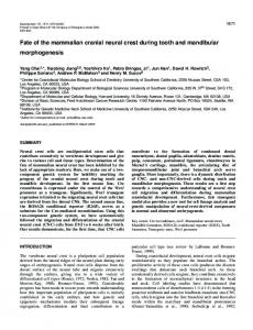
Fig. 3. Skeletal alterations in injected tadpoles. (A) Percent distribution of normal and abnormal skeletal phenotypes observed in stage 49 tadpoles co-injected with XHoxa2 and GFP RNAs, showing low (GFP–, B) or high (GFP+, C) GFP expression in the branchial region. (D,E) The migration pathways (D, modified from Sadaghiani and Thiebaud, 1987) and contribution to the visceral skeleton (coupled to cartilage staining of the actual structures, E) of individual cranial NC segments in Xenopus. Infrarostral (I) and basihyal (BH) are mesodermal derivatives. (F-L) Flat-mounts of Alcian Blue-stained skeletal preparations from stage 49 tadpoles. Uninjected side always on the left. (F,G,K) Segmentation phenotype of XHoxa2 injected tadpoles showing fusion of mandibular-hyoid arch (F,G), and 3rd-hyoid arch (K) elements. In K, branchial rays (arrow) characteristic of the 3rd (first gill; G1) arch are fused on the posterior surface of the ceratohyal cartilage. (H,L) Homeotic phenotype showing serial duplication of the ceratohyal cartilage (C2) replacing 1st (H) and 3rd (L) arch elements, respectively. (I,J) Mirror-image homeotic phenotype of palatoquadrate (Q) and proximal portion of Meckel’s (M) cartilage in embryos injected with mouse Hoxa2 and XHoxa2, respectively. The broken line represents the axis of symmetry. aBCS, anterior branchial crest segment; C, ceratohyal; Et, ethmoid; G, gill; HCS, hyoid crest segment; Mes and Mes MCS, mesencephalic and metencephalic mandibular crest segment, respectively; pBCS, posterior branchial crest segment.
mandibular-hyoid fusion, and mirror-image duplication, respectively. The jaw-opening muscles hyoangularis and quadratoangularis were selectively lacking (Fig. 4B,C). Moreover, the orbitohyoideus was shortened and altered in the region normally occupied by the palatoquadrate (star, Fig. 4B,C). In contrast, the 1st arch intermandibular and levator mandibulae and 2nd arch interhyoideus muscles were not affected (Fig. 4B,C). Instead, in embryos showing 3rd arch elements with ceratohyal morphology (Fig. 4D), some fibres of the 2nd arch interhyoid muscle navigated to the posterior edge of the ectopic ceratohyal cartilage (arrowheads). Moreover, full duplications of the subarcuales rectus were observed (arrow). Finally, the genohyoideus muscle was selectively lacking (but not when only 1st arch proximal elements were affected; compare Fig. 4B with 4C). In summary, XHoxa2 widespread overexpression results in selective alterations of only those muscles that normally insert
on the affected skeletal elements, supporting the idea of identity changes of subsets of 1st and 3rd arch NC. Patterns of neural crest segmentation and migration To visualise cranial NC in experimental embryos, we used two pan neural crest markers, XSlug (Mayor et al., 1995) and XAP2 (Winning et al., 1991) (Fig. 5A-E). At stage 18, the neural crest has formed three premigratory blocks (MCS, HCS, BCS; Sadaghiani and Thiebaud, 1987; Fig. 5A). In contrast, this segregation process was clearly affected in the injected embryos (100%; n=43) (Fig. 5B). However, at stage 28 not all the analysed embryos were found to be affected at the same extent. In fact, 14 (47%) displayed severe NC abnormalities mainly involving 1st-2nd arch fusion (Fig. 5E), 7 (23%) showed a mild segmentation phenotype (Fig. 5D) and 9 (30%) showed an essentially normal NC pattern (not shown). Thus, the molecular analysis suggests that NC cells tend to recover
Hoxa2 patterns postmigratory cranial neural crest 5373
Fig. 4. Selective alterations of skeletomuscular connectivity in injected tadpoles. (A-D) Flat mounts showing stage 49 wild-type- (A) and XHoxa2-injected (B-D) embryos double stained for cartilage and muscles. (B,C) Segmentation and homeotic mirror-image phenotypes, respectively, showing the lack of hyoangularis (Ha) and quadratoangularis (Qa) muscles and the shortening of the orbitohyoideus (Oh, asterisks), as compared with wild type (A) or the uninjected (left) side. Intermandibular (Im, arrow) and levator mandibulae (Lm) muscles appear unaffected. (D) 3rd arch homeotic phenotype showing defasciculation of the interhyoid (Ih) muscle and insertion onto the transformed skeletal element (arrowheads), and full duplication (Sr2) of the subarcuales rectus (Sr) muscle (arrows). Note that the genohyoideus (Gh) muscle is selectively missing on the injected side.
early segmentation/migration defects proceeding through late developmental stages. This is likely to be due to the timecourse of exogenous Hoxa2 dilution and spatial distribution following cell division. Interestingly, such molecular differences reflect the variable distribution and penetrance of the skeletal phenotypes observed at stage 49 (Fig. 3). Altogether, these data underscore the remarkable plasticity of hindbrain NC. Stage-specific inducible Hoxa2 gain-of-function To allow temporal control of exogenous Hoxa2 activity, we used an hormone-inducible system (Kolm and Sive, 1995). A chimeric protein (XHoxa2:GR) was generated by fusion of XHoxa2 to the ligand binding domain of the glucocorticoid receptor. The XHoxa2:GR inducibility and transcriptional activity were tested in cell cotransfection experiments with a reporter driven by a Hoxa2 target sequence (Di Rocco et al., 1997) (Fig. 6A). In the absence of dexamethasone (DEX), the fusion protein is inactive. In contrast, in the presence of DEX a XHoxa2:GR-dependent transcriptional activity was observed that could be enhanced by cotransfection of the Hox cofactor Pbx1 (Di Rocco et al., 1997), as also seen in cells cotransfected with mouse, fish and frog wild-type Hoxa2. Thus, XHoxa2:GR displays similar transcriptional activity and biochemical properties (including cooperative interaction with cofactors) as wild-type Hoxa2 proteins. XHoxa2:GR RNA was injected in Xenopus embryos (100 pg; n=3383). DEX treatment was performed at distinct stages of NC development, namely pre- (15), early (20) and postmigratory (26,28,30) stages. Following induction, which takes place within 2-4 hours of first treatment (Kolm and Sive, 1995), DEX was maintained throughout development up to stage 49 when embryos were fixed for analysis. DEX-treated uninjected embryos did not display skeletal alterations, neither did untreated XHoxa2:GR-injected embryos (0%; n=530). In contrast, skeletal analysis of injected DEX-treated tadpoles revealed a panel of abnormalities identical to those obtained by injecting wild type Hoxa2 RNAs (see Figs 3, 4, 6; data not shown). However, a striking qualitative distribution of
phenotypes could be observed depending on the stage of the induction, uncovering distinct temporal effects of Hoxa2 overexpression (Fig. 6B,C). The graph in Fig. 6B summarizes
Fig. 5. Neural crest segregation and migration in XHoxa2injected embryos. (A,B) Lateral views of a whole-mount stage 18 embryo hybridised with XSlug. (A) Pre-migratory NC blocks are clearly segregated on the uninjected side (arrows), though not on the injected side (B, asterisks). (C-E) Lateral views of stage 28 embryos hybridized with XAP-2 showing mild (D) and severe (E) alterations in NC stream segregation and migration, as compared with wild type (C). BCS, branchial crest segment; CG, cement gland; E, eye; HCS, hyoid crest segment; MCS, mandibular crest segment; PA, pharyngeal arch.
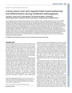
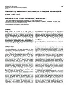
5374 M. Pasqualetti and others Fig. 6. XHoxa2 induction after NC migration results in homeosis of mandibular arch skeletal elements. (A) Luciferase activity from COS cell extracts transiently transfected with the pAdML ARE reporter, β-gal, and Pbx1 together with either Xenopus (XHoxa2), mouse (mHoxa2), zebrafish (zHoxa2) or DEX inducible (XHoxa2:GR) cDNA constructs. Bars represent the mean luciferase activity±s.e.m. of six independent experiments after normalising to β-gal activity. (B) Distribution of normal (N), segmentation (S) and homeotic (H) phenotypes in XHoxa2:GR-injected tadpoles after DEX treatment at stages (st) 15, 20, 26, 28 and 30. In C, embryos have been co-injected with GFP and selected at stage 49 for high levels of GFP expression in the branchial region. Note the significant enrichment of homeotic phenotypes in stage 26 and 28, but not stage 30, in DEX-treated GFP-selected embryos, when compared with (B). (D) Dorsal view of a whole-mount Alcian Blue-stained skeleton of a stage 49 injected tadpole, DEX-treated at stage 28, showing the loss (vertical arrow) of the muscular process (MP) of the palatoquadrate (Q) on the injected side (right); the broken red line marks the palatoquadrate edge. (E) Skeletal flat-mount of a stage 49 tadpole, treated with DEX at stage 28, showing a mirror-image homeotic transformation of the palatoquadrate and the proximal portion of the Meckel’s (M) cartilage into ceratohyal (C2) on the injected side. The red and black broken lines indicate the distal part of the Meckel’s and the axis of symmetry, respectively. (F,G) Lateral views of the uninjected (F) and injected (G) sides of a stage 37 embryo, treated with DEX at stage 28 and hybridised with Xbap. In F, expression is in the precursors of palatoquadrate and proximal part of the Meckel’s cartilage (arrow, Q+M). In G, a selective loss of Xbap expression (arrow) at the level of these precursors is observed. (H,I) Lateral views of uninjected (H) and injected (I) sides of a stage 37 embryo, treated with DEX at stage 28, and hybridised with Xgsc. Fusion of 1st and 2nd arch expression domains is indicated in I and J (arrows). (J) Ventral view of the embryo in H,I after dissection of the cement gland. Spatial reorganisation of Xgsc transcripts is evident in 1st arch (PA1, arrowhead). C, ceratoyal; E, eye; Et, ethmoid; I, infrarostral; PA2, 2nd pharyngeal arch.
the overall distribution of skeletal phenotypes obtained in a very large panel of DEX-treated tadpoles (n=2853). DEX-mediated induction of XHoxa2:GR activity at pre(15) and early (20) migratory stages resulted in both segmentation and homeotic classes of phenotypes (Fig. 6B, phenotypes S and H) similar to the injection of wild-type Hoxa2 RNAs (Fig. 3F-L). However, two important differences were found after DEX-mediated induction at stage 20, when compared with induction at stage 15. First, segmentation phenotypes were less frequent. Second, 3rd-to-2nd arch serial duplications could not be induced any longer (not shown; see Fig. 3L), suggesting that the 3rd arch NC patterning program could not be modified by Hoxa2 after the start of NC migration. XHoxa2:GR induction at these early stages resulted in variable NC stream fusions and migration defects similar to those observed in embryos injected with wild-type Hoxa2 RNAs, as assessed by XAP-2 hybridisation (not shown; see Fig. 5B,D,E). By stage 26, mandibular NC has already fully migrated to the 1st arch, which is physically separated from the 2nd arch by the 1st pharyngeal pouch (Nieuwkoop and Faber, 1967; Sadaghiani and Thiebaud, 1987). Strikingly, out of 549 embryos showing skeletal alterations following DEX treatment
at postmigratory stages (i.e. 26, 28 and 30), 526 were of the homeotic class and mandibular arch-specific (Fig. 6B, phenotype H). Importantly, such alterations selectively involved r1+r2-derived 1st arch structures (i.e. the palatoquadrate and the proximal portion of the Meckel’s cartilage; Kontges and Lumsden, 1996; Sadaghiani and Thiebaud, 1987), while midbrain-derived structures (i.e. the distalmost portion of the Meckel’s and ethmoid plate; Kontges and Lumsden, 1996; Sadaghiani and Thiebaud, 1987) were spared (Fig. 6D,E). The penetrance and expressivity of these alterations were variable, probably due to quantitative requirement of XHoxa2:GR and/or cell community effects. Nevertheless, from the analysis of the phenotypes a clear stepwise progression towards mirror image transformation to hyoid morphology could be identified. The most frequent structure to be affected was the muscular process (MP) of the palatoquadrate (Q) (Fig. 6D), on which insert the jaw-opening muscles, which was either highly reduced or lacking in 52% of the abnormal embryos (n=277) (Fig. 6D). The next step of the transformation involved the fusion of the Meckel’s to the altered palatoquadrate into a single element with consequent loss of the jaw joint (n=173; 33% of the affected embryos).
Hoxa2 patterns postmigratory cranial neural crest 5375 Finally, progressive thickening and shape changes of this element were observed resembling a partial ceratohyal cartilage with a reverse polarity, thus representing a mirror image homeotic transformation of r1+r2– towards r4-derived NC skeletal derivatives (n=76; 15% of the affected embryos) (Fig. 6E). Moreover, molecular analysis with XAP-2 showed that, as expected, the segmental organisation of NC in the pharyngeal arches was no longer affected after DEX-mediated induction at stages 26 and later (not shown). A total of 1841 embryos were DEX treated at stage 26 (n=1060), 28 (n=479) and 30 (n=302). The overall percentage of affected tadpoles after DEX-mediated induction at stage 30 dropped to less than 10%, when compared with 27% and 35% in stage 28 and stage 26 induced embryos, respectively (Fig. 6B; compare N and H phenotypes). Thus, between stage 28 and 30 XHoxa2:GR levels may become limiting, owing to its spatial distribution and dilution as cell divisions proceed. Interestingly, it is also possible that around stage 30 mandibular cells start to lose competence to respond to XHoxa2:GR activity. This may be indirectly suggested by the observation that the relative percentage of stage 30-induced embryos displaying mandibular abnormalities could not be enriched by GFP selection at stage 49, unlike that of embryos induced between stage 26 (47%) and 28 (50%) (Fig. 6C). Altogether, these results show different qualitative readouts of XHoxa2:GR activity depending on the stage of induction, providing a rationale for the interpretation of the phenotypes obtained injecting Hoxa2 wild-type RNAs (Fig. 3). The main finding is that mandibular cells are not committed about their pattern of skeletogenesis before migration. Moreover, XHoxa2:GR induction at postmigratory stages results in a reorganisation of the rhombencephalic 1st arch NC pattern towards a 2nd arch ceratohyal morphology with a reverse polarity. Molecular evidence of mandibular fate changes A group of 206 embryos were co-injected with XHoxa2:GR and GFP, and induced using DEX at postmigratory stage 28. 48 of them were GFP selected (GFP+) at stage 37 and fixed for in situ hybridisation analysis. The rest (n=158) was allowed to develop up to stage 49, GFP selected and stained for skeletal analysis. In situ hybridisation was performed using the bagpipe-related gene, Xbap. In the wild-type 1st arch, Xbap is selectively expressed in the NC precursors of the palatoquadrate and of the proximal part of the Meckel’s cartilage (Newman et al., 1997) (Q+M, Fig. 6F). Strikingly, 1st arch Xbap expression was severely downregulated or lacking in GFP+ embryos (n=27; 56%) whereas the remainder of the pattern was unchanged (compare Fig. 6F,G). Moreover, analysis at stage 49 revealed that the percentage of GFP+ selected embryos carrying mandibular arch-specific skeletal phenotypes (n=88; 56%) was the same as that of embryos displaying a molecular phenotype. In another set of experiments, XHoxa2:GR-injected embryos (induced with DEX at stage 28) were GFP selected at stage 37 (n=105) and in situ hybridisation analysis was performed to compare expression of Xbap (n=57) to that of goosecoid (Xgsc) (n=48). As in mouse and zebrafish, Xgsc is expressed in both mandibular and hyoid arch NC derivatives, though with different levels (Newman et al., 1997; Fig. 6H). Interestingly, 1st arch Xgsc expression levels appeared similar
to 2nd arch levels in 65% of injected embryos (n=31) (Fig. 6J), in good correlation with the percentage of injected embryos lacking 1st arch Xbap expression (62%; n=35). Moreover, Xgsc transcript distribution appeared spatially reorganised, prefigurating the morphological changes evident at the skeletal level later on. Altogether, these findings indicate a direct correlation between molecular and skeletal phenotypes in postmigratory 1st arch NC. They also provide conclusive evidence of fate changes of palatoquadrate and proximal Meckel’s NC precursors towards hyoid identity and support the idea that the variable expressivity of 1st arch skeletal phenotypes represents different degrees of severity of the same homeotic (H) phenotype. DISCUSSION By performing gain-of-function experiments in Xenopus, we show that vertebrate Hoxa2 proteins share a conserved role in conferring a hyoid identity to rhombencephalic NC. By exploiting the advantage of an inducible system, we provide the first temporal analysis of the patterning activity of a vertebrate Hox gene on the NC contributing to the craniofacial and visceral skeleton. These results have both developmental and evolutionary implications for the understanding the genetic control of vertebrate craniofacial morphogenesis.
Hoxa2 is a selector of hyoid morphology We show that widespread overexpression of distinct vertebrate Hoxa2 proteins in frog embryos is sufficient to transform subsets of NC derivatives selectively towards hyoid morphology (Fig. 3). This indicates phylogenetic conservation of Hoxa2 function and underscores its fundamental role as a selector of hyoid identity in vertebrates. Our results suggest that Hoxa2 performs a unique function among Hox proteins. In fact, overexpression of the Hoxa2 paralogue, Hoxb2, in zebrafish embryos resulted in fusion of 1st and 2nd arch cartilage without obvious morphological transformation (Yan et al., 1998), underscoring qualitative differences between paralogue group 2 proteins in the control of hyoid crest patterning. In contrast, zebrafish Hoxa1 gainof-function (Alexandre et al., 1996) resulted in lack of palatoquadrate and Meckel’s cartilages as well as in broadened ceratohyals. This phenotype partially resembles those obtained inducing Hoxa2 activity at pre- and early NC migratory stages, as well as injecting wild-type Hoxa2 RNAs (Fig. 3G; data not shown). In Hoxa1-injected zebrafish embryos (Alexandre et al., 1996), early changes of r2-r3 towards r4 identity resulted in fusion of 1st and 2nd arch NC into a single stream, which developed as an enlarged hyoid arch. Thus, in light of our results, one possibility is that early Hoxa1 overexpression results in ectopic Hoxa2 activation in premigratory NC. Hoxa2 overexpression within its own domain of expression also leads to morphological transformation of 3rd arch elements towards hyoid morphology (Fig. 3L; see below). This finding further supports the idea that the qualitative readout of Hox protein activity is also dependent on quantitative parameters (e.g. Greer et al., 2000). In this respect, it is noteworthy that XHoxa2 expression levels are higher in the 2nd
5376 M. Pasqualetti and others than in the 3rd arch, where expression is downregulated at late stages (Fig. 2).
Hoxa2 and the control of branchial skeletomuscular connectivity NC populations from single rhombomeres form not only the connective tissue of individual branchial muscles but also the skeletal domain onto which they attach (Kontges and Lumsden, 1996; Noden, 1986). Moreover, the deployment of individual NC populations on a given skeletal element strictly respects the anteroposterior order of origin in the hindbrain. Thus, the axial origin of individual NC populations defines the precise pattern of skeletomuscular connectivity. Such a highly patterned mechanism prompts the question of its genetic control and evolutionary conservation. Our results indicate that normal levels and spatial distribution of Hoxa2 are required for a normal branchial skeletomuscular pattern in Xenopus embryos. In particular, as the morphological changes of 1st and 3rd arch NC derivatives are always associated to the selective alteration of their inserting muscles (Fig. 4), our findings argue for a phylogenetic conservation of the relationships between parts of elements, muscles and attachment points, even though the shape of skeletal derivatives has changed during vertebrate evolution. Temporal analysis of Hoxa2 patterning function Hox gene expression is established at early NC developmental stages and is maintained through late stages of pharyngeal arch morphogenesis (e.g. Barrow and Capecchi, 1999; Davenne et al., 1999; Kanzler et al., 1998; Prince and Lumsden, 1994) (Fig. 2). While Hox genes are required for normal pharyngeal arch patterning, no information is available about their temporal activity. Our present work allow to conclude about distinct temporal effects of Hoxa2 gain-of-function. Segmentation phenotypes could be obtained by inducing XHoxa2:GR activity at early stages (stage 15 and 20), though not after NC migration in the arches (i.e. from stage 26 onwards) (Fig. 6). Specific combinations of Hox genes are expressed in rhombomeres and NC subpopulations migrating in adjacent arches, with the exception of r1+r2-derived NC, which is devoid of Hox expression. One possibility is that the combinatorial segmental domains of Hox gene expression in the neuroepithelium and NC contribute to set out a molecular system of repulsive-attractive cues that prevent NC cells from mixing, segregate them into distinct streams and target them to the correct destination (O’Leary and Wilkinson, 1999). Thus, unrestricted expression of a single Hox gene at pre- or early migratory stages may be sufficient to disrupt such interactions and result in NC segregation and pathfinding defects. A few molecules involved in the pathfinding of cranial NC have been recently identified (e.g. Eickholt et al., 1999; Golding et al., 2000; Helbling et al., 1998; Smith et al., 1997). Among them, members of the Eph receptor and ephrin gene families are involved in mediating cell-contact-dependent repulsive cues restricting intermingling and targeting NC cells to correct destination in the arches (Smith et al., 1997). Some of these genes operate downstream of the Hox genes, including Hoxa2 (Gavalas et al., 1997; Taneja et al., 1996). Interestingly, Hoxbinding sites were found in the promoter of Epha2 (Chen and Ruley, 1998). Indeed, in Hoxa2 injected Xenopus embryos alterations in the segmental expression pattern of Eph receptors
and ephrins were observed (data not shown). Thus, early NC segmentation and migration defects could be partially mediated through Eph and ephrin abnormal signalling both in NC and mesoderm. The main finding of this study is that molecular and morphological changes of 1st arch NC derivatives could be selectively obtained following widespread induction of XHoxa2:GR activity after crest migration (stages 26, 28 and 30). Proximal r1+r2-derived jaw elements, the palatoquadrate and the proximal part of the Meckel’s cartilage (Fig. 6E), lose their joint and acquire a morphology resembling a partial duplication of the 2nd arch cartilage, albeit with a reverse polarity (Fig. 6E). These results can be interpreted as a homeotic transformation of a specific subpopulation of postmigratory 1st arch mandibular crest cells towards 2nd arch hyoid morphology, i.e. the reverse of the knockout phenotype whereby Hoxa2 is inactivated in 2nd arch NC. Thus, the absence or the presence of Hoxa2 expression in 1st or 2nd arch, respectively, is crucial to determine the final morphology of NC derivatives. Indeed, in the normal embryo rhombencephalic 1st and 2nd arch NC cells appear preprogrammed with respect to Hoxa2 expression. When r4 premigratory NC is grafted in the place of r2, r4-derived cells migrating to the 1st arch maintain Hoxa2 expression provided that the size of the cell community being relocated is sufficiently large (Couly et al., 1998; Prince and Lumsden, 1994; Trainor and Krumlauf, 2000). In contrast, Hoxa2 is selectively downregulated in 1st arch NC migrating from the r2 level, although it is expressed in the r2 neuroepithelium (Prince and Lumsden, 1994). Moreover, r2-derived 1st arch NC transposed posteriorly at the level of 2nd arch does not become Hoxa2-positive (Couly et al., 1998; Trainor and Krumlauf, 2000). Our gain-of-function analysis in Xenopus provides an explanation for such a tight control of this premigratory mechanism of Hoxa2 regulation. In fact, ectopic expression of Hoxa2 in the 1st arch is sufficient to suppress development of proximal jaw elements with their articulation, and re-direct their pattern of skeletogenesis towards hyoid morphology. Thus, preprogramming of rhombomeric mandibular crest not to express Hoxa2 is a prerequisite to allow normal jaw development. But are these cells indeed committed to their skeletal pattern before migration? The results of XHoxa2:GR induction at postmigratory stages are striking and provide for the first time the demonstration that the morphogenesis of Hoxnegative r1+r2-derived NC can be redirected by forced Hoxa2 expression, well after cells reached their final destination in the 1st arch. These data reconcile a number of observations showing either stability or plasticity of Hox-negative mandibular NC under different experimental conditions (e.g. Couly et al., 1998; Hunt et al., 1998). The present findings also confirm and further extend the observation of Couly et al. (1998) that 2nd arch NC is not able to support jaw development when transplanted at the place of 1st arch NC in chick. Interestingly, such experiments did not result in ectopic hyoid structures either. Rather, r4/r6-derived NC cells relocated in the 1st arch were not able to differentiate into skeletal derivatives (Couly et al., 1998). This suggests that, while permissive for their migration, the 1st arch environment does not allow differentiation of 2nd arch Hoxa2-expressing skeletogenic NC cells. Thus, in our gain-of-function experiment, widespread Hoxa2 expression is likely to provide not only specific
Hoxa2 patterns postmigratory cranial neural crest 5377 patterning information to mandibular NC, but also alter accordingly the 1st arch environment to allow development of hyoid skeletal structures. Specific interactions with environmental signals are in fact required during and after migration to pattern cranial NC (e.g. Ferguson et al., 2000; Hall, 1990; Saldivar et al., 1996; Trainor and Krumlauf, 2000). Finally, it is noteworthy that 3rd-to-2nd arch transformations, already observed after injection of wild type Hoxa2 (Fig. 3L), could only be obtained after XHoxa2:GR induction at premigratory stage 15 (Fig. 6B; data not shown), but not at later stages. It is likely that high Hoxa2 levels in premigratory 3rd arch NC are necessary to overcome the molecular prepatterning conferred by Hox paralogue group 3 genes, whereas maintenance of high Hoxa2 levels through late stages is further required for full homeosis of skeletal structures (see Fig. 3L; see below). These findings underscore fundamental differences in the timing of rostrocaudal specification of rhombencephalic NC, depending on preprogramming of Hox expression. Mirror-image duplication and late patterning role of Hoxa2 Our study provide indirect information about the temporal requirement of normal Hoxa2 activity in the morphogenesis of 2nd arch elements. In fact, we show that the mirror-image duplication obtained in DEX-induced tadpoles is induced in NC after migration, and is reversed as compared with the phenotype obtained in Hoxa2 mutants. This provides evidence of a late patterning role of Hoxa2 and its likely involvement in controlling condensation size and shape of skeletal structures. Thus, it is tempting to speculate that the Hoxa2 mutant phenotype also results from lack of Hoxa2 function in postmigratory 2nd arch NC, as also suggested from the analysis of its late expression pattern (Fig. 2E,G). The symmetrical arrangement of the duplicated skeletal elements in Hoxa2 mutant mice led to the hypothesis that a zone of polarising activity might be present at the interface between mandibular and hyoid arch to which posterior 1st (Hoxa2-negative) and 2nd (Hoxa2-positive) arch NC would normally respond (Rijli et al., 1993). In Hoxa2 mutant mice, 1st and 2nd arch NC (both Hoxa2-negative) would similarly interpret the putative signal resulting in the mirror-image duplication. This hypothesis is strongly supported by the gainof-function experiment in frog, where 1st and 2nd arch NC cells (both Hoxa2-positive) develop a reverse mirror image transformation. However, it is noteworthy that the duplication was never complete, lacking the caudal portion of the ectopic ceratohyal (Figs 3I,J, 5E). As mesencephalic NC cells segregate in the anterior portion of the mandibular arch and are not repatterned by ectopic Hoxa2 expression, they likely constitute a physical constraint to the development of a full ectopic ceratohyal. Interestingly, the mirror-image duplication in Hoxa2 mutants is also partial and appears spatially constrained (Gendron-Maguire et al., 1993; Rijli et al., 1993). Further studies will be required to address the nature of the signal(s) involved in the mirror-image duplication. Ground patterns and evolution of jaws A key evolutionary innovation in the history of vertebrates has been the appearance of the gnathostome biting jaws and of their
articulation, the primary jaw joint. It is still controversial whether upper and lower jaws derive from a transformation of a mouth structure (e.g. the velum of lampreys) or a modification of the anterior gill-bearing visceral arches of an agnathan ancestor (reviewed in Forey and Janvier, 1993). The results of this study are not only relevant for the understanding of patterning of jaws and their articulation, but they may also provide a conceptual framework to investigate jaw evolution in the transition between agnathans and gnathostomes. The existence of a default ‘ground pattern’ programme for rhombencephalic NC was suggested corresponding to the Hoxnegative r1+r2 programme and shared by r4-derived hyoid Hoxa2-expressing NC (Kontges and Lumsden, 1996; Rijli et al., 1993). The present work conclusively demonstrates the existence of such a default developmental pathway for rostral rhombencephalic NC and that Hoxa2 is sufficient to modify it, acting as a true selector gene. Most interestingly, our results provide further support for the proposal that the mandibular arch does not represent an homogeneous default state of morphogenetic specification (Kontges and Lumsden, 1996). In fact, Hoxa2 is not able to re-pattern mesencephalic NC contributing to the distal portion of the lower jaw and the ethmoidal plate. These structures appear to be genetically specified by Otx2 (e.g. Kuratani et al., 1997). The mandibular arch is therefore a complex patterning system in which NC cells of different axial origins are under the control of distinct genetic programs that differentially segregate them within the arch, yet they are integrated in a combinatorial fashion on the jaw elements (Kontges and Lumsden, 1996). In particular, the articulation between palatoquadrate and lower jaw (primary jaw joint) is confined to the skeletal regions derived from rhombomeric 1st arch crest (Kontges and Lumsden, 1996). Strikingly, the expression of Hoxa2 alone is sufficient to suppress the primary jaw joint and results in the fusion of upper and lower jaw cartilages into a single element. Conversely, the lack of Hoxa2 leads to the appearance of a second set of articulated jaw structures in the 2nd arch. In this context, the study of the Hoxa2 expression pattern in the mandibular and branchial arches of lampreys, as compared with that of Otx2 (Horigome et al., 1999; Tomsa and Langeland, 1999), will be particularly informative about the agnathan to gnathostome transition. This analysis will show whether the downregulation of Hoxa2 in r2-derived NC may have been a prerequisite for the acquisition of a palatoquadrate and a primary jaw joint, providing insights into the evolution of jaws. We thank S. Kuratani, R. Neun and R. Vignali for valuable discussions and comments on the manuscript. M. Andreazzoli, U. Strähle, B. Thisse and C. Thisse are also gratefully acknowledged for discussions and expertise about injection procedures. We also thank the following for kind gifts of reagents: A. Fainsod (GR cassette), C. Fromental-Ramain, J.M. Garnier and R. Harland (zebrafish, mouse and Xenopus cDNA libraries, respectively), P.A. Krieg (Xbap and gsc probes), D. Wilkinson (Eph and ephrin probes), V. Zappavigna (pAdMLARE and pSGpbx1 constructs). The 12/101 antibody was from Developmental Studies Hybridoma Bank under contract NO1HD-7-3263. M. P. was supported by EMBO, EEC TMR, and Association pour la Recherche sur le Cancer fellowships. Work in F. M. R.’s laboratory was supported by CNRS, INSERM, Hôpital Universitaire de Strasbourg, Ligue Nationale Contre le Cancer, Association pour la Recherche sur le Cancer and Programme Génome du CNRS.
5378 M. Pasqualetti and others REFERENCES Alexandre, D., Clarke, J. D., Oxtoby, E., Yan, Y. L., Jowett, T. and Holder, N. (1996). Ectopic expression of Hoxa-1 in the zebrafish alters the fate of the mandibular arch neural crest and phenocopies a retinoic acid-induced phenotype. Development 122, 735-746. Barrow, J. R. and Capecchi, M. R. (1999). Compensatory defects associated with mutations in Hoxa1 restore normal palatogenesis to Hoxa2 mutants. Development 126, 5011-5026. Chen, J. and Ruley, H. E. (1998). An enhancer element in the EphA2 (Eck) gene sufficient for rhombomere- specific expression is activated by HOXA1 and HOXB1 homeobox proteins. J. Biol. Chem. 273, 24670-24675. Couly, G. F., Coltey, P. M. and Le Douarin, N. M. (1993). The triple origin of skull in higher vertebrates: a study in quail- chick chimeras. Development 117, 409-429. Couly, G., Grapin-Botton, A., Coltey, P. and Le Douarin, N. M. (1996). The regeneration of the cephalic neural crest, a problem revisited: the regenerating cells originate from the contralateral or from the anterior and posterior neural fold. Development 122, 3393-3407. Couly, G., Grapin-Botton, A., Coltey, P., Ruhin, B. and Le Douarin, N. M. (1998). Determination of the identity of the derivatives of the cephalic neural crest: incompatibility between Hox gene expression and lower jaw development. Development 125, 3445-3459. Davenne, M., Maconochie, M. K., Neun, R., Pattyn, A., Chambon, P., Krumlauf, R. and Rijli, F. M. (1999). Hoxa2 and Hoxb2 control dorsoventral patterns of neuronal development in the rostral hindbrain. Neuron 22, 677-691. Di Rocco, G., Mavilio, F. and Zappavigna, V. (1997). Functional dissection of a transcriptionally active, target-specific Hox-Pbx complex. EMBO J 16, 364454. Eickholt, B. J., Mackenzie, S. L., Graham, A., Walsh, F. S. and Doherty, P. (1999). Evidence for collapsin-1 functioning in the control of neural crest migration in both trunk and hindbrain regions. Development 126, 2181-2189. Ferguson, C. A., Tucker, A. S. and Sharpe, P. T. (2000). Temporospatial cell interactions regulating mandibular and maxillary arch patterning. Development 127, 403-412. Forey, P. and Janvier, P. (1993). Agnathans and the origin of jawed vertebrates. Nature 361, 129-134. Fraser, S., Keynes, R. and Lumsden, A. (1990). Segmentation in the chick embryo hindbrain is defined by cell lineage restrictions. Nature 344, 431-435. Fritz, A. F., Cho, K. W., Wright, C. V., Jegalian, B. G. and De Robertis, E. M. (1989). Duplicated homeobox genes in Xenopus. Dev. Biol. 131, 584-588. Gavalas, A., Davenne, M., Lumsden, A., Chambon, P. and Rijli, F. M. (1997). Role of Hoxa-2 in axon pathfinding and rostral hindbrain patterning. Development 124, 3693-3702. Gendron-Maguire, M., Mallo, M., Zhang, M. and Gridley, T. (1993). Hoxa2 mutant mice exhibit homeotic transformation of skeletal elements derived from cranial neural crest. Cell 75, 1317-1331. Golding, J. P., Trainor, P., Krumlauf, R. and Gassmann, M. (2000). Defects in pathfinding by cranial neural crest cells in mice lacking the neuregulin receptor ErbB4. Nat. Cell Biol. 2, 103-109. Greer, J. M., Puetz, J., Thomas, K. R. and Capecchi, M. R. (2000). Maintenance of functional equivalence during paralogous Hox gene evolution [see comments]. Nature 403, 661-665. Hall, B. K. (1990). Evolutionary issues in craniofacial biology. Cleft Palate J. 27, 95-100. Hanken, J., Klymkowsky, M. W., Alley, K. E. and Jennings, D. H. (1997). Jaw muscle development as evidence for embryonic repatterning in directdeveloping frogs. Proc. R. Soc. Lond. B Biol. Sci. 264, 1349-1354. Harland, R. M. (1991). In situ hybridization: an improved whole-mount method for Xenopus embryos. Methods Cell Biol. 36, 685-695. Helbling, P. M., Tran, C. T. and Brandli, A. W. (1998). Requirement for EphA receptor signaling in the segregation of Xenopus third and fourth arch neural crest cells. Mech. Dev. 78, 63-79. Horigome, N., Myojin, M., Ueki, T., Hirano, S., Aizawa, S. and Kuratani, S. (1999). Development of cephalic neural crest cells in embryos of Lampetra japonica, with special reference to the evolution of the jaw. Dev. Biol. 207, 287-308. Hunt, P., Gulisano, M., Cook, M., Sham, M. H., Faiella, A., Wilkinson, D., Boncinelli, E. and Krumlauf, R. (1991). A distinct Hox code for the branchial region of the vertebrate head. Nature 353, 861-864. Hunt, P., Clarke, J. D., Buxton, P., Ferretti, P. and Thorogood, P. (1998). Stability and plasticity of neural crest patterning and branchial arch Hox code after extensive cephalic crest rotation. Dev. Biol. 198, 82-104. Kanzler, B., Kuschert, S. J., Liu, Y. H. and Mallo, M. (1998). Hoxa-2 restricts the chondrogenic domain and inhibits bone formation during development of the branchial area. Development 125, 2587-2597.
Kolm, P. J. and Sive, H. L. (1995). Efficient hormone-inducible protein function in Xenopus laevis. Dev. Biol. 171, 267-272. Kontges, G. and Lumsden, A. (1996). Rhombencephalic neural crest segmentation is preserved throughout craniofacial ontogeny. Development 122, 3229-3242. Kuratani, S., Matsuo, I. and Aizawa, S. (1997). Developmental patterning and evolution of the mammalian viscerocranium: genetic insights into comparative morphology. Dev. Dyn. 209, 139-155. Lumsden, A., Sprawson, N. and Graham, A. (1991). Segmental origin and migration of neural crest cells in the hindbrain region of the chick embryo. Development 113, 1281-1291. Maconochie, M., Krishnamurthy, R., Nonchev, S., Meier, P., Manzanares, M., Mitchell, P. J. and Krumlauf, R. (1999). Regulation of Hoxa2 in cranial neural crest cells involves members of the AP-2 family. Development 126, 1483-1494. Mayor, R., Morgan, R. and Sargent, M. G. (1995). Induction of the prospective neural crest of Xenopus. Development 121, 767-777. Newman, C. S., Grow, M. W., Cleaver, O., Chia, F. and Krieg, P. (1997). Xbap, a vertebrate gene related to bagpipe, is expressed in developing craniofacial structures and in anterior gut muscle. Dev. Biol. 181, 223-233. Nieuwkoop, P. D. and Faber, J. (1967). Normal Table of Xenopus laevis (Daudin). Amsterdam: North Holland. Noden, D. M. (1983). The role of the neural crest in patterning of avian cranial skeletal, connective, and muscle tissues. Dev. Biol. 96, 144-165. Noden, D. M. (1986). Patterning of avian craniofacial muscles. Dev. Biol. 116, 347-356. O’Leary, D. D. and Wilkinson, D. G. (1999). Eph receptors and ephrins in neural development. Curr. Opin. Neurobiol. 9, 65-73. Pasqualetti, M., Ori, M., Castagna, M., Marazziti, D., Cassano, G. B. and Nardi, I. (1999). Distribution and cellular localization of the serotonin type 2C receptor messenger RNA in human brain. Neuroscience 92, 601-611. Prince, V. and Lumsden, A. (1994). Hoxa-2 expression in normal and transposed rhombomeres: independent regulation in the neural tube and neural crest. Development 120, 911-923. Prince, V. E., Moens, C. B., Kimmel, C. B. and Ho, R. K. (1998). Zebrafish hox genes: expression in the hindbrain region of wild-type and mutants of the segmentation gene, valentino. Development 125, 393-406. Rijli, F. M., Mark, M., Lakkaraju, S., Dierich, A., Dolle, P. and Chambon, P. (1993). A homeotic transformation is generated in the rostral branchial region of the head by disruption of Hoxa-2, which acts as a selector gene. Cell 75, 1333-1349. Sadaghiani, B. and Thiebaud, C. H. (1987). Neural crest development in the Xenopus laevis embryo, studied by interspecific transplantation and scanning electron microscopy. Dev. Biol. 124, 91-110. Saldivar, J. R., Krull, C. E., Krumlauf, R., Ariza-McNaughton, L. and Bronner-Fraser, M. (1996). Rhombomere of origin determines autonomous versus environmentally regulated expression of Hoxa-3 in the avian embryo. Development 122, 895-904. Saldivar, J. R., Sechrist, J. W., Krull, C. E., Ruffins, S. and Bronner-Fraser, M. (1997). Dorsal hindbrain ablation results in rerouting of neural crest migration and changes in gene expression, but normal hyoid development. Development 124, 2729-2739. Sechrist, J., Serbedzija, G. N., Scherson, T., Fraser, S. E. and BronnerFraser, M. (1993). Segmental migration of the hindbrain neural crest does not arise from its segmental generation. Development 118, 691-703. Smith, A., Robinson, V., Patel, K. and Wilkinson, D. G. (1997). The EphA4 and EphB1 receptor tyrosine kinases and ephrin-B2 ligand regulate targeted migration of branchial neural crest cells. Curr Biol 7, 561-570. Tan, D. P., Ferrante, J., Nazarali, A., Shao, X., Kozak, C. A., Guo, V. and Nirenberg, M. (1992). Murine Hox-1.11 homeobox gene structure and expression. Proc. Natl. Acad. Sci. USA 89, 6280-6284. Taneja, R., Thisse, B., Rijli, F. M., Thisse, C., Bouillet, P., Dolle, P. and Chambon, P. (1996). The expression pattern of the mouse receptor tyrosine kinase gene MDK1 is conserved through evolution and requires Hoxa-2 for rhombomere-specific expression in mouse embryos. Dev. Biol. 177, 397-412. Tomsa, J. M. and Langeland, J. A. (1999). Otx expression during lamprey embryogenesis provides insights into the evolution of the vertebrate head and jaw. Dev. Biol. 207, 26-37. Trainor, P. and Krumlauf, R. (2000). Plasticity in mouse neural crest cells reveals a new patterning role for cranial mesoderm. Nat. Cell Biol. 2, 96-102. Winning, R. S., Shea, L. J., Marcus, S. J. and Sargent, T. D. (1991). Developmental regulation of transcription factor AP-2 during Xenopus laevis embryogenesis. Nucleic Acids Res. 19, 3709-1374. Yan, Y. L., Jowett, T. and Postlethwait, J. H. (1998). Ectopic expression of hoxb2 after retinoic acid treatment or mRNA injection: disruption of hindbrain and craniofacial morphogenesis in zebrafish embryos. Dev. Dyn. 213, 370-385.