three positions of otherwise cysteineless HtrI, a photo- taxis transducer found in
Halobacterium salinarum that transmits signals from the photoreceptor sensory.
THE JOURNAL OF BIOLOGICAL CHEMISTRY © 1998 by The American Society for Biochemistry and Molecular Biology, Inc.
Vol. 273, No. 31, Issue of July 31, pp. 19722–19728, 1998 Printed in U.S.A.
HtrI Is a Dimer Whose Interface Is Sensitive to Receptor Photoactivation and His-166 Replacements in Sensory Rhodopsin I* (Received for publication, April 17, 1998, and in revised form, May 20, 1998)
Xue-Nong Zhang and John L. Spudich‡ From the Department of Microbiology and Molecular Genetics, University of Texas Medical School, Houston, Texas 77030
HtrI is a transducer protein found in the archaeon Halobacterium salinarum (1, 2), which is homologous to eubacterial methyl-accepting chemotaxis proteins (MCPs),1 such as the aspartate receptor (Tar) in Escherichia coli (3–5). It contains two transmembrane segments, connected by a 220-residue portion to methylation regions and a His-kinase binding domain that share high sequence identity with those of the MCP family. Together with its membrane partner, sensory rhodopsin I (SRI), HtrI mediates phototaxis. HtrI and SRI physically interact and form a tight complex in the membrane (6 – 8). Signaling by the complex starts from the photoactivation of SRI, which is structurally similar to the visual pigment rhodopsin. The E. coli Tar also contains two transmembrane segments and exists as a dimer both in its ligand-free state and activated, * This work was supported by National Institutes of Health Grant R01-GM27750 (to J. L. S.). The costs of publication of this article were defrayed in part by the payment of page charges. This article must therefore be hereby marked “advertisement” in accordance with 18 U.S.C. Section 1734 solely to indicate this fact. ‡ To whom correspondence should be addressed. Tel.: 713-500-5458; Fax: 713-500-5499; E-mail:
[email protected]. 1 The abbreviations used are: MCP, methyl-accepting chemotaxis protein; SRI, sensory rhodopsin I; PCR, polymerase chain reaction.
ligand-occupied state (9). Extensive site-directed disulfide cross-linking studies of Tar and related MCPs have revealed a four-helix bundle of the transmembrane helices in the membrane and details of helix packing (10 –12). Activation of these types of receptors occurs when a new conformation is assumed either within a single subunit (13–15) or between subunits within a dimer (11, 12, 16 –18). In this study, we applied site-directed disulfide cross-linking to monocysteine HtrI mutants to examine whether HtrI exists as a dimer, to detect a conformational change in HtrI during SRI activation, and to assess the effects of SRI signaling mutants on the HtrI conformational change. The results indicate that HtrI is a dimer both in the presence and absence of SRI and both in the dark and in the light. The conformation of HtrI, as probed by the cross-linking behavior of I64C, is sensitive to SRI binding, photoactivation of SRI, and mutations in SRI. Secondary structure analysis predicts the region in the cytoplasmic part following TM2 to be coiled coil, a motif responsible for dimerization in many other proteins. MATERIALS AND METHODS
Chemical Reagents and Enzymes—Polyethylene glycol 600, 1,10phenanthroline, formamide, and N-ethylmaleimide were purchased from Sigma; AG 501-X8 and Bio-Rex MSZ 501 (D) mixed bed resin (20 –50-mesh) were from Bio-Rad; ECL Western blotting kit was from Amersham Pharmacia Biotech; and Pfu DNA polymerase was from Stratagene (La Jolla, CA). Bacterial Strains, Culture Conditions, and Transformation— Halobacteria salinarum strain Pho81Wr2(SRI2HtrI2) (1) and its transformants were grown in the dark at 37 °C in flasks on a rotary shaker at 240 RPM. Polyethylene glycol-mediated spheroplast transformation of halobacteria was performed as described (19) with the following two modifications. Polyethylene glycol 600 was purified by absorbing to ion exchange resin AG 501-X8 according to the instructions provided by the manufacturer. Spheroplasts were made from freshly grown cultures at A600 5 0.4; DNA (200 ng/ml in 10 mM Tris-HCl, 1 mM EDTA, pH 8.0) was used directly for transformation without first mixing with spheroplast solution. Site-directed Mutagenesis and Plasmid Construction—Site-specific mutagenesis was carried out by PCR according to Chen and Przybyla (20). A 529-base pair SpeI/SalI fragment from the native htrI gene (1), which encodes the two transmembrane segments and a part of the cytoplasmic portion, and a 630-base pair BamHI/NotI fragment from the synthetic sopI gene (21) were first cloned into pBluescript KS2 (Stratagene, La Jolla, CA) and used as the template for the PCR reaction. T3 and T7 primers and synthetic oligonucleotides (Bioserve, Laurel, MD) containing the desired mutations were used as PCR primers. Reactions were performed in a Programmable Thermal Controller100 (MJ Research, Watertown, MA) at 94 °C for 1 min, 55 °C for 1 min, and 72 °C for 1 min for 31 cycles. To optimize the reaction, 1– 6% formamide was included in some of the PCR reactions (22). PCR fragments were purified from agarose gel using a glass powder method (23). After digestion by appropriate enzymes, the fragment was replaced into pVJY1 (1) or pTR2 (6). The mutations were confirmed by sequencing. Escherichia coli strain DH5a (Stratagene) was used for plasmid manipulation and amplification. Motion Analysis—Motility responses to SRI photoactivation were assayed by computer-assisted cell tracking and motion analysis as described (21). Pulse durations were controlled by a Uniblitz electronic
19722
This paper is available on line at http://www.jbc.org
Downloaded from http://www.jbc.org/ by guest on October 2, 2017
Single cysteine substitutions were introduced into three positions of otherwise cysteineless HtrI, a phototaxis transducer found in Halobacterium salinarum that transmits signals from the photoreceptor sensory rhodopsin I (SRI) to a cytoplasmic pathway controlling the cell’s motility. Oxidative cross-linking of the monocysteine HtrI mutants in membrane suspensions resulted in dimer forms evident in SDS-polyacrylamide gels. The rate of cross-linking of I64C on the cytoplasmic side of HtrI was accelerated by SRI binding in the dark and further increased by SRI photoactivation. Several residue replacements of His-166 in SRI accelerated the cross-linking rate of I64C in the dark and His-166 mutants that exhibit “inverted signaling” (mediating repellent instead of the normally attractant response to orange light) inverted the light effect on the cross-linking rate of I64C. Secondary structure prediction of HtrI indicates a coiled coil structure in the cytoplasmic region following TM2, a dimerization domain found in a diverse group of proteins. We conclude that 1) HtrI exists as a dimer both in the absence of SRI and in the SRI-HtrI complex, 2) binding of SRI in the dark increases reactivity of the two cysteines at position 64 in the dimer by increasing their proximity or mobility, 3) light activation of wild-type SRI further increases their reactivity, 4) His-166 replacements in the SRI receptor have conformational effects on the structure of HtrI at position 64, and 5) inverted signaling by His-166 mutants likely results from an inverted conformational change at this region induced by SRI photoactivation.
HtrI Is a Dimer Sensitive to SRI Conformation
19723
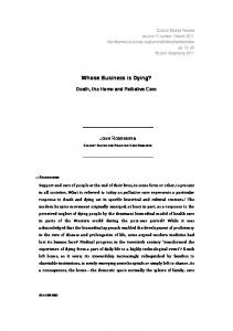
FIG. 1. Positions of single cysteine substitutions in HtrI. Cylinders represent a-helical transmembrane regions predicted by the PHDsec algorithm (50). N, N terminus of HtrI; C, C terminus.
FIG. 2. Phototaxis responses of wild type and single cysteinecontaining HtrI mutants. Two stimuli were delivered: an orange light step down stimulus, consisting of 4-s removal of 600 6 20-nm light (open bars), and a near-UV stimulus consisting of a 20-ms, 400-nm pulse under continuous orange light background illumination (filled bars). The phototaxis index is calculated in s21 as the integral of the swimming reversal frequency measured by motion analysis over the first 2 s after the stimulus minus the integral over 2 s starting from 6 s after the stimuli were initiated. Values plotted represent the mean of at least two independent measurements, and the error bar represents 1 S.D. shutter (Vincent Associates, Rochester, NY). Phototaxis stimuli were delivered through an epiiluminator from a Nikon 100-W Hg/Xe or from a 150-watt tungsten/halogen lamp. Oxidation Procedure and Western Blot Analysis—Membranes were isolated from sonicated stationary phase cells as described (24) and suspended in 4 M NaCl, 25 mM Tris-HCl, pH 6.8. Membrane samples in sonication buffer (4 M NaCl 25 mM Tris, pH 6.8); low salt membrane dilution buffer (250 mM KCl, 20 mM Tris-HCl, pH 8.0), which previously has been shown to maintain HtrI and SRI in a molecular complex as assessed by spectroscopic criteria (7); and all other solutions were allowed to equilibrate to reaction temperature (10 or 25 °C) prior to mixing. The oxidation reaction was initiated by the addition of 65 ml of membrane suspension at a protein concentration of 4 mg/ml to 10 ml of 300 mM 1,10-phenanthroline (in ethanol) and 10 ml of 150 mM CuSO4 (in H2O) or other concentrations noted in the Fig. 5 legend, diluted into 915 ml of low salt membrane dilution buffer. Oxidation of the membrane by Cu(II)-(1,10-phenanthroline)3 in the sonication buffer, which contains 4 M NaCl, is impractical due to the chelation of 1,10-phenanthroline by heavy metal ions present as impurities in NaCl. To quench the reaction, 20-ml aliquots were transferred to 60 ml of SDS sample buffer containing 10 mM N-ethylmaleimide and 5 mM EDTA and left on ice. Samples were heated at 65 °C for 5 min before loading on to a 7% SDS-PAGE gel for separation. Proteins were electrotransferred to polyvinylidene diflu-
oride membrane at 4 °C, 200 mA for 3 h. HtrI was detected using HtrI-specific multiclonal antibody, and immunoblots were developed using the ECL Western blotting kit. Linearity of the signals was tested by comparing bands of serially diluted samples. Reactions were performed in a removable sample chamber taken from an SLM Aminco DW-2000 spectrophotometer (SLM Instruments, Urbana, IL) connecting to a Haake K15 refrigerated water bath circulator (Haake Mess-Technik GmbH u. Co., Karlsruhe, Germany) for temperature control. A magnetic stirring bar was used throughout the experiment for efficient mixing of reactants. Illumination was from a 100-watt tungsten/halogen lamp focused on the sample after passing through a 5% CuSO4 H2O solution (path length 5 3 cm), two heatabsorbing filters, and a long pass orange light filter (Corion LG530, Corion Corp., Holliston, MA); The final light intensity was 1.5 3 106 ergszcm22zs21 at the position of the sample. The temperature was continuously monitored with a thermocouple probe inserted in the sample and controlled to within 60.25 °C. RESULTS
Phototaxis Responses of Cysteine-substituted HtrI Mutants— Single cysteine substitutions were introduced into cysteineless HtrI at three different positions. Ala-4 is near the N-terminal side of the predicted TM1, Asn-33 is at the periplasmic end of TM1, and Ile-64 is located in the cytoplasmic part following the predicted TM2 (Fig. 1). All three mutants, like wild type, mediated attractant responses to orange light and repellent responses to UV light (Fig. 2). However, HtrI cells carrying I64C exhibited smaller responses to both 600-nm step down and 400-nm step up stimuli. The reduced responses were not due to a reduction in the expression level of the mutant HtrI, since identical amounts of protein were obtained for all the HtrI mutants as assayed by immunoblotting (data not shown). Nor were the smaller responses due to reduction of SRI expression: laser flash photolysis of SRI in membranes isolated from these
Downloaded from http://www.jbc.org/ by guest on October 2, 2017
FIG. 3. Immunoblot analysis of membranes containing wild type (WT) and single cysteine-substituted HtrI mutants. A, oxidative cross-linking catalyzed by Cu(II)-(1,10-phenanthroline)3 was performed with membrane suspensions at room temperature for 2 h. Pho81Wr2 membrane that does not contain HtrI was used as a control. In the case of HtrI I64C, oxidized sample was reduced by the addition of 0.5 mM dithiothreitol and 10% b-mercaptoethanol to the SDS-polyacrylamide gel electrophoresis sample buffer. 2, no catalyst added; 1, catalyst added; R, reduced. B, time course of the oxidative cross-linking reaction of cysteine-substituted HtrI mutant membrane at 10 °C. Samples at time 0 were not treated with catalyst.
19724
HtrI Is a Dimer Sensitive to SRI Conformation
transformants revealed identical yields (65%) of the S373 photoproduct. These observations indicate that Ile-64 is in a sensitive position but not vital for signaling by the SRI-HtrI complex. Oxidative Cross-linking of Cysteine-substituted HtrI Mutants—To address the question of whether HtrI exists as a homodimer, like its eubacterial counterparts, intermolecular disulfide bond formation between pairs of homologous cysteine residues (i.e. from corresponding residues of the two monomers within the same dimer) was studied by oxidative cross-linking. Since there are no cysteines in wild-type HtrI, any dimer formed will be derived from the experimentally introduced substitutions. Samples were analyzed by nonreducing SDSpolyacrylamide gel electrophoresis and immunoblotted with anti-HtrI antibody. HtrI monomer, which has a molecular mass of 54 kDa, runs abnormally at a 97-kDa position, which has been attributed to its low pI of 3.9 (1). All three cysteinecontaining HtrI proteins exhibited dimer forms evident in the presence of bands at ;200 kDa before catalyst is added, while the wild-type HtrI migrated exclusively as a monomer (Fig. 3A). The extent of cross-linking without added catalyst was greater in the case of A4C and N33C than in that of I64C. When catalyst was added to the membrane suspensions and the reaction was incubated at room temperature for 2 h, however, only I64C was found to cross-link completely (Fig. 3A). Very little effect of the catalyst addition was observed for A4C and N33C. Cross-linking of I64C was completed within minutes at 10 °C (Fig. 3B). The complete cross-linking of I64C indicates there is an even number of HtrI monomers in the tight HtrISRI complex. To rule out the possibility that random collisions of free monomers are responsible for HtrI cross-linking, a plasmid that overexpresses the I64C-HtrI-SRI complex was constructed and transformed into Pho81Wr2. The cross-linking rate of the highly expressed I64C in this membrane was found to be the
FIG. 5. Extent of oxidation of HtrI I64C at different catalyst concentrations in the dark (filled squares) and in 600-nm light (open circles). The percentage of monomer remaining at 2 min after the initiation of the reaction, relative to the amount of monomer at time 0 as 100%, was plotted as a function of catalyst concentration. 1*, 6 min at 150 mM catalyst. Reactions were at 10 °C.
same as that of the I64C membrane in which HtrI was 4-fold less concentrated (data not shown). In the case of I64C, the cross-linked dimer can be reduced to monomer by adding 0.5 mM dithiothreitol and 10% b-mercaptoethanol to the SDS-polyacrylamide gel electrophoresis loading buffer (Fig. 3A), confirming the disulfide linkage. However, extended reduction (room temperature, 2 h) did not give complete dissociation to monomers. Incomplete reduction after oxidation and preexisting dimers have been observed in other transducers (25–27). Effects of SRI on the Cross-linking Pattern of HtrI—Previous studies have shown that HtrI interacts with SRI in its native membrane and changes SRI properties. This is based on the observations that removal of HtrI (6 – 8), some deletion constructs of HtrI (28), and mutations in HtrI (29) affect the kinetics of the SRI photocycle. However, SRI effects on the conformation of HtrI have not been reported. To test for a possible effect of SRI on the cross-linking rate of I64C, plasmids were constructed to contain either only I64C/htrI or the I64C/ htrI-sopI pair and transformed into strain Pho81Wr2. The kinetics of cross-linking in the isolated membranes was monitored at 10 °C. HtrI dimers were observed independent of the presence of SRI (Fig. 4A). However, complete cross-linking of the I64C monomer occurred only in the presence of SRI (Fig. 4A). When HtrI is expressed alone, the reaction is slow (Fig. 4A) and does not reach completion even at room temperature for 2 h (data not shown). Furthermore, in the absence of SRI, more HtrI preexists as dimers than when it is complexed with SRI (Fig. 4A). These data show that the cross-linking reaction at position 64 is sensitive to the presence of SRI. Effects of Light on the Cross-linking Pattern of HtrI—Orange light converts SRI (lmax 5 587 nm) into its attractant signaling conformation, which is believed to alter the conformation of HtrI by protein-protein interaction (2). We tested whether the cross-linking behavior of I64C is sensitive to the putative HtrI
Downloaded from http://www.jbc.org/ by guest on October 2, 2017
FIG. 4. Oxidative cross-linking of HtrI I64C in the presence of SRI (A, 1SRI; B, a and b), absence of SRI (A, 2SRI; B, c and d), and in the dark (A; B, b and c) and 600-nm light (B, a and d). The amount of monomer at time 0 was taken as 100%. Reactions were done at 10 °C. Each point represents the mean 6 S.D. of at least three parallel measurements.
HtrI Is a Dimer Sensitive to SRI Conformation
19725
conformational change induced by SRI photoexcitation. Orange light moderately accelerated the reaction rate (Fig. 4B, a and b), indicating that I64C is in a sensitive position. Light accelerated the reaction rate only in the presence of SRI (Fig. 4B, c and d) and was evident at various catalyst concentrations (Fig. 5). Cysteine Cross-linking as a Probe for the Conformational Changes Introduced by SRI Mutations—SRI mediates attractant phototaxis to orange light stimulation. Previously, it was found that certain substitutions at either of two positions in SRI, Asp-201 and His-166, result in inverted (i.e. repellent) responses to orange light (24, 30). Three SRI mutants that mediate inverted responses to orange light (D201N, H166A, and H166Y) and one that eliminates phototactic responses to orange light (H166R) were expressed together with HtrI I64C in Pho81Wr2. In order to test for correlation between the phototaxis signaling and the cross-linking behavior, both the motion analysis and cross-linking reaction were carried out at 25 °C. Wild type, H166R, H166A, and H166Y SRI expressed with HtrI I64C mediate similar phototaxis responses as when they are expressed together with wild type HtrI. However, D201N, which has an inverted response phenotype when expressed with wild type HtrI (24), had a normal response in the presence of the I64C mutation (Fig. 6A). Evidently, I64C is an extragenic suppressor of D201N, which is an additional indication that position 64 is a sensitive position in HtrI. The cross-linking rates of I64C in HtrI complexed to the various SRI mutants were compared. To obtain a measurable rate at 25 °C, the catalyst was diluted to 1⁄50 of that used at 10 °C. Under these conditions, HtrI I64C with wild type SRI maintained the same dark/light relationship in terms of crosslinking rate, i.e. light moderately accelerated the reaction. Two of the mutants, SRI D201N-HtrI I64C and SRI H166R-HtrI I64C, exhibited cross-linking rates comparable with that of the wild type in the dark (Fig. 6B), and light accelerated the crosslinking rate of SRI D201N-HtrI I64C (Fig. 6B), which mediated essentially wild type phototaxis responses (Fig. 6A), but not of SRI H166R-HtrI I64C (Fig. 6B), which did not mediate phototaxis responses to orange light (Fig. 6A). For the two inverted
signaling mutants, SRI H166A-HtrI I64C and SRI H166Y-HtrI I64C, greatly accelerated reaction rates were observed in the dark (complete oxidation by 20 s). To slow down the reaction to allow comparison of the rates in the dark and light, catalyst was further diluted. Orange light was found to retard the cross-linking reaction of I64C (Fig. 6B). Light retarded the rate in the inverted mutant membranes also at 10 °C (data not shown). DISCUSSION
HtrI, an Archaeal Transducer, Like Its Eubacterial Counterparts, Exists as a Dimer—The dimeric nature of Tar was first suggested from site-directed cysteine cross-linking experiments (9) and was confirmed by observation of a dimeric ligand binding domain in Tar crystals (31). The data presented here show that HtrI is also oligomeric and are consistent with it being a dimer. All three introduced cysteines cross-link HtrI into dimer forms that are resistant to SDS in nonreducing electrophoresis. In the case of I64C, complete oxidation within 20 s in some conditions was observed. The unusual behavior of A4C and N33C, i.e. more preexisting cross-linked dimer and slow reaction rate, resembles that of R4C in the Tar homodimer (32). The high reactivity observed for I64C may reflect a flexible structure in the cytoplasmic region following TM2 or proximity of the two cysteine residues in the dimer. An Extended Coiled Coil Dimerization Domain in HtrI Is Identified by Sequence Analysis—In addition to the efficient cross-linking of I64C, protein sequence analysis supports dimerization of the cytoplasmic regions adjacent to TM2 in HtrI. A region of 71 residues (Fig. 7A, from residue 90 to 160) corresponding to a6 in Tar is predicted to assume a coiled coil structure by two prediction algorithms COILS (33) and PAIRECOIL (34). From position 96 to 154, using a window of 21, the probability is assessed as .99% by both algorithms, whereas the generally accepted coiled coil methylation regions in Tar, K1, and R1 (33) are assessed at about 70 and 80%, respectively, by these programs. Two-stranded a-helical coiled coil is found as a dimerization domain in a diverse group of proteins (35) and is defined as two a-helices that are wound into a superhelix
Downloaded from http://www.jbc.org/ by guest on October 2, 2017
FIG. 6. A, phototaxis behavior of cells carrying HtrI I64C expressed under different SRI mutant backgrounds at 25 °C. Two stimuli were delivered: a 600-nm step down stimulus (open bar) that is the same as in Fig. 2 and a step up stimulus consisting of a 4-s pulse of 600 6 20-nm light (hatched bar). SRI H166A and SRI H166Y exhibit inverted (i.e. repellent) orange light responses. The dashed and dotted line shows the level of detectable responses above the measurement noise. B, in vitro cross-linking of membranes containing HtrI I64C in complex with different SRI mutants at 25 °C. The fraction of monomer remaining was plotted against time. The reactions were done in complete darkness (filled circles) or with orange light illumination (open circles). The amount of monomer at time 0 was taken as 100%, and each tick indicates 20%. Catalyst concentration for wild type (WT), H166R, and D201N was 3 mM, and that for H166A and H166Y was 0.03 mM.
19726
HtrI Is a Dimer Sensitive to SRI Conformation
Downloaded from http://www.jbc.org/ by guest on October 2, 2017
FIG. 7. A, sequence alignment of HtrI and eight other methyl-accepting transducers. The predicted secondary structure is represented by cylinders (a-helical), lines (loop), or an open arrow (b-sheet). Predicted transmembrane segments and conserved loop regions are boxed; positions a and d in the predicted coiled coil structure (see “Discussion”) are shaded. The arrow from residue 147 on HtrI shows the shortest region sufficient for SRI interaction according to a deletion study (28), and HtrI/HtrII chimera analysis indicates that only the first 60 residues of HtrI are required (see “Discussion”). HtrI_H and HtrII_H, HtrI and HtrII from H. salinarum; HtrII_N and HtrII_V, HtrII from N. pharaonis and H. vallismortis, respectively; Htc_H, Htd_H, and Htf_H, putative MCPs from H. salinarum; Tar_E and Tar_S, Tar from E. coli and Salmonella typhimurium, respectively. B, helix wheel representation of the predicted coiled coil region in HtrI. Positions in the heptad repeat are represented by circles with hydrophobic a and d positions shaded. The sequence at each position reads from the center to the outside. Orientation of the helices is as viewed from the periplasmic space. Clustered alanines and charged residues in possible interacting faces of the helices (designated by open double-headed arrows) are in a larger font and boxed.
through the “knobs-into-holes” packing of amino acid side chains (36). A heptad repeat denoted as abcdefg is characteristic, in which hydrophobic residues are found mainly at a and d positions, while the other positions are more hydrophilic.
Frequently, opposite charged residues are found at the e and g positions, which have been suggested to stabilize the coiled coil structure (37). The coiled coil motif is more evident in a helix wheel model representation of the 71 residues in HtrI (Fig. 7B);
HtrI Is a Dimer Sensitive to SRI Conformation
2
X.-N. Zhang, J. Zhu, and J. L. Spudich, manuscript in preparation.
energetically unfavorable. Rotation of subunits in the plane of the lipid bilayer (45), which would result in slight winding or unwinding (46) of the a6 coiled coil, is an interesting possibility. The putative coiled coil of HtrI (Fig. 7B), unlike the GCN4 leucine zipper in which the hydrophobic core is exclusively formed by leucines (47), contains a large number of small chain residues. In particular, the a and d positions between residue 123 and residue 148 are exclusively occupied by alanine residues, which have been shown to increase the flexibility of coiled coils (48). A similar feature has been noted in CM-tropomyosin and has been suggested to facilitate transmission of a conformational change along its long axis (49) as might occur also in the HtrI dimer. The a6 coiled coil is bounded by short flexible regions, which are present in all transducer sequences so far examined (this study and Ref. 3). This feature might allow for the divergence (viewed from the cytoplasm) of the two TM2 helices in the membrane and of the methylation helices at the distal end of the coiled coil. Acknowledgments—We thank Elena Spudich and Bastianella Perazzona for help with immunoblot analysis and Kwang-Hwan Jung for stimulating discussions about the inverted signaling mutants. REFERENCES 1. Yao, V. J., and Spudich, J. L. (1992) Proc. Natl. Acad. Sci. U. S. A. 89, 11915–11919 2. Hoff, W. D., Jung, K. H., and Spudich, J. L. (1997) Annu. Rev. Biophys. Biomol. Struct. 26, 223–258 3. Le Moual, H., and Koshland, D. E., Jr. (1996) J. Mol. Biol. 261, 568 –585 4. Stock, J. B., and Surette, M. (1996) in Escherichia coli and Salmonella typhimurium: Cellular and Molecular Biology (Neidhardt, F., ed) 2nd Ed., pp. 123–145, American Society for Microbiology Press, Washington, D. C. 5. Falke, J. J., Bass, R. B., Butler, S. L., Chervitz, S. A., and Danielson, M. A. (1997) Annu. Rev. Cell Dev. Biol. 13, 457–512 6. Spudich, E. N., and Spudich, J. L. (1993) J. Biol. Chem. 268, 16095–16097 7. Olson, K. D., and Spudich, J. L. (1993) Biophys. J. 65, 2578 –25858 8. Krah, M., Marwan, W., Vermeglio, A., and Oesterhelt, D. (1994) EMBO J. 13, 2150 –2155 9. Milligan, D. L., and Koshland, D. E., Jr. (1988) J. Biol. Chem. 263, 6268 – 6275 10. Pakula, A. A., and Simon, M. I. (1992) Proc. Natl. Acad. Sci. U. S. A. 89, 4144 – 4148 11. Chervitz, S. A., and Falke, J. J. (1996) Proc. Natl. Acad. Sci. U. S. A. 93, 2545–2550 12. Hughson, A. G., and Hazelbauer, G. L. (1996) Proc. Natl. Acad. Sci. U. S. A. 93, 11546 –11551 13. Milligan, D. L., and Koshland, D. E., Jr. (1991) Science 254, 1651–1654 14. Gardina, P. J., and Manson, M. D. (1996) Science 274, 425– 426 15. Tatsuno, I., Homma, M., Oosawa, K., and Kawagishi, I. (1996) Science 274, 423– 425 16. Chervitz, S. A., Lin, C. M., and Falke, J. J. (1995) Biochemistry 34, 9722–9733 17. Lee, G. F., Lebert, M. R., Lilly, A. A., and Hazelbauer, G. L. (1995) Proc. Natl. Acad. Sci. U. S. A. 92, 3391–3395 18. Cochran, A. G., and Kim, P. S. (1996) Science 271, 1113–1116 19. Cline, S. W., and Doolittle, W. F. (1987) J. Bacteriol. 169, 1341–1344 20. Chen, B., and Przybyla, A. E. (1994) BioTechniques 17, 657– 659 21. Krebs, M. P., Spudich, E. N., Khorana, H. G., and Spudich, J. L. (1993) Proc. Natl. Acad. Sci. U. S. A. 90, 3486 –3490 22. Sarkar, G., Kapelner, S., and Sommer, S. S. (1990) Nucleic Acids Res. 18, 7465 23. Boom, R., Sol, C. J. A., Salimans, M. M. M., Jansen, C. L., Wertheim-van Dillen, P. M. E., and van der Noordaa, J. (1990) J. Clin. Microbiol. 28, 495–503 24. Olson, K. D., Zhang, X. N., and Spudich, J. L. (1995) Proc. Natl. Acad. Sci. U. S. A. 92, 3185–3189 25. Lynch, B. A., and Koshland, D. E., Jr. (1991) Proc. Natl. Acad. Sci. U. S. A. 88, 10402–10406 26. Lee, G. F., Burrows, G. G., Lebert, M. R., Dutton, D. P., and Hazelbauer, G. L. (1994) J. Biol. Chem. 269, 29920 –29927 27. Chervitz, S. A., and Falke, J. J. (1995) J. Biol. Chem. 270, 24043–24053 28. Perazzona, B., Spudich, E. N., and Spudich, J. L. (1996) J. Bacteriol. 178, 6475– 6478 29. Jung, K. H., and Spudich, J. L. (1996) Proc. Natl. Acad. Sci. U. S. A. 93, 6557– 6561 30. Zhang, X. N., and Spudich, J. L. (1997) Biophys. J. 73, 1516 –1523 31. Yeh, J. I., Biemann, H. P., Prive, G. G., Pandit, J., Koshland, D. E., Jr., and Kim, S. H. (1996) J. Mol. Biol. 262, 186 –201 32. Stoddard, B. L., Bui, J., and Koshland, D. E., Jr. (1992) Biochemistry 31, 11978 –11983 33. Lupas, A., Van Dyke, M., and Stock, J. (1991) Science 252, 1162–1164 34. Berger, B., Wilson, D. B., Wolf, E., Tonchev, T., Milla, M., and Kim, P. S. (1995) Proc. Natl. Acad. Sci. U. S. A. 92, 8259 – 8263 35. Hodges, R. S. (1996) Biochem. Cell Biol. 74, 133–154 36. Lupas, A. (1996) Trends Biochem. Sci. 21, 375–382 37. McLachlan, A. D., and Stewart, M. (1975) J. Mol. Biol. 98, 293–304 38. Zhang, W., Brooun, A., Mueller, M. M., and Alam, M. (1996) Proc. Natl. Acad.
Downloaded from http://www.jbc.org/ by guest on October 2, 2017
a and d positions define hydrophobic faces, position e is highly positively charged, position g is highly negatively charged, and other positions are hydrophilic. The corresponding region in HtrIIs from H. salinarum (38), Natronobacterium pharaonis (39), and Haloarcula vallismortis (39) and three other unclassified MCPs from H. salinarum (40) are also predicted to contain coiled coil a6 region with varied lengths. Although coiled coil structure is not predicted for the corresponding region of Tar by these two algorithms (probability , 2%), the presence of a shortened version of this coiled coil region seems likely when Tar.E and Tar.S are aligned with the seven halobacterial transducers (Fig. 7A). Indirect experimental evidence supporting the existence and the importance of such a coiled coil region in both Tar and HtrI is substantial. 1) A recent disulfide cross-linking study of this region in Tar (residue 259 –290) indicates an a-helical conformation, a close juxtaposition, and a parallel alignment at the interface between two subunits within a dimer, consistent with it being a coiled coil (41). 2) Both biochemical and genetic studies of hybrids consisting of a full-length and truncated Tar proteins demonstrated that the a6 region 248 –259 in Tar is required for efficient signaling, suggesting its involvement in subunit interaction (13–15). 3) When a leucine zipper, an extensively studied coiled coil structure, was fused to the cytoplasmic portion (257– 553) of Tar, CheA activation was observed (18, 42). 4) A deletion study with HtrI showed that the N-terminal 147-residue fragment of HtrI interacts with SRI, whereas the N-terminal 97-residue fragment that is lacking most of the predicted coiled coil does not (28). Dimerization of HtrI through this region may be important for its stability. Consistent with this a chimeric transducer containing the cytoplasmic portion of HtrI and the N-terminal 60 residues of HtrI is produced in amounts comparable with wild type and fully interacts with SRI.2 The 71-residue region in HtrI is unusually long for a coiled coil domain, most known coiled coils spanning ;40 amino acids (36). Possibly, the high salt concentrations found in H. salinarum, N. pharaonis, and H. vallismortis cells may effectively shield electrostatic interactions between side chains, and hence a longer coiled coil region and a more stringent hydrophobic core are needed for stabilizing the dimer. Implications for the Signaling Mechanism—Light accelerates the oxidation rate of I64C, suggesting that the reaction rate can be used as a probe for the conformation of HtrI in the light-activated SRI-HtrI complex. The effect of light on the reaction rate in several mutants supports this suggestion. Light has little or no effect on the rate in membranes from the double mutant SRI H166R-HtrI I64C, which does not show phototaxis responses. The mutants SRI H166A-HtrI I64C and SRI H166Y-HtrI I64C exhibit inverted (repellent) behavioral responses to normally attractant orange light, which has been explained in terms of an inverted conformational change in the SRI-HtrI complex (43). Consistent with this interpretation, the oxidation rates observed here are inverted; the rate is much higher than that of wild type in the dark, and the rate is decreased by light in the inverted mutant membranes. The oxidation rate of I64C therefore correlates closely with the conformational state of the complex deduced from behavioral measurements. The strong prediction of a6 as a long coiled coil structure has implications for the transmission of the signal from the membrane to the His-kinase binding domain, a current important question also for eubacterial MCPs (44). Models involving significant sliding of one HtrI monomer with respect to the other (e.g. piston-like, scissors-like, or see-saw movements) will be
19727
19728
HtrI Is a Dimer Sensitive to SRI Conformation
Sci. U. S. A. 93, 8230 – 8235 39. Seidel, R., Scharf, B., Gautel, M., Kleine, K., Oesterhelt, D., and Engelhard, M. (1995) Proc. Natl. Acad. Sci. U. S. A. 92, 3036 –3040 40. Zhang, W., Brooun, A., McCandless, J., Banda, P., and Alam, M. (1996) Proc. Natl. Acad. Sci. U. S. A. 93, 4649 – 4654 41. Chen, X., and Koshland, D. E., Jr. (1997) Biochemistry 36, 11858 –11864 42. Surette, M. G., and Stock, J. B. (1996) J. Biol. Chem. 271, 17966 –17973 43. Jung, K.-H., and Spudich, J. (1998) J. Bacteriol. 180, 2033–2042 44. Stoddard, B. L., Biemann, H. P., and Koshland, D. E., Jr. (1992) Cold Spring Harbor Symp. Quant. Biol. 57, 1–15
45. Maruyama, I. N., Mikawa, Y. G., and Maruyama, H. I. (1995) J. Mol. Biol. 253, 530 –546 46. Kim, S. H., Prive, G. G., Yeh, J., Scott, W. G., and Milburn, M. V. (1992) Cold Spring Harbor Symp. Quant. Biol. 57, 17–24 47. O’Shea, E. K., Klemm, J. D., Kim, P. S., and Alber, T. (1991) Science 254, 539 –544 48. Hodges, R. S., Saund, A. K., Chong, P. C., St.-Pierre, S. A., and Reid, R. E. (1981) J. Biol. Chem. 256, 1214 –1224 49. Talbot, J. A., and Hodges, R. S. (1982) Acc. Chem. Res. 15, 224 –230 50. Rost, B., and Sander, C. (1993) J. Mol. Biol. 232, 584 –599
Downloaded from http://www.jbc.org/ by guest on October 2, 2017
HtrI Is a Dimer Whose Interface Is Sensitive to Receptor Photoactivation and His-166 Replacements in Sensory Rhodopsin I Xue-Nong Zhang and John L. Spudich J. Biol. Chem. 1998, 273:19722-19728. doi: 10.1074/jbc.273.31.19722
Access the most updated version of this article at http://www.jbc.org/content/273/31/19722 Alerts: • When this article is cited • When a correction for this article is posted Click here to choose from all of JBC's e-mail alerts Downloaded from http://www.jbc.org/ by guest on October 2, 2017
This article cites 49 references, 31 of which can be accessed free at http://www.jbc.org/content/273/31/19722.full.html#ref-list-1