INFECTION AND IMMUNITY, Nov. 2007, p. 5425–5433 0019-9567/07/$08.00⫹0 doi:10.1128/IAI.00261-07 Copyright © 2007, American Society for Microbiology. All Rights Reserved.
Vol. 75, No. 11
Human Monoclonal Antibodies against Anthrax Lethal Factor and Protective Antigen Act Independently To Protect against Bacillus anthracis Infection and Enhance Endogenous Immunity to Anthrax䌤 Mark T. Albrecht,1* Han Li,2 E. Diane Williamson,3 Chris S. LeButt,3 Helen C. Flick-Smith,3 Conrad P. Quinn,2 Hans Westra,4 Darrell Galloway,5 Alfred Mateczun,1 Stanley Goldman,1 Herman Groen,4,6 and Les W. J. Baillie7 Biological Defense Research Directorate, Naval Medical Research Center, Silver Spring, Maryland 20910-75001; MPIR Laboratory, Meningitis & Vaccine Preventable Diseases Branch, Division of Bacterial Diseases, National Center for Immunization and Respiratory Diseases, Centers for Disease Control & Prevention, Atlanta, Georgia2; Defense Science Technology Laboratories, Porton Down, Salisbury, Wiltshire SP4 0JQ, United Kingdom3; IQ Corporation, Rozenburglaan 13a, 9727 DL Groningen, The Netherlands4; The Ohio State University, Department of Microbiology, Columbus, Ohio 432105; University of Groningen, University Medical Center, Department of Pathology and Laboratory Medicine, Medical Biology Section, Laboratory for Tumor Immunology, Groningen, The Netherlands6; and Cardiff University, Welsh School of Pharmacy, Cardiff CF10 3NB, Wales, United Kingdom7 Received 16 February 2007/Returned for modification 6 April 2007/Accepted 7 June 2007
The unpredictable nature of bioterrorism and the absence of real-time detection systems have highlighted the need for an efficient postexposure therapy for Bacillus anthracis infection. One approach is passive immunization through the administration of antibodies that mitigate the biological action of anthrax toxin. We isolated and characterized two protective fully human monoclonal antibodies with specificity for protective antigen (PA) and lethal factor (LF). These antibodies, designated IQNPA (anti-PA) and IQNLF (anti-LF), were developed as hybridomas from individuals immunized with licensed anthrax vaccine. The effective concentration of IQNPA that neutralized 50% of the toxin in anthrax toxin neutralization assays was 0.3 nM, while 0.1 nM IQNLF neutralized the same amount of toxin. When combined, the antibodies had additive neutralization efficacy. IQNPA binds to domain IV of PA containing the host cell receptor binding site, while IQNLF recognizes domain I containing the PA binding region in LF. A single 180-g dose of either antibody given to A/J mice 2.5 h before challenge conferred 100% protection against a lethal intraperitoneal spore challenge with 24 50% lethal doses [LD50s] of B. anthracis Sterne and against rechallenge on day 20 with a more aggressive challenge dose of 41 LD50s. Mice treated with either antibody and infected with B. anthracis Sterne developed detectable murine anti-PA and anti-LF immunoglobulin G antibody responses by day 17 that were dependent on which antibody the mice had received. Based on these results, IQNPA and IQNLF act independently during prophylactic anthrax treatment and do not interfere with the establishment of endogenous immunity.
Bacillus anthracis, the causative agent of anthrax, is a sporeforming, gram-positive bacterium. Following inhalation, spores are phagocytosed by alveolar macrophages and transported through the lymphatic channels to hilar and tracheobronchial lymph nodes, where the spores germinate, leading to the multiplication and systemic circulation of vegetative bacilli (13, 43). Critical to the virulence of B. anthracis is the secretion of a tripartite exotoxin consisting of two enzymatically active subunits: lethal factor (LF) and edema factor (EF). These proteins bind to protective antigen (PA), the cell-binding component, to form lethal toxin (LeTx) and edema toxin, respectively (53). The biological activities of LeTx and edema toxin are analogous to those of other A-B toxin systems (14). PA initially
binds to a cell surface receptor, including human capillary morphogenesis protein 2 and tumor endothelial marker 8 (4, 45), and undergoes furin-like mediated cleavage of the Nterminal domain. This event yields an amino-terminal 20-kDa fragment and a carboxyl-terminal 63-kDa activated PA63 protein with exposed LF/EF binding domains. The PA63 conformer assembles to form a ring-shaped heptamer with the capacity to bind up to three copies of LF or EF (22, 31, 32). At this point the toxin complex is endocytosed. Subsequent acidification of the endosome causes the PA63 heptamer to insert into the membrane, forming a transmembrane channel that traffics LF and EF to the cytosol (29). LF endopeptidase activity with the MEK family of signal transduction proteins down-regulates both the innate and acquired immune responses by inhibiting cytokine responses, dendritic cell responses, and B- and T-cell immunity (1, 30). EF, an adenylate cyclase, incapacitates phagocytes and cytokine pathways through cyclic AMP induction and up-regulates the PA63 receptor on target cells (17, 36). Given the central role of the
* Corresponding author. Mailing address: Biological Defense Research Directorate, Naval Medical Research Center, 12300 Washington Ave., Silver Spring, MD 20910-7500. Phone: (301) 231-6716. Fax: (301) 231-6799. E-mail:
[email protected]. 䌤 Published ahead of print on 23 July 2007. 5425
5426
ALBRECHT ET AL.
toxins in anthrax pathology, the ability to neutralize their effects is of value at all stages of infection. The credentials of B. anthracis as an aerosolized bioterror agent were confirmed by the 2001 postal attacks in the United States, which resulted in five deaths (20). These events underscored the need for postexposure medical countermeasures that are effective, particularly during middle to advanced stages of infection, when bacteremia and toxemia ensue. Animal studies have previously suggested that early treatment of anthrax is essential since the disease reaches a point when antibiotics are no longer effective due to the accumulation of a lethal level of toxin (48, 49, 56). In order to counteract the limitations of antibiotics, several groups have been pursuing various therapeutic strategies that evoke rapid protection against anthrax by targeting PA, LF, or capsular antigen (7, 18, 21, 23, 28, 33, 35, 37, 46, 59, 63). The most promising approach has been administration of antitoxin antibodies to generate a state of immediate passive immunity. This therapy involves the transfer of serum from an immunized donor or monoclonal antibodies (MAbs) to an exposed or at risk recipient. The efficacy of this treatment has been demonstrated in an anthrax guinea pig challenge model using polyclonal anti-PA serum from immunized guinea pigs (26). A murine MAb specific for LF has also exhibited protective efficacy during experimental LeTx challenge of athymic nude mice (63). One of the major concerns with this therapeutic agent is the immunogenicity of the antibody as a foreign protein. This concern has been partially circumvented by the generation of an affinity-enhanced, humanized, anti-PA MAb that was developed from a murine immunoglobulin G (IgG) (33). More recently, human peripheral blood lymphocytes from immunized humans have been used in hybridomas as progenitors of prophylactic anti-PA antibodies (44, 58). The use of human IgG eliminates the risk of adverse reactions associated with nonhuman serum and antibodies while the immune system recall response is utilized to produce highaffinity toxin-neutralizing antibodies. Here we report the isolation and characterization of two protective PA- and LF-specific MAbs obtained from human donors. This is the first reported example of a protective human-derived LF MAb. Each antibody was tested for its individual capacity to neutralize LeTx activity in vitro. The antibodies were also assayed together in various combinations to assess the additive neutralizing potential. The efficacy of the anti-PA and anti-LF MAbs as a prechallenge treatment for mice infected intraperitoneally (i.p.) with a lethal dose of B. anthracis Sterne spores was evaluated. We also examined the ability of the protected mice to survive a second lethal challenge 20 days after the initial infection. Endogenous murine anti-PA and anti-LF IgG responses were measured, and their protective efficacy was determined. MATERIALS AND METHODS Ethics. Human blood samples were obtained with appropriate ethical permission (IQ Therapeutics BV, The Netherlands). The experiments reported here were conducted in compliance with the Animal Welfare Act and in accordance with the principles set forth in the Guide for the Care and Use of Laboratory Animals (19). Development and identification of the anti-PA and anti-LF hybridomas. Blood samples were drawn from healthy individuals immunized with the United Kingdom-licensed anthrax vaccine following annual booster immunizations. Samples
INFECT. IMMUN. demonstrating anthrax LeTx-neutralizing activity in cytotoxicity assays (15, 40) were selected for hybridoma development using a polyethylene glycol-based variant of the hybridoma electrofusion technology described by H. Groen and H. H. Westra (U.S. patent applications USSN 60/710,626 and USSN 11/072,102). Hybridoma fusions were screened for expression of anti-PA- and anti-LF-specific antibodies by enzyme-linked immunosorbent assays (ELISAs) (2). Hybridoma clones producing anti-PA and anti-LF MAb IgG were expanded and stabilized, and the antibodies were evaluated for anthrax LeTx neutralization (18). Candidate anti-PA and anti-LF antibodies were isotyped using a human Ig subclass ELISA kit (Invitrogen, Carlsbad, CA). Anthrax LeTx neutralization activity assay. The capacity of the candidate anti-PA and anti-LF MAbs to neutralize LeTx was measured by the toxin neutralization activity (TNA) assay, which has been described in detail elsewhere (41, 54). Prior to testing, recombinant PA (rPA) and recombinant LF (rLF) were titrated for toxin potency with J774A.1 cells (TIB-67; American Type Culture Collection, Manassas, VA). The protein concentrations of rPA and rLF that resulted in more than 95% cell lysis at a fixed cell density of 3 ⫻ 104 cells/well in this assay were 50 and 24 ng/ml, respectively. Antibodies were assayed individually and in combination at various ratios to determine their individual and combined neutralization capacities. Test antibodies for individual analysis were prepared in a separate 96-well microtiter plate as twofold dilutions in triplicate. Combination assays were conducted in triplicate with two sets of MAbs. The first set of samples was prepared with a fixed concentration of anti-PA (30.6 ng/ml; 144 pM) mixed with anti-LF at concentrations of 9, 18, and 36 ng/ml. The second set of samples was prepared with a fixed concentration of anti-LF (9 ng/ml; 60 pM) mixed with anti-PA at concentrations of 21.6, 43.3, and 86.3 ng/ml. Assay endpoints were calculated using SAS statistical analysis software, version 9.0 (SAS Institute Inc., Cary, NC), running an endpoint calculation algorithm developed at the CDC (54). The primary endpoint calculated from the four-parameter logistic log fit curve is the 50% effective antibody dilution (ED50). This value is the reciprocal of the antibody dilution corresponding to the inflection point (“c” parameter) of the four-parameter logistic log fit of the serum neutralization curve (antibody dilution factor versus optical density). The final endpoint value used in the present study is the effective molar concentration required for 50% toxin neutralization (EC50), which was calculated from the ED50 using a nominal molecular mass of 150 kDa for human IgG. Domain mapping of PA- and LF-specific MAbs. Two different approaches were utilized to map the specific antigen domains recognized by the candidate anti-PA and anti-LF antibodies. The target domain recognized by the anti-PA MAb was identified using glutathione S-transferase (GST) fusions of the four PA domains in modified ELISAs. The development of these fusions has been described previously (10). Equimolar concentrations (6.02 ⫻ 10⫺11 mol/ml, equivalent to 5 g/ml of rPA) of the GST fusions and rPA were used in ELISAs, where the assay cutoff was defined as the dilution resulting in an optical density at 414 nm that was 0.1 U above the nonspecific background (61). The candidate anti-PA antibody was assayed in duplicate using a starting dilution of 1:100. The results of this assay were confirmed in native and denaturing Western blots (55) with rPA and PA domain IV resolved on a 4 to 20% Tris-HCl Ready Gel (Bio-Rad, Hercules, CA). Domain mapping of the LF-specific MAb was accomplished with rLF domains purified by cobalt affinity chromatograph. LF domains I (amino acids 1 to 262), II (amino acids 263 to 552), III (amino acids 303 to 385), IV (amino acids 552 to 776), and II to IV (amino acids 263 to 776) (6) were PCR amplified and cloned downstream of and in frame with the T5 promoter and seven-His tag sequence of the Escherichia coli expression vector pQE30 (QIAGEN, Valencia, CA). Constructs were confirmed via dideoxy sequencing. Domains were expressed from either E. coli XL-1Blue or E. coli M15. Host strains were grown in LB medium (BD-Difco, Franklin Lakes, NJ) with the appropriate antibiotics to an optical density at 600 nm of 0.55 to 0.65 and induced with 1 mM isopropyl--Dthiogalactoside (IPTG) (Sigma Fine Chemicals, St. Louis, MO) for 4 to 24 h at 25 to 37°C. Cells were pelleted, washed, and lysed with a French press at 16,000 lb/in2. The resulting lysates were cleared by centrifugation at 45,000 ⫻ g for 15 min. A cleared lysate was batch bound to TALON resin (Clontech, Mountain View, CA) and then washed with 10 column volumes of 300 mM NaCl, 50 mM Na2HPO4, 20 mM imidazole (pH 7.0). Proteins were eluted in 5 column volumes of 300 mM NaCl, 50 mM Na2HPO4, 150 mM imidazole (pH 7.0). Fractions containing a protein of the correct size (determined by sodium dodecyl sulfate [SDS]-polyacrylamide gel electrophoresis with Coomassie blue staining) were pooled and dialyzed into 10 mM HEPES, 50 mM NaCl (pH 7.5). Recognition of the target domain by the candidate anti-LF MAb was determined by native and denaturing Western blotting (55) using rLF and the domains resolved using 50 ng/lane on a 4 to 20% Tris-HCl Criterion precast gel (Bio-Rad). The proteins were also resolved on a 4 to 12% bis-Tris Criterion XT precast gel (Bio-Rad) and
VOL. 75, 2007
PASSIVE PROTECTION ENHANCES ENDOGENOUS IMMUNITY
5427
FIG. 1. Toxin neutralization by IQNPA and IQNLF. The abilities of both antibodies to neutralize a given amount of toxin were evaluated using the TNA assay (‚ and 䡺). In order to determine if combining IQNPA and IQNLF had a positive or negative effect on neutralization, the two MAbs were mixed in various ratios prior to use in the assay (Œ and 䡵). The results are expressed as the effective concentration of antibody that protected 50% of the cells in the assay (EC50).
silver stained using an Invitrogen SilverQuest kit according to the manufacturer’s instructions. Mouse passive protection studies. B. anthracis Sterne (34F2) spores were prepared in sporulation broth (containing [per liter] 23 g of nutrient broth, 0.025 g of MnSO4 䡠 H2O, and 0.25 g of KH2PO4). After a 72-h incubation at 37°C, spores were harvested by centrifugation, washed with phosphate-buffered saline (PBS), and enumerated by viable plate counting. Immediately prior to challenge the spores were diluted to obtain the appropriate concentration in endotoxinfree PBS and administered to the mice by i.p. injection. The 50% lethal dose (LD50) of this strain in A/J mice is ⬃2 ⫻ 104 spores (Helen Flick-Smith, unpublished results). The A/J mouse model was selected for this study because it is an established model for i.p. challenge (3, 39) and A/J mice are sensitive to the B. anthracis strain used in this investigation (39, 60). Groups of 10 6- to 8-week-old female A/J mice (The Jackson Laboratory, Bar Harbor, ME) were inoculated i.p. with 180 g of either the anti-PA or anti-LF antibody in 100 l of sterile endotoxin-free PBS (Invitrogen-Gibco, Carlsbad, CA). After 2.5 h the mice were challenged i.p. with an average of 4.8 ⫻ 105 CFU per mouse (approximately 24 LD50s). Surviving mice were rechallenged i.p. 20 days later with the same stock of spores at a level of 8.3 ⫻ 105 CFU per mouse (approximately 41 LD50s). The infection dose was increased in this challenge to determine if immunity to the anthrax toxins could develop in the presence of the toxin-specific antibodies. Both challenges included a control group of five mice that were inoculated i.p. with 100 l of sterile endotoxin-free PBS prior to challenge. Following challenge, mice were monitored four times daily for evidence of morbidity or mortality during the first week and twice daily thereafter. Blood samples were collected from healthy mice via tail bleeding using capillary microvette tubes (Sarstedt, Germany) on days ⫺4 (pretreatment and prechallenge), 10, 17, 27 (after the second challenge), and 34 (terminal bleed). Symptomatic mice were euthanized with CO2 prior to blood and tissue collection. Hearts were collected postmortem from all mice, swabbed onto 5% (vol/vol) sheep blood agar, and incubated at 37°C for 24 to 72 h. Analysis of endogenous mouse IgG responses. Murine anti-PA, anti-LF, and anti-human MAb IgG concentrations were quantified by ELISAs. Briefly, Immulon-IV HBX microtiter plates (Thermo Labsystems, Franklin, MA) were coated overnight at 4°C with rPA, rLF, or the anti-PA and anti-LF human MAbs at a concentration of 1 g/ml in 0.2 M carbonate buffer (pH 7.4). Each plate included a standard curve comprising doubling dilutions of purified mouse anti-PA IgG (provided by S. Park, BioPort Corp., Lansing, MI) or nonspecific human IgG (Sigma, St. Louis, MO) at concentrations ranging from 1,000 to 1.95 ng/ml. Plates were blocked with 5% (wt/vol) nonfat milk in PBS plus 0.1% (vol/vol) Tween 20 (pH 7.4) (PBST) for 1 h at 37°C and washed three times with PBST. Serum samples were serially diluted twofold in PBST and captured on the
antigen-coated wells for 1 h at 37°C. Following washing, bound antibodies were detected using species-specific horseradish peroxidase-conjugated antibody. The secondary antibody was incubated in the wells for 1 h at 37°C. Plates were developed with the SureBlue Reserve substrate (Kirkegaard and Perry Laboratories, Gaithersburg, MD). The reaction was stopped by addition of 1 N HCl, and the absorbance at 450 nm was read with a standard ELISA plate reader. Optical density values in the linear portion and range of the standard curve were used to interpolate the IgG concentrations (g/ml) in the samples. Results were expressed as geometric mean concentrations and geometric standard deviations (SD). All statistical analyses were conducted in Excel (Microsoft, Redmond, WA) using t tests assuming normality, and a P value of ⱕ0.05 was considered significant. The abilities of the murine anti-PA and anti-LF antibodies in the terminal bleed sera to neutralize LeTx were measured in J774A.1 mouse macrophage cytotoxicity assays as described previously (15, 40). These assays were conducted using 66.8 ng/ml of rPA and 53.5 ng/ml of rLF to achieve more than 95% cell lysis at a fixed cell density of 3 ⫻ 104 cells/well as determined by toxin potency testing. Prior to analysis, human MAbs were absorbed out of the terminal bleed sera using BioMag particles covalently attached to Fc-specific goat anti-human IgG (Bangs Laboratories, Fishers, IN). Briefly, 250 g of BioMag particles was washed four times with PBS and suspended in 270 l (final volume) of PBS. The washed beads were mixed with 30 l of terminal bleed serum and incubated for 30 min at room temperature with agitation (table top vortex mixer at the lowest setting). This step was repeated two more times with 250 g of fresh beads each time. Reacted beads were magnetically removed using a Dynal neodymium iron-boron magnet (Invitrogen, Carlsbad, CA). At the end of the third incubation the BioMag particles were separated from the serum by centrifugation and by using the magnet. This procedure was found to have no adverse effect on the mouse IgG titer in the sample (data not shown). Absorbed serum was stored at 4°C until it was analyzed. Pharmacokinetic analysis of human MAb in mice. The pharmacokinetics of the anti-PA and anti-LF antibodies in mice were determined using the ELISA protocol described above. This assay measured the residual human IgG present in the mice using rPA and rLF as the target antigens and an horseradish peroxidase-conjugated rabbit anti-human IgG antibody (Jackson ImmunoResearch Laboratories, West Grove, PA) to detect the bound human antibodies. The IgG concentration data were modeled with Data Fit 8.1 (Oakdale Engineering, Oakdale, PA) using a nonlinear exponential curve defined by the following equation: t1/2 ⫽ 共t2 ⫺ t1兲 ⫻ 关ln(2)/ln共关Ct1/Ct2兴兲兴, where t1 and t2 are times 1 and 2, respectively, Ct1 and Ct2 are the human MAb concentrations at t1 and t2, respectively, and t1/2 is the half-life.
5428
ALBRECHT ET AL.
INFECT. IMMUN.
TABLE 1. Domain mapping of IQNPA by ELISA endpoint titer Fusion(s)a
IQNPA endpoint titer
GSTI ..................................................................................⬍1:100 GSTI-II..............................................................................⬍1:100 GSTI-III ............................................................................⬍1:100 GSTI-IV ............................................................................ 1:128,000b GSTIII-IV ......................................................................... 1:256,000b GSTIV ............................................................................... 1:128,000b a Equimolar concentrations of fusions were used based on the requirement for 6.02 ⫻ 10⫺11 mol/ml (equivalent to 5 m/ml rPA). b Endpoint titers greater than 1:12,800 were defined as positive reactions and indicated that IQNPA bound within the domain.
RESULTS Isolation of human MAbs. Electrofusion hybridoma technology generated 250 stable hybridoma cell lines expressing PA-specific MAbs, five of which (2%) were able to neutralize anthrax toxin activity (data not shown). Ten stable LF-specific hybridoma cell lines were obtained, two of which (20%) produced anti-LF MAbs capable of neutralizing anthrax toxin (data not shown). From these pools two MAbs, an anti-PA IgG1 MAb designated IQNPA and an anti-LF IgG1 MAb designated IQNLF, were selected for further study based on their performance in the preliminary cytotoxicity assays. Toxin neutralization by IQNPA and IQNLF. In the TNA assay reported here, MAb IQNLF (EC50, 0.1 nM) was 3-fold more effective than IQNPA (EC50, 0.3 nM) and IQNPA and IQNLF were approximately 4- and 11-fold more effective than the human polyclonal reference standard AVR801 (EC50, 1.1 nM), respectively. Individually, increasing the concentrations of IQNPA and IQNLF in the TNA assay had a minimal effect on the EC50 (Fig. 1). This is indicative of the independent neutralizing activities of the two antibodies. Increasing the concentration of IQNLF in the presence of a constant IQNPA concentration or increasing the concentration of IQNPA in the presence of a constant IQNLF concentration resulted in overall neutralization per total protein (nM) that was intermediate between that of the more potent IQNLF antibody and that of the IQNPA antibody. The parallel shifts of the ED50s for these combinations (data not shown) suggested that the neutralization activities of IQNPA and IQNLF have independent roles in toxin neutralization. Had the slopes been steeper, this would
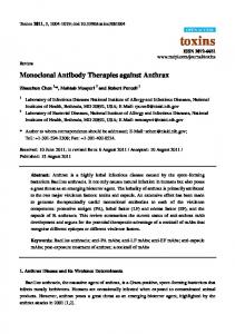
have indicated a synergistic effect. The EC50s demonstrate that there is a linear one-to-one relationship between the toxin neutralization capacities of IQNPA and IQNLF when they are combined (Fig. 1), confirming that their neutralizing activities are not synergistic but additive. Domain mapping of toxin-neutralizing antibodies. Six GST fusions of the individual PA domains and combinations of these domains were produced and tested for IQNPA binding/ recognition. Each fusion was named based on the PA domain(s) that it contains. IQNPA strongly recognized GSTI-IV, GSTIII-IV, and GSTIV but not GSTI, GSTI-II, or GSTI-III, indicating that the neutralizing epitope is within domain IV (Table 1). This binding recognition of domain IV was confirmed in a denatured and native protein Western blot analysis (data not shown). The four recombinant domains of LF plus a fifth construct encompassing domains II to IV were probed with IQNLF via native and denatured protein Western blot analysis. Domain I was consistently recognized in both blots by IQNLF, while domains II, III, IV, and II to IV were not recognized, demonstrating that the neutralizing epitope is located within domain I (Fig. 2). In vivo protective efficacy against B. anthracis spore challenge. The ability of a single 180-g dose of either IQNPA or IQNLF to confer passive protection in A/J mice against 24 LD50s of B. anthracis Sterne spores was determined. Antibody administered 2.5 h prior to i.p. challenge protected 100% of the mice over the course of the study with no overt clinical signs of illness (Fig. 3). The mice in the untreated control group had a mean time to death (MTD) of 87 ⫾ 27 h (MTD ⫾ SD), and B. anthracis was cultured from the heart tissue of these mice at the time of death. The protected mice were subjected to a second lethal spore challenge (41 LD50s) 20 days later. Once again, these animals survived until the end of the experiment on day 34 without overt illness (Fig. 3). Necropsy and tissue cultures from these mice failed to detect the presence of B. anthracis. In contrast, the second set of untreated controls had a MTD ⫾ SD of 69 ⫾ 18 h and were bacteremic at the time of death. Stimulation of mouse anti-PA and anti-LF antibody responses. Analysis of the collected serum samples indicated that mice protected by the MAbs mounted an antitoxin antibody response following the initial infection that increased signifi-
FIG. 2. rLF domain recognition by IQNLF. (A and B) SDS-denatured and native Western blots probed with IQNLF. (C) Silver-stained SDS-denaturing gel showing the approximate size of each domain. The gels were loaded with markers (left and right lanes) and the same concentration (50 ng) of each of the following constructs: full-length rLF (lane 1), LF domain I (lane 2), LF domain II (lane 3), LF domain III (lane 4), LF domain IV (lane 5), and LF domains II to IV (lane 6).
VOL. 75, 2007
PASSIVE PROTECTION ENHANCES ENDOGENOUS IMMUNITY
5429
FIG. 3. Protection of mice from an i.p. anthrax spore challenge by prechallenge injection of IQNPA and IQNLF. Mice were inoculated i.p. with 180 g of either IQNPA (n ⫽ 10) or IQNLF (n ⫽ 10) 2.5 h prior to an i.p. spore challenge consisting of 24 LD50s. The MTD ⫾ SD for the untreated controls was 87 ⫾ 27 h. Twenty days later surviving mice were rechallenged with 41 LD50s. In this second challenge the MTD ⫾ SD for the controls was 69 ⫾ 18 h.
cantly (P ⱕ 0.05) after the second challenge (Fig. 4). These murine responses were specific to the antigen recognized by the human MAb that the mice received prior to challenge. The animals receiving IQNPA produced significantly more anti-PA IgG than anti-LF IgG by days 17 and 27 (P ⱕ 0.05), while the mice that received IQNLF produced significantly more anti-LF IgG than anti-PA IgG by days 17, 27, and 35 (P ⱕ 0.001) (Fig. 4). ELISAs confirmed that the murine antibody concentrations were not due to the exogenous human IgG in the sera crossreacting with the secondary anti-mouse IgG used in the assays.
Mouse anti-IQNPA and anti-IQNLF IgG responses also increased from a background geometric mean titer (GMT) of 0.7 g/ml prior to treatment to 12.9 ⫾ 2.4 and 8.7 ⫾ 1.6 g/ml, respectively, by day 34 (data not shown). The toxin-neutralizing capacity of the endogenous mouse IgG response produced after the second challenge was evaluated. Terminal bleed sera from the mice receiving IQNPA had a geometric mean ED50 of 100 ⫾ 2 (n ⫽ 6), while the geometric mean ED50 of the terminal bleed sera from mice receiving IQNLF was 213 ⫾ 3 (n ⫽ 7). This neutralization activity had a
FIG. 4. Mouse specific anti-toxin IgG titers after B. anthracis Sterne strain spore challenge. Anti-PA and anti-LF IgG titers were measured in five sample bleeds taken during the course of the study. Solid symbols (Œ and F) indicate GMTs obtained for mice treated with IQNPA, while open symbols (‚ and E) indicate GMTs obtained for mice treated with IQNLF. Mice were challenged on days 0 and 20, as indicated by the vertical dashed lines. Mice produced a modest amount of anti-toxin IgG 10 days after the first challenge, which increased following the second challenge on day 20. Mice treated with IQNPA produced a greater anti-PA IgG response than anti-LF IgG response, while the converse was true for mice receiving IQNLF.
5430
ALBRECHT ET AL.
INFECT. IMMUN.
FIG. 5. Clearance of IQNPA and IQNLF in vivo. The levels of IQNPA and IQNLF asymptotically decreased by 77.5 and 91.3%, respectively, over the course of the 34-day study. The half-life (t1/2) of each antibody was calculated using the formula: t1/2 ⫽ 共t2 ⫺ t1兲 ⫻ ln共2兲/ln共关Ct1/Ct2兲兴, where t1 and t2 are times 1 and 2, respectively, and Ct1 and Ct2 are the MAb titers at t1 and t2, respectively. Using this equation, IQNPA had a half-life of 11.2 days, while IQNLF had a half-life of 6.8 days.
positive correlation with the murine anti-PA and anti-LF ELISA titers (R2 ⫽ 0.58 and R2 ⫽ 0.51, respectively), but it could not be attributed to nonspecific serum protection, since pooled naı¨ve serum from the same mice prior to treatment failed to protect macrophages (data not shown). Nor can neutralization be attributed to the presence of residual IQNPA or IQNLF, since serum samples that had detectable concentrations of these antibodies in ELISA after absorption were excluded from the analysis. Clearance of human MAbs. Both antibodies were retained in the bloodstream of all mice; however, IQNPA persisted at a higher level than IQNLF throughout the course of the 34-day study (Fig. 5). IQNPA was detected in the mice at a GMT of 85.8 ⫾ 1.4 g/ml on day 10 (the first posttreatment/challenge bleed), and the level decreased 77.5% to 19.3 ⫾ 1.5 g/ml by day 34. IQNLF was initially detected at a level of 73.6 ⫾ 1.3 g/ml, and the level decreased 91.3% to 6.4 ⫾ 1.8 g/ml by the end of the study. The half-life of IQNPA in mice is 11.2 days, while that of IQNLF is 6.8 days. DISCUSSION The currently recommended postexposure therapeutic treatment for anthrax is a combination of antibiotics (ciprofloxacin or doxycycline), licensed human vaccine (AVA), and, in severe cases, intravenously administered preformed human polyclonal anthrax immunoglobulin (AIGIV) derived from immunized donors (9; http://www2.niaid.nih.gov/Biodefense/Research/products .htm#a1). The advantages of using AIGIV are that it provides instant protection, is likely to be effective during mid- to advanced-stage disease, is equally effective against antibiotic-resistant strains, results in minimal adverse reactions, has a prolonged serum half-life, and targets multiple epitopes, making it difficult to subvert its efficacy (52; L. Price, A. J. Vogler, S. James, and P. Keim, presented at the 4th International Conference on Anthrax, 10 to 13 June, 2001, Annapolis, MD). Despite these advantages AIGIV does suffer from the need to maintain and constantly renew stocks of antibodies with sufficiently high toxin neutraliza-
tion potency from an immunologically diverse population of donors. An alternative approach would be to develop protective human MAbs with neutralizing specificity for PA and LF that could be used individually or in combination and can be manufactured on a large scale (http://www2.niaid.nih.gov/Biodefense /Research/products.htm#a1). Here two fully human MAbs, IQNPA and IQNLF, with the ability to neutralize separate components of the anthrax LeTx and to passively protect mice upon spore challenge are described. Most importantly, these antibodies did not interfere with the development of endogenous anti-PA and anti-LF immune responses. Both antibodies were specific for their cognate antigens and independently had a greater capacity to neutralize LeTx than the reference polyclonal serum. Isolation of hybridomas producing potent toxin-neutralizing antibodies is indicative of germ line antibodies that have matured through genetic recombination, somatic hypermutation, and extensive selection. This structural plasticity plays a critical role in the ability of the immune system to adapt and produce highly specific, highaffinity neutralizing antibodies (62). When combined in vitro, IQNPA and IQNLF were found to have a composite effect on LeTx neutralization intermediate between their individual effects, suggesting that they function independent of each other and noncompetitively by neutralizing unrelated processes during LeTx intoxication. Importantly, this MAb combination did not have a deleterious effect on LeTx neutralization. In order to plausibly explain the ability of IQNPA and IQNLF to neutralize LeTx without interfering with each other, their antigen binding domains were identified. The specific recognition of PA domain IV by IQNPA suggests that this MAb impedes the interaction of PA with the host cell. PA domain IV has been implicated in binding to cell surface receptors (5, 47, 57); therefore, it is reasonable to propose that IQNPA probably interferes with the attachment of PA to capillary morphogenesis protein 2 and/or tumor endothelial marker 8. Several LF-neutralizing antibodies have also been identified in other studies (25, 63), one of which binds domain
VOL. 75, 2007
PASSIVE PROTECTION ENHANCES ENDOGENOUS IMMUNITY
III and exerts its protective effect by a mechanism that is not clear yet (25). In contrast, the binding of IQNLF to LF domain I indicates that it may obstruct the formation of the LeTx complex by disrupting or preventing the interaction of LF with proteolytically active PA63, which occurs between amino acids in PA domains I and II and LF domain I (8, 24). The fact that IQNPA and IQNLF bind to protein domains that are involved in separate yet related processes during intoxication logically explains why the MAb combination did not have a negative effect on LeTx neutralization. Prophylactic administration of 180 g of IQNPA or IQNLF as a single, stand-alone treatment resulted in 100% survival. To our knowledge, IQNLF is the first fully human LF-specific MAb capable of conferring complete protection against such a challenge. Analysis of the postmortem heart swabs revealed that antibody-treated animals not only were protected during both challenges but were able to clear the infecting organism. The observation that both antibodies persisted in the mice over the course of the study is indicative of their IgG1 subclass, which is reported to have an in vivo half-life of 7 to 21 days (34, 50, 51). This is a desirable quality for a prophylactic or therapeutic antibody considering that B. anthracis spores can persist in the lungs and the environment long after a biological attack (11, 12). Although the calculated half-lives of IQNPA and IQNLF were 11.2 and 6.8 days, respectively, we have been able to demonstrate that 45 g of IQNPA and 22 g of IQNLF are able to fully protect mice against a lethal spore challenge (unpublished data). In the present study, these concentrations were detected in mouse serum on day 21 for IQNPA and on day 17 for IQNLF. Based on these observations, it is quite possible that survival after the second challenge was due to both the residual human MAb in circulation and the endogenous murine anti-LeTx antibodies produced after challenge. Analysis of the endogenous murine antibody response prior to the second challenge and at the end of the study revealed that in addition to the human MAbs the serum also contained mouse anti-PA- and anti-LF-specific polyclonal IgGs. The concentrations of these endogenous IgGs were substantially boosted following the second challenge and included antibodies with toxin-neutralizing activity. This was not unexpected and is consistent with previous studies using human or humanized PA-specific MAbs (33, 37). Although the endogenous IgG concentrations reported here were below previously identified serological correlates of protection (27, 38, 42) and the ED50s and IgG concentrations are weakly correlated, their contribution to protection cannot be ruled out. It is quite possible that the titers would have become more robust if there had been more time between the two challenges. Interestingly, the magnitudes of the endogenous PA- and LF-specific antibody responses were dependent on which human antibody the mice received prior to challenge. Mice given IQNPA generated a significantly higher IgG response to PA than to LF (P ⱕ 0.05), while mice inoculated with IQNLF generated a significantly higher IgG response to LF than to PA (P ⱕ 0.001). These findings support the hypothesis that the toxin-neutralizing capacities of IQNPA and IQNLF not only allow the development of a protective endogenous antibody response against PA and LF but also may in fact stimulate this humoral response. One possible explanation for this immunostimulatory effect
5431
is the formation of antigen-antibody immune complexes and their subsequent binding to Fc receptors on macrophages and natural killer cells. This process would specifically enhance PA and LF antigen uptake and presentation on major histocompatibility complex molecules to stimulate T- and B-cell expansion and the subsequent production of endogenous murine polyclonal antibodies to PA and LF, as was observed. During application in humans (or higher-order mammals) IQNPA and IQNLF may also fix complement, a feature of IgG1; however, since the A/J mouse is C5 deficient, the full effect of complement cannot be discussed in terms of the present study (http: //jaxmice.jax.org/strain/000646.html). Recent work examining the potential role of IQNPA and IQNLF in antibody-dependent cell-mediated cytotoxicity (ADCC) indicates that these MAbs can also function opsonically (Alan Cross, personal communication) to clear vegetative bacilli. This may explain the absence of bacteremia in the surviving mice. As a natural consequence of ADCC a variety of anthrax antigens are released that aid in the establishment of a robust polyclonal antibody response to the vegetative bacilli. Due to the observed phenomena it is reasonable to infer that IQNPA and IQNLF not only passively protected the mice but primed and immunized them as a result of the antibody treatment in combination with the challenge organism. The ability of therapeutic antibodies to confer protection while at the same time enabling or even stimulating an individual to generate his or her own protective immune response is an extremely desirable property. This observation may have implications for enhancing responses to the existing licensed anthrax vaccine when it is used as a postexposure therapy. We concluded, based on the data presented here, that passive protection and immunostimulation against B. anthracis by IQNPA and IQNLF occur through toxin neutralization to inhibit separate processes of anthrax intoxication. This may be due to inhibition of PA binding to the host cell surface and inhibition of LeTx formation. Once PA and LF are bound by IQNPA and IQNLF, it is likely that they will be delivered to antigen-presenting cells for antigen processing and presentation, thus initiating the observed B-cell responses. T-cell responses may also have been elicited, but this was not evaluated. The clearance of the circulating vegetative bacilli by ADCC may have also contributed to protection. Evaluation of these mechanisms during IQNPA- and IQNLF-mediated passive protection is ongoing. While the use of one of these MAbs is likely to be effective during passive protection, there are concerns about the deliberate circumvention of epitope binding sites within PA or LF resulting in a protein whose biological activity is no longer inhibited by a previously protective antibody. Indeed, the feasibility of just such an event has been reported for PA (16). As a consequence, the NIAID “Expert Consultation on Monoclonal Antibodies for Anthrax” has recommended that researchers consider developing a cocktail of MAbs which target different regions of the anthrax LeTx (http://www2.niaid.nih.gov /Biodefense/Research/products.htm#a1). To this end, a treatment composed of both IQNPA and IQNLF would be valuable. This treatment would broaden the spectrum of protection by ensuring that toxin formation and activity are blocked at two independent levels, namely, cellular attachment and LF binding, with two distinct antibodies.
5432
ALBRECHT ET AL.
We propose that administration of a single dose of antibody comprising a cocktail of human MAbs targeting PA and LF would provide immediate and broad-spectrum protection to naı¨ve populations while facilitating the establishment of an endogenous protective immune response in exposed individuals. The use of multiple antibodies specific to different toxins should be of particular value in the event of exposure to novel anthrax-causing strains in which PA has been altered by nature or genetic engineering to abolish epitopes incorporated in the currently licensed vaccines.
INFECT. IMMUN.
16.
17.
18.
ACKNOWLEDGMENTS This study was financially supported by The Ministries of Defense of The Netherlands and Italy under EUCLID contract 99/EF13.09/013, by The Dutch Ministry of Defense (contract 909.18.8390), by the Biological Defense Research Directorate, U.S. Naval Medical Research Center, under a CRADA, and by U.S. Defense Threat Reduction Agency grant Baillie 2.10018_05_NM_B. The views expressed in this article are those of the authors and do not necessarily reflect the official policy or position of the Department of the Navy, the Department of Defense, or the U.S. Government. We thank Tatiana Pervaia, Karen Brenneman, Alison Freeman, Patty Wilkins, Tom Taylor, Ineke van der Gun, Kunja Slopsema, Christine Cool, Wilma Steeman, and John Daniels for their technical contributions and HB Consultancy and N. van Xanten for project management. M. T. Albrecht, H. Li, D. Galloway, A. Mateczun, C. P. Quinn, and S. Goldman are employees of the U.S. Government. This work was prepared as part of their official duties. REFERENCES 1. Agrawal, A., J. Lingappa, S. H. Leppla, S. Agrawal, A. Jabbar, C. Quinn, and B. Pulendran. 2003. Impairment of dendritic cells and adaptive immunity by anthrax lethal toxin. Nature 424:329–334. 2. Baillie, L. W., K. Fowler, and P. C. Turnbull. 1999. Human immune responses to the UK human anthrax vaccine. J. Appl. Microbiol. 87:306–308. 3. Beedham, R. J., P. C. Turnbull, and E. D. Williamson. 2001. Passive transfer of protection against Bacillus anthracis infection in a murine model. Vaccine 19:4409–4416. 4. Bradley, K. A., J. Mogridge, M. Mourez, R. J. Collier, and J. A. Young. 2001. Identification of the cellular receptor for anthrax toxin. Nature 414:225–229. 5. Brossier, F., M. Levy, A. Landier, P. Lafaye, and M. Mock. 2004. Functional analysis of Bacillus anthracis protective antigen by using neutralizing monoclonal antibodies. Infect. Immun. 72:6313–6317. 6. Brossier, F., and M. Mock. 2001. Toxins of Bacillus anthracis. Toxicon 39: 1747–1755. 7. Cirino, N. M., D. Sblattero, D. Allen, S. R. Peterson, J. D. Marks, P. J. Jackson, A. Bradbury, and B. E. Lehnert. 1999. Disruption of anthrax toxin binding with the use of human antibodies and competitive inhibitors. Infect. Immun. 67:2957–2963. 8. Cunningham, K., D. B. Lacy, J. Mogridge, and R. J. Collier. 2002. Mapping the lethal factor and edema factor binding sites on oligomeric anthrax protective antigen. Proc. Natl. Acad. Sci. USA 99:7049–7053. 9. Enserink, M. 2002. Anthrax. ‘Borrowed immunity’ may save future victims. Science 295:777. 10. Flick-Smith, H. C., N. J. Walker, P. Gibson, H. Bullifent, S. Hayward, J. Miller, R. W. Titball, and E. D. Williamson. 2002. A recombinant carboxyterminal domain of the protective antigen of Bacillus anthracis protects mice against anthrax infection. Infect. Immun. 70:1653–1656. 11. Friedlander, A. M., S. L. Welkos, M. L. Pitt, J. W. Ezzell, P. L. Worsham, K. J. Rose, B. E. Ivins, J. R. Lowe, G. B. Howe, P. Mikesell, et al. 1993. Postexposure prophylaxis against experimental inhalation anthrax. J. Infect. Dis. 167:1239–1243. 12. Grinberg, L. M., F. A. Abramova, O. V. Yampolskaya, D. H. Walker, and J. H. Smith. 2001. Quantitative pathology of inhalational anthrax I: quantitative microscopic findings. Mod. Pathol. 14:482–495. 13. Guidi-Rontani, C., M. Weber-Levy, E. Labruyere, and M. Mock. 1999. Germination of Bacillus anthracis spores within alveolar macrophages. Mol. Microbiol. 31:9–17. 14. Hanna, P. C., S. Kochi, and R. J. Collier. 1992. Biochemical and physiological changes induced by anthrax lethal toxin in J774 macrophage-like cells. Mol. Biol. Cell 3:1269–1277. 15. Hermanson, G., V. Whitlow, S. Parker, K. Tonsky, D. Rusalov, M. Ferrari, P. Lalor, M. Komai, R. Mere, M. Bell, K. Brenneman, A. Mateczun, T. Evans, D. Kaslow, D. Galloway, and P. Hobart. 2004. A cationic lipidformulated plasmid DNA vaccine confers sustained antibody-mediated pro-
19. 20.
21.
22.
23.
24.
25.
26. 27. 28.
29. 30. 31. 32. 33.
34. 35. 36. 37.
tection against aerosolized anthrax spores. Proc. Natl. Acad. Sci. USA 101: 13601–13606. Hoffmaster, A. R., J. Ravel, D. A. Rasko, G. D. Chapman, M. D. Chute, C. K. Marston, B. K. De, C. T. Sacchi, C. Fitzgerald, L. W. Mayer, M. C. Maiden, F. G. Priest, M. Barker, L. Jiang, R. Z. Cer, J. Rilstone, S. N. Peterson, R. S. Weyant, D. R. Galloway, T. D. Read, T. Popovic, and C. M. Fraser. 2004. Identification of anthrax toxin genes in a Bacillus cereus associated with an illness resembling inhalation anthrax. Proc. Natl. Acad. Sci. USA 101:8449– 8454. Hoover, D. L., A. M. Friedlander, L. C. Rogers, I. K. Yoon, R. L. Warren, and A. S. Cross. 1994. Anthrax edema toxin differentially regulates lipopolysaccharide-induced monocyte production of tumor necrosis factor alpha and interleukin-6 by increasing intracellular cyclic AMP. Infect. Immun. 62: 4432–4439. Hull, A. K., C. J. Criscuolo, V. Mett, H. Groen, W. Steeman, H. Westra, G. Chapman, B. Legutki, L. Baillie, and V. Yusibov. 2005. Human-derived, plant-produced monoclonal antibody for the treatment of anthrax. Vaccine 23:2082–2086. Institute of Laboratory Animal Resources Commission on Life Sciences. 1996. Guide for the care and use of laboratory animals. National Academy Press, Washington, DC. Jernigan, J. A., D. S. Stephens, D. A. Ashford, C. Omenaca, M. S. Topiel, M. Galbraith, M. Tapper, T. L. Fisk, S. Zaki, T. Popovic, R. F. Meyer, C. P. Quinn, S. A. Harper, S. K. Fridkin, J. J. Sejvar, C. W. Shepard, M. McConnell, J. Guarner, W. J. Shieh, J. M. Malecki, J. L. Gerberding, J. M. Hughes, and B. A. Perkins. 2001. Bioterrorism-related inhalational anthrax: the first 10 cases reported in the United States. Emerg. Infect. Dis. 7:933– 944. Kim, C., N. Gajendran, H. W. Mittrucker, M. Weiwad, Y. H. Song, R. Hurwitz, M. Wilmanns, G. Fischer, and S. H. Kaufmann. 2005. Human alpha-defensins neutralize anthrax lethal toxin and protect against its fatal consequences. Proc. Natl. Acad. Sci. USA 102:4830–4835. Klimpel, K. R., S. S. Molloy, G. Thomas, and S. H. Leppla. 1992. Anthrax toxin protective antigen is activated by a cell surface protease with the sequence specificity and catalytic properties of furin. Proc. Natl. Acad. Sci. USA 89:10277–10281. Kozel, T. R., W. J. Murphy, S. Brandt, B. R. Blazar, J. A. Lovchik, P. Thorkildson, A. Percival, and C. R. Lyons. 2004. mAbs to Bacillus anthracis capsular antigen for immunoprotection in anthrax and detection of antigenemia. Proc. Natl. Acad. Sci. USA 101:5042–5047. Lacy, D. B., H. C. Lin, R. A. Melnyk, O. Schueler-Furman, L. Reither, K. Cunningham, D. Baker, and R. J. Collier. 2005. A model of anthrax toxin lethal factor bound to protective antigen. Proc. Natl. Acad. Sci. USA 102: 16409–16414. Lim, N. K., J. H. Kim, M. S. Oh, S. Lee, S. Y. Kim, K. S. Kim, H. J. Kang, H. J. Hong, and K. S. Inn. 2005. An anthrax lethal factor-neutralizing monoclonal antibody protects rats before and after challenge with anthrax toxin. Infect. Immun. 73:6547–6551. Little, S. F., B. E. Ivins, P. F. Fellows, and A. M. Friedlander. 1997. Passive protection by polyclonal antibodies against Bacillus anthracis infection in guinea pigs. Infect. Immun. 65:5171–5175. Little, S. F., B. E. Ivins, P. F. Fellows, M. L. Pitt, S. L. Norris, and G. P. Andrews. 2004. Defining a serological correlate of protection in rabbits for a recombinant anthrax vaccine. Vaccine 22:422–430. Maynard, J. A., C. B. Maassen, S. H. Leppla, K. Brasky, J. L. Patterson, B. L. Iverson, and G. Georgiou. 2002. Protection against anthrax toxin by recombinant antibody fragments correlates with antigen affinity. Nat. Biotechnol. 20:597–601. Milne, J. C., D. Furlong, P. C. Hanna, J. S. Wall, and R. J. Collier. 1994. Anthrax protective antigen forms oligomers during intoxication of mammalian cells. J. Biol. Chem. 269:20607–20612. Moayeri, M., and S. H. Leppla. 2004. The roles of anthrax toxin in pathogenesis. Curr. Opin. Microbiol. 7:19–24. Mogridge, J., K. Cunningham, and R. J. Collier. 2002. Stoichiometry of anthrax toxin complexes. Biochemistry 41:1079–1082. Mogridge, J., K. Cunningham, D. B. Lacy, M. Mourez, and R. J. Collier. 2002. The lethal and edema factors of anthrax toxin bind only to oligomeric forms of the protective antigen. Proc. Natl. Acad. Sci. USA 99:7045–7048. Mohamed, N., M. Clagett, J. Li, S. Jones, S. Pincus, G. D’Alia, L. Nardone, M. Babin, G. Spitalny, and L. Casey. 2005. A high-affinity monoclonal antibody to anthrax protective antigen passively protects rabbits before and after aerosolized Bacillus anthracis spore challenge. Infect. Immun. 73:795– 802. Morell, A., W. D. Terry, and T. A. Waldmann. 1970. Metabolic properties of IgG subclasses in man. J. Clin. Investig. 49:673–680. Mourez, M., R. S. Kane, J. Mogridge, S. Metallo, P. Deschatelets, B. R. Sellman, G. M. Whitesides, and R. J. Collier. 2001. Designing a polyvalent inhibitor of anthrax toxin. Nat. Biotechnol. 19:958–961. O’Brien, J., A. Friedlander, T. Dreier, J. Ezzell, and S. Leppla. 1985. Effects of anthrax toxin components on human neutrophils. Infect. Immun. 47:306– 310. Peterson, J. W., J. E. Comer, D. M. Noffsinger, A. Wenglikowski, K. G.
VOL. 75, 2007
38. 39.
40.
41.
42. 43. 44.
45. 46. 47. 48. 49.
PASSIVE PROTECTION ENHANCES ENDOGENOUS IMMUNITY
Walberg, B. M. Chatuev, A. K. Chopra, L. R. Stanberry, A. S. Kang, W. W. Scholz, and J. Sircar. 2006. Human monoclonal anti-protective antigen antibody completely protects rabbits and is synergistic with ciprofloxacin in protecting mice and guinea pigs against inhalation anthrax. Infect. Immun. 74:1016–1024. Pitt, M. L., S. F. Little, B. E. Ivins, P. Fellows, J. Barth, J. Hewetson, P. Gibbs, M. Dertzbaugh, and A. M. Friedlander. 2001. In vitro correlate of immunity in a rabbit model of inhalational anthrax. Vaccine 19:4768–4773. Popov, S. G., T. G. Popova, E. Grene, F. Klotz, J. Cardwell, C. Bradburne, Y. Jama, M. Maland, J. Wells, A. Nalca, T. Voss, C. Bailey, and K. Alibek. 2004. Systemic cytokine response in murine anthrax. Cell. Microbiol. 6:225– 233. Price, B. M., A. L. Liner, S. Park, S. H. Leppla, A. Mateczun, and D. R. Galloway. 2001. Protection against anthrax lethal toxin challenge by genetic immunization with a plasmid encoding the lethal factor protein. Infect. Immun. 69:4509–4515. Quinn, C. P., P. M. Dull, V. Semenova, H. Li, S. Crotty, T. H. Taylor, E. Steward-Clark, K. L. Stamey, D. S. Schmidt, K. W. Stinson, A. E. Freeman, C. M. Elie, S. K. Martin, C. Greene, R. D. Aubert, J. Glidewell, B. A. Perkins, R. Ahmed, and D. S. Stephens. 2004. Immune responses to Bacillus anthracis protective antigen in patients with bioterrorism-related cutaneous or inhalation anthrax. J. Infect. Dis. 190:1228–1236. Reuveny, S., M. D. White, Y. Y. Adar, Y. Kafri, Z. Altboum, Y. Gozes, D. Kobiler, A. Shafferman, and B. Velan. 2001. Search for correlates of protective immunity conferred by anthrax vaccine. Infect. Immun. 69:2888–2893. Ross, J. M. 1957. The pathogenesis of anthrax following the administration of spores by the respiratory route. J. Clin. Pathol. 73:485–494. Sawada-Hirai, R., I. Jiang, F. Wang, S. M. Sun, R. Nedellec, P. Ruther, A. Alvarez, D. Millis, P. R. Morrow, and A. S. Kang. 2004. Human anti-anthrax protective antigen neutralizing monoclonal antibodies derived from donors vaccinated with anthrax vaccine adsorbed. J. Immune Based Ther. Vaccines 2:5. Scobie, H. M., G. J. Rainey, K. A. Bradley, and J. A. Young. 2003. Human capillary morphogenesis protein 2 functions as an anthrax toxin receptor. Proc. Natl. Acad. Sci. USA 100:5170–5174. Sellman, B. R., M. Mourez, and R. J. Collier. 2001. Dominant-negative mutants of a toxin subunit: an approach to therapy of anthrax. Science 292:695–697. Singh, Y., K. R. Klimpel, C. P. Quinn, V. K. Chaudhary, and S. H. Leppla. 1991. The carboxyl-terminal end of protective antigen is required for receptor binding and anthrax toxin activity. J. Biol. Chem. 266:15493–15497. Smith, H., and J. Keppie. 1954. Observations on experimental anthrax: demonstration of a specific lethal factor produced in vivo by Bacillus. anthracis. Nature 173:869–870. Smith, H., J. Keppie, J. L. Stanley, and P. W. Harris-Smith. 1955. The chemical basis of the virulence of Bacillus anthracis. IV. Secondary shock as
Editor: D. L. Burns
50.
51. 52.
53. 54.
55.
56.
57.
58.
59.
60. 61.
62.
63.
5433
a major factor in the death of guinea pigs from anthrax. Br. J. Exp. Pathol. 36:323–327. Spiegelberg, H. L., and B. G. Fishkin. 1972. The catabolism of human G immunoglobulins of different heavy chain subclasses. 3. The catabolism of heavy chain disease proteins and of Fc fragments of myeloma proteins. Clin. Exp. Immunol. 10:599–607. Spiegelberg, H. L., and W. O. Weigle. 1965. Studies on the catabolism of gamma-G subunits and chains. J. Immunol. 95:1034–1040. Stepanov, A. V., L. I. Marinin, A. P. Pomerantsev, and N. A. Staritsin. 1996. Development of novel vaccines against anthrax in man. J. Biotechnol. 44: 155–160. Stephen, J. 1986. Anthrax toxin, p. 381–395. In Pharmacology of bacterial toxins. Pergamon Press, Oxford, United Kingdom. Taylor, T. H., C. Quinn, D. Schmidt, et al. 2003. Novel mathematical approach to TNA endpoints, abstr. P624. Program Abstr. 5th Int. Conf. Anthrax and 3rd Int. Workshop Mol. Biol. Bacillus cereus, B. anthracis, and B. thuringiensis, Nice, France. Towbin, H., T. Staehelin, and J. Gordon. 1979. Electrophoretic transfer of proteins from polyacrylamide gels to nitrocellulose sheets: procedure and some applications. Proc. Natl. Acad. Sci. USA 76:4350–4354. Turnbull, P. C. B. 1990. Terminal bacterial and toxin levels in the blood of guinea pigs dying of anthrax. Proceedings of the International Workshop on Anthrax. Salisbury Med. Bull. Spec. Suppl. 68:53–55. Varughese, M., A. V. Teixeira, S. Liu, and S. H. Leppla. 1999. Identification of a receptor-binding region within domain 4 of the protective antigen component of anthrax toxin. Infect. Immun. 67:1860–1865. Wang, F., P. Ruther, I. Jiang, R. Sawada-Hirai, S. M. Sun, R. Nedellec, P. R. Morrow, and A. S. Kang. 2004. Human monoclonal antibodies that neutralize anthrax toxin by inhibiting heptamer assembly. Hum. Antib. 13:105–110. Wang, H., M. N. Griffiths, D. R. Burton, and P. Ghazal. 2000. Rapid antibody responses by low-dose, single-step, dendritic cell-targeted immunization. Proc. Natl. Acad. Sci. USA 97:847–852. Welkos, S. L., T. J. Keener, and P. H. Gibbs. 1986. Differences in susceptibility of inbred mice to Bacillus anthracis. Infect. Immun. 51:795–800. Williamson, E. D., I. Hodgson, N. J. Walker, A. W. Topping, M. G. Duchars, J. M. Mott, J. Estep, C. Lebutt, H. C. Flick-Smith, H. E. Jones, H. Li, and C. P. Quinn. 2005. Immunogenicity of recombinant protective antigen and efficacy against aerosol challenge with anthrax. Infect. Immun. 73:5978–5987. Yin, J., A. E. Beuscher, S. E. Andryski, R. C. Stevens, and P. G. Schultz. 2003. Structural plasticity and the evolution of antibody affinity and specificity. J. Mol. Biol. 330:651–656. Zhao, P., X. Liang, J. Kalbfleisch, H. M. Koo, and B. Cao. 2003. Neutralizing monoclonal antibody against anthrax lethal factor inhibits intoxication in a mouse model. Hum. Antib. 12:129–135.