Human monocyte-derived dendritic cells differentiated in the presence of IL-2 produce proinflammatory cytokines and prime Th1 immune response Nunzia Sanarico,*,† Antonio Ciaramella,*,‡ Alessandra Sacchi,§ Daniela Bernasconi,¶ Paola Bossu`,‡ Francesca Mariani,† Vittorio Colizzi,*,ⱍⱍ and Silvia Vendetti§,¶,1 *Department of Biology, University of Rome Tor Vergata, Italy; †Institute of Neurobiology and Molecular Medicine, Italian National Council of Research, Rome; ‡Department of Clinical and Behavioural Neurology, IRCCS Santa Lucia Foundation, Rome, Italy; and §National Institute for Infectious Disease “L. Spallanzani,” IRCCS, Rome, Italy; ¶ Department of Infectious, Parasitic and Immune-Mediated Diseases, Istituto Superiore di Sanita`, Rome, Italy; and ⱍⱍ Centre International “Chantal Biya” Yaounde`, Cameroun
Abstract: Interleukin (IL)-2 plays an important role in the control of the immune responses, and it is released in a variety of tissues in response to inflammatory stimuli. As monocytes and mature dendritic cells (DCs) express CD25, the high-affinity subunit of IL-2 receptor, we examined the effect of exogenous IL-2 on the in vitro generation and maturation of DCs from monocytes. Human monocyte-derived DCs (MDDCs) were generated by culturing monocytes with granulocyte macrophage-colony stimulating factor (GM-CSF) and IL-4 in the presence or absence of IL-2. The cytokine was added at the beginning and after 5 days of culture. Our findings indicate that IL-2 does induce monocytes to differentiate into DCs with the same morphology and phenotype of that obtained in the presence of GM-CSF and IL-4 alone, but with some distinctive functional properties. DCs differentiated in the presence of IL-2 secreted significantly more IL-1, TNF-␣, and IL-12 p70 in response to lipopolysaccharide stimulation and induced allogeneic, naı¨ve T cells to release a significantly higher amount of interferon-␥ if compared with DCs obtained by culturing monocytes with GM-CSF and IL-4. These results indicate unrecognized effects of IL-2 on human MDDCs and suggest that an IL-2rich environment during differentiation and maturation of DCs can modify their T helper cell-inducing properties. J. Leukoc. Biol. 80: 555–562; 2006. Key Words: antigen-presenting cell differentiation
䡠
T cell polarization
䡠
cell
in virtually all tissues, where they serve as sentinel to collect and process antigens [3]. Inflammatory signals and microbial products such as lipopolysaccharide (LPS) and bacterial DNA can activate immature DCs (imDCs) inducing maturation [4]. Mature DCs (mDCs) up-regulate costimulatory molecules and move to lymphoid tissues, where they bind T cells and sustain their activation. The interaction of T cells with DCs is crucial for directing T helper (Th) cell differentiation toward the Th1, Th2, or T regulatory phenotype [1, 5]. It is well established that monocytes differentiate into DCs in vitro and in vivo [6, 7]. Besides granulocyte macrophagecolony stimulating factor (GM-CSF) combined with interleukin (IL)-4 are commonly used for the in vitro generation of myeloid DCs from CD14⫹ precursors, new strategies of DC differentiation have been described, and a diverse array of cytokines has been shown to be able to induce or alter DC generation [4, 8]. According to recent reports, human monocytes cultured in the presence of IL-3 and IL-4 differentiate into DCs, which produce less IL-12 and shift Th cell responses toward a Th2 cytokine pattern [9]. Monocytes incubated for 24 h with GMCSF and IL-4 and with proinflammatory mediators for another 24 h develop into fully differentiated DCs [10]. Furthermore, IL-15 alone induces conversion of monocytes into mDCs [11], and IL-15 has been found to skew monocyte differentiation into DCs with features of Langerhans cells when added to GM-CSF [12]. All these data suggest that alternative activation pathways may produce different types of DCs from the same monocyte precursors and that different cytokines may contribute to the initiation of immune responses through their ability to stimulate DCs. IL-2 is important in regulating immune responses, as it has a pleiotropic effect on T and B lymphocytes, natural killer (NK) cells, as well as others hematopoietic cells [13]. The effects of this cytokine are mediated through an IL-2-specific cell sur-
INTRODUCTION Dendritic cells (DCs) are the most potent antigen-presenting cells (APC) and act as the first line of defense, initiating specific immune response to novel antigens through the interaction and activation of naı¨ve T cells [1, 2]. DCs are distributed 0741-5400/06/0080-555 © Society for Leukocyte Biology
1
Correspondence: Department of Infectious, Parasitic and Immune-Mediated Diseases, Istituto Superiore di Sanita`, Viale regina Elena 299, 00161 Rome, Italy. E-mail:
[email protected] Received November 25, 2005; revised March 17, 2006; accepted April 20, 2006; doi: 10.1189/jlb.1105690.
Journal of Leukocyte Biology Volume 80, September 2006 555
face receptor (IL-2R), which includes three subunits encoded by distinct genes, the ␣-, -, and ␥-chains [14]. Monocytes express IL-2R, and IL-2 stimulation is known to result in the up-regulation of proinflammatory cytokines and in the phosphorylation of selected kinases [15]. However, whether IL-2 could influence the differentiation of monocytes into DCs has not been fully investigated. It has also been shown that the IL-2R␣ (CD25) subunit is up-regulated on DCs during maturation in mouse and human DCs [16 –18], and this raises the questions of whether CD25 expressed on mDCs is functional and whether the presence of IL-2 during DC differentiation and maturation influences their capacity to stimulate T cells. Therefore, in this study, we asked whether monocytes developing into DCs and LPS-matured DCs would receive instructional signals through IL-2R, and we evaluated their phenotype and functions. By comparing monocytes differentiated and matured into DCs in the presence or absence of IL-2, DCs, and IL-2DCs, respectively, we found that IL-2DCs, upon LPS stimulation, produced a higher amount of proinflammatory cytokines, such as IL-1 and TNF-␣, and regulatory cytokines, such as IL-12, priming naı¨ve T cells to produce interferon-␥ (IFN-␥). These data show that IL-2, which is known to have an important role on T lymphocytes, also plays a role in cells of the innate immune system, influencing their capacity to drive the adaptive response.
MATERIALS AND METHODS Reagents Cultures were maintained in RPMI-1640 medium (EuroClone, Milan, Italy) supplemented with 10% heat-inactivated fetal bovine serum (Hyclone, Logan, UT), 2 mM L-glutamine, 10 mM HEPES buffer, 1% sodium pyruvate, 2 mM penicillin, and 50 g/ml streptomycin (all EuroClone), hereafter referred to as complete medium. Recombinant GM-CSF and IL-4 were purchased from EuroClone; LPS and brefeldin A were from Sigma-Aldrich (St. Louis, MO).
Generation of DC Peripheral blood mononuclear cells were isolated by Lympholyte-H (Cederlane, Hornby, Canada) gradient centrifugation from buffy coats drawn from healthy donors provided by “La Sapienza” University Transfusion Service (Rome, Italy). Monocytes were positively selected by anti-CD14-coated magnetic beads (Miltenyi Biotec, Bergisch Gladbach, Germany). The purity of monocytes was ⬎98%, and no T cell contamination was observed. Cells were plated at 1.5 ⫻ 106 cell/ml in complete medium supplemented with GM-CSF (50 ng/ml) and IL-4 (10 ng/ml). After 5 days of culture, part of the cells was stimulated to mature for 2 more days by adding LPS (200 ng/ml). Preliminary LPS dose-response experiments were performed to evaluate the optimal concentration with high capacity to confer maturation to DCs and low toxicity. IL-2 (100 U/ml; R&D Systems, Minneapolis, MN) was added to the culture at Days 1 and 5. These DCs will hereafter be called IL-2DCs, as opposed to the DCs differentiated in the presence of GM-CSF and IL-4 alone. On Day 7, immature and mature cells were collected and analyzed for membrane phenotype. Supernatants were stored at – 80°C.
15 min at ⫹4°C, washed, and then analyzed by a flow cytometer. Intracellular staining was performed to determine the production of IL-12, IL-10, TNF-␣, IL-6, and IL-1. After 1 h from LPS stimulation, 10 g/ml brefeldin A was added to cells to inhibit cytokine secretion. DCs were incubated for 2–7 h, then washed with PBS, fixed with 2% paraformaldehyde for 15 min at room temperature (RT), washed again, and stained with mAb in PBS-0.5% saponin for 30 min at RT. Samples were washed twice with PBS 0.5% saponin and analyzed by flow cytometry.
The cDNA arrays Total RNA was extracted from DCs and IL-2DCs after 9 h of LPS stimulation. RNA was extracted with a 4 M guanidin isothiocyanate single-step method [19]. The extraction was performed on an RNase-free bench in a separate room. A total RNA sample (1.5 g) was reverse-transcripted using the Ampolabelig kit (Superarray Bioscience Corp., Frederick, MD) according to the manufacturer’s instruction in the presence of [␣-33]P for the generation of radiolabeled cDNA probes. ␣-dCT33P-labeled cDNA produced for each sample was hybridized to human dendritic and APC gene arrays (GEArray, S series, Supearray Bioscience Corp.) according to the manufacturer’s instruction, and membranes were exposed to a Phosphor-Imager (Typhoon, Amersham Biosciences Europe GMBH, Germany). The acquired files were analyzed using the Array Vision 7.0 software (Imaging Research Inc., St. Catharines, Canada), and data were expressed as normalized density (nDens) of each spot, corresponding to the density value of the spot minus background density and expressed as a multiple of the reference density value. Genes with a nDens level of less than 0.2 were excluded as unexpressed. The genes showing a change of expression of 2.5-fold or greater were considered differentially modulated in DCs versus IL-2DCs.
Cytokine determination by enzyme-linked immunosorbent assay (ELISA) Supernatants from DCs and IL-2DCs in their immature state or LPS-stimulated for the last 48 h were collected, and the levels of IL-6, IL-1, TNF-␣, IL-10, and IL-12 p70 were evaluated by ELISA kits (Pierce Endogen, Woburn, MA), according to the manufacturer’s instruction.
DC-T cell coculture The ability of DCs and IL-2DCs to polarize naı¨ve T cells was evaluated. Cord blood mononuclear cells (CBMC) were isolated by Lympholyte-H, and monocytes were depleted by adherence. In parallel, naı¨ve CD45RA⫹ T cells were negatively selected by naı¨ve CD4⫹ T cell isolation kits (magnetic cell sorter, Miltenyi Biotec). DCs or IL-2DCs (2⫻105 cells), immature or LPS-stimulated for 48 h, were washed and cultured with CBMC or with CBMC-purified, naı¨ve T cells (1⫻106 cells). After 8 days, culture supernatants were harvested, and the level of IFN-␥ was quantified by ELISA (Pierce Endogen). To perform intracellular staining after 7 days of coculture, cells were stimulated with ␣-CD3 (0.5 g/ml) for 1 h, and then 10 g/ml brefeldin A was added to the cell to inhibit cytokine secretion. Cells were incubated overnight and stained first with anti-CD3 on the membrane and then for intracellular IFN-␥ (all from BD Biosciences) as described above.
Graphical, statistical, and flow cytometry analysis Microsoft Excel (Microsoft Corp., Redmond, WA) was used for graphical and statistical analysis. Data were expressed as mean ⫾ SD, and statistical significance was determined by two-tailed, paired Student’s t-test. P ⬍ 0.05 was considered statistically significant. The flow cytometry analysis was performed on a FACSCalibur flow cytometer (Becton Dickinson, San Jose, CA), and data were analyzed using CellQuest software (Becton Dickinson).
Flow cytometry The following monoclonal antibodies (mAb), directly fluorocrome-conjugated, were used for fluorescein-activated cell sorter analysis: anti-CD1a, CD25, CD80, CD86, CD83, human leukocyte antigen (HLA)-DR, CD14, CD11c, HLA-A,B,C, IL-12, IL-10, IL-1, TNF-␣, IL-6, and their isotype-matched mAb were from BD Biosciences (Milan, Italy). To determine surface cell phenotype, cells were washed in assay buffer [phosphate-buffered saline (PBS), 0.5% bovine serum albumin, and 0.1% sodium azide], incubated with mAb for
556
Journal of Leukocyte Biology Volume 80, September 2006
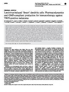
RESULTS DC phenotype is not affected by treatment with IL-2 Many investigators observed that CD25 is expressed on monocytes and DCs upon maturation, and it has been regarded as an http://www.jleukbio.org
activation marker [14, 15, 18, 20]. To evaluate whether CD25 expression on monocytes and mDCs was functional and whether the presence of IL-2 during DC differentiation and maturation could have any effects on DCs, monocytes were cultured for 7 days with GM-CSF and IL-4 in the absence (DCs) or in the presence of IL-2 (IL-2DCs), which was added to the cultures on Days 1 and 5. The purity of monocytes was higher than 98%, and no T cell contamination was observed. DCs and IL-2DCs were induced to mature by incubating them with LPS (200 ng/ml) for the last 48 h. It has been reported that the method used to isolate monocytes has an impact on the capacity of the resulting DCs to respond to endotoxin [21]. To address this point, we performed dose-response experiments to identify the optimal concentration of LPS for conferring DC maturation with low toxicity of cells derived from monocytes isolated by anti-CD14-coated microbeads. We found that 200 ng/ml LPS was the optimal concentration, as it induced a good up-regulation of the maturation markers CD83, CD25, and HLA-ABC and relative low percentage of annexin V-positive cells (data not shown). mDCs were then examined, by flow cytometry, for the typical marker profiles. By comparing DCs and IL-2DCs in their immature or mature state, we did not find significant differences in DC phenotypes. Cells were equally able to undergo maturation following stimulation with LPS, as assessed by the up-regulation of activation markers such as HLA-ABC, HLA-DR, CD80, CD86, and CD83, and the expression of CD25 was not affected by the presence of IL-2 (Fig. 1, A, C, and D). Different doses of IL-2, from 0 up to 500 U/ml, were used, and no differences in the phenotype of monocyte-derived DCs (MDDCs) were observed (data not shown). Conversely, we also examined whether the treatment with IL-2 could affect the viability of IL-2DCs, compared with DCs, by the expression of phosphatidylserine on the cell membrane and the percentage of hypodiploid nuclei. The results indicate that no induced cell death was observable in consequence of IL-2 treatment in all cell types analyzed (data not shown). As it has been described that other cytokines such as IL-15, TNF-␣, or IFN-␣ could replace IL-4 during the differentiation of monocytes into DCs, giving rise to different DC populations [12, 22, 23], we cultured monocytes in the presence of GMCSF and IL-2 (GM-CSF⫹IL-2) to analyze whether IL-2 could induce the differentiation of DCs in the absence of IL-4. We found that these monocytes gave rise to a population, which was adherent for the most part and did express CD14, resembling a macrophage-like phenotype. On the contrary, the expression of HLA-ABC, HLA-DR, and CD86 resulted higher in their immature state if compared with DCs and IL-2DCs (Fig. 1B). Following LPS treatment, they up-regulated Class I, Class II, and CD80 molecules, whereas they did not express CD83 molecules, which are known to be expressed mDCs selectively. These results indicate that IL-2 could not replace IL-4 in the differentiation of DCs from human monocytes.
Changes in gene expression profile in DCs and IL-2DCs As a first approach to test whether IL-2 could affect the overall gene expression of DCs, a cDNA array analysis was performed. We compared the gene expression profile of DCs and IL-2DCs
Fig. 1. Flow cytometry analysis of DCs and IL-2DCs. Human monocytes were cultured for 7 days in the presence of GM-CSF and IL-4 alone (DCs) or in combination with IL-2 (100 U/ml) (IL-2DCs). IL-2 was added to the culture on Days 1 and 5. DCs and IL-2DCs were induced to mature by incubating them with LPS (200 ng/ml) for the last 48 h. Cells were analyzed for their immature and mature phenotypes by flow cytometry (A). To determine whether IL-2 could replace IL-4 during the differentiation of monocytes into DCs, monocytes were cultured for 7 days with GM-CSF and IL-2. Unstimulated and LPS-treated cells were analyzed as described above (B). Empty histograms show the isotype-matched control immunoglobulin (Ig) staining. The panels are representative of one donor of 20 analyzed. The mean fluorescence intensity (C) and the percentages of positive cells (D) for the different markers analyzed ⫾ SE of 20 independent experiments are shown.
Sanarico et al. IL-2-conditioned DCs enhance a Th1-like immune response
557
stimulated with LPS for 9 h. The expression of 165 genes involved in DC activation and maturation was analyzed, and 77 genes were identified to be expressed in reliable amounts (nDens higher than 0.2) in DCs and IL-2DCs. Among these genes, we identified a group of transcripts as IL-1␣, IL-1, and IL-6, significantly up-regulated in IL-2DCs as compared with DCs (cut-off: 2.5-fold induction), and the rest of the transcripts resulted similarly modulated in both cell types (Fig. 2). These results suggest that IL-2DCs preferentially produce inflammatory cytokines as compared with DCs.
IL-2DCs increase the production of IL-1, TNF␣, and IL-12 upon LPS stimulation To further analyze the effect of IL-2 on the production of proinflammatory cytokines by DCs, we performed an intracellular staining for IL-1, IL-6, and TNF-␣ of cells differentiated and maturated in the presence or in the absence of IL-2. Furthermore, we tested culture supernatants by ELISA. Figure 3A shows the histogram plots of DCs and IL-2DCs expressing IL-1, IL-6, and TNF-␣ 6 h after LPS stimulation and reveals that the production of IL-1 and TNF-␣ resulted in increased cells differentiated and stimulated in the presence of IL-2 compared with DCs. On the contrary, the production of IL-6 resulted, unaffected by IL-2 treatment. Of note, DCs and IL-2DCs, at an immature state, did not produce any of the cytokines analyzed (data not shown). Intracellular staining performed at 3 h and 9 h after the maturation stimulus was similar to that observed at 6 h (data not shown). Furthermore, to test whether the augmented production of IL-1 and TNF-␣ in IL-2DCs was only a transient phenomenon, we evaluated by ELISA the presence of these cytokines on supernatants after
48 h of LPS stimulation. We also analyzed the amount of IL-6 to assess if this cytokine also could be affected in a longer period. Figure 3B shows that IL-2DCs released significantly higher levels of IL-1 (191⫾38 vs. 169⫾57; n⫽5; P⫽0.007) and TNF-␣ (1514⫾106 vs. 1226⫾90; n⫽5; P⫽0.01) after LPS stimulation as compared with DCs. Conversely, the amount of these cytokines in unstimulated cells was comparable between DCs and IL-2DCs. In accordance with intracellular analysis, the production of IL-6 did not differ significantly between DCs and IL-2DCs. We next asked whether IL-2 treatment could affect the production of regulatory cytokines. The accumulation of IL-10 and IL-12, which are pivotal cytokines in directing the polarization of the immune response, was evaluated. We found that after LPS stimulation, the production and release of IL-12 p70 resulted in increased IL-2DCs compared with DCs, as shown by intracellular staining (Fig. 4A) and ELISA assay (2808⫾465 vs. 2024⫾221; P⫽0.009; n⫽5; Fig. 4B). On the contrary, the production and release of IL-10 were unaffected by IL-2 stimulation (737⫾68 vs. 753⫾60; P⫽0.66; n⫽5; Fig. 4, A and B). Different doses of IL-2, from 0 up to 500 U/ml, were used, and we found that the production of IL-12 by MDDC and their capacity to induce IFN-␥ by CD4⫹ T isolated from cord blood were dose-dependent. However, statistically significant differences were not observed between the 100 and 500 U/ml (data not shown). To assess whether the increase of IL-12 could influence the levels of IL-10 during DC maturation, we evaluated the accumulation of both cytokines in culture supernatants at different timepoints (9, 24, and 48 h) after LPS stimulation. We found that IL-10 and IL-12 were increased after 9 h of LPS stimulation in
Fig. 2. Changes in gene expression profile in DCs and IL-2DCs stimulated with LPS (200 ng/ml) for 9 h. Total RNA was extracted, reverse-transcripted in cDNA, and used to hybridize human DCs and APC gene arrays. Dots represent 77 genes out of 165, which were considered to be expressed in reliable amounts (nDens was higher than 0.2).
558
Journal of Leukocyte Biology Volume 80, September 2006
http://www.jleukbio.org
Fig. 3. LPS-stimulated IL-2DCs produce more IL-1 and TNF-␣ if compared with DCs. (A) DCs and IL-2DCs, differentiated for 5 days, were stimulated with LPS (200 ng/ml) for 1 h. Brefeldin A (10 g/ml) was added for the last 5 h to stop cytokine secretion. Cells were harvested and stained for intracellular IL-1, IL-6, and TNF-␣. Empty histograms show the isotype-matched control Ig staining. (B) Supernatants from DCs and IL2DCs in their immature state or stimulated for 48 h with LPS (200 ng/ml) were examined by ELISA for the presence of IL-1, IL-6, and TNF-␣. The graphs show the mean of triplicate values ⫾ SD of one representing experiment of five performed.
DCs and IL-2DCs as compared with untreated immature cells. Furthermore, mature IL-2DCs produced a higher amount of IL-12 than DCs already after 9 h of LPS stimulation and reached a plateau after 24 h (Fig. 4C). These data suggest that there is no modulation of IL-10 levels by the increase of IL-12. Taken together, these results indicated that IL-2DCs were able to produce and release significantly higher amounts of inflammatory (IL-1, TNF-␣) or regulatory (IL-12) cytokines, suggesting that they might have a different capacity in directing immune response.
IL-2DCs induce naı¨ve T cells to produce a higher amount of IFN-␥ as compared with DCs DCs have the unique capacity to stimulate naı¨ve T lymphocytes, driving them into distinct classes of effectors cells. As IL-2DCs resulted more efficiently at producing and releasing IL-12, which is a well-known Th1-inducing factor, we speculated that these cells could differ in their capacity to polarize the immune response. To address this question, we cultured DCs and IL-2DCs with allogeneic CBMC for 8 days. The supernatants of cocultures were analyzed by ELISA for the presence of IFN-␥. As shown in Fig. 5A, CBMC cultured in the presence of mature IL-2DCs differentiate into cells pro-
ducing significantly higher levels of IFN-␥ as compared with CBMC cultured with mDCs (1200⫾186 vs. 901⫾214; P⫽0.023; n⫽5). It has been reported that NK cells from cord blood are able to produce high levels of IFN-␥ in comparison with adult NK cells [24]. To assess whether naive T cells produced IFN-␥, or different cord blood populations were involved, we performed two different sets of experiments. First, CBMC cocultered for 7 days with DCs or IL-2DCs were stimulated with ␣-CD3 (0.5 g/ml) for 1 h, incubated with brefeldin A (10 g/ml) overnight, and then stained on the membrane with anti-CD3 and intracellularly with anti-IFN-␥. In this way, we found that the anti-CD3-gated CBMC population produced a higher amount IFN-␥ when they were cocultured with IL-2DCs as compared with DCs (Fig. 5B). Second, we purified CD45RA⫹ T cells from cord blood and cocultured them with DCs or IL-2DCs for 8 days. The levels of IFN-␥ in the culture supernatants were evaluated by ELISA. We found that naive CBMC-derived CD45RA⫹ T cells still produced IFN-␥ in response to mDCs or IL-2DCs, and consistently, the production of IFN-␥ was increased when naı¨ve, CBMC-derived CD45RA⫹ T cells were cocultured with IL-2DCs (Fig. 5C; 1242⫾37 vs. 1061⫾72; n⫽5; P⫽0.017). These data suggest that IL-2DCs were able to
Sanarico et al. IL-2-conditioned DCs enhance a Th1-like immune response
559
Fig. 4. LPS-stimulated IL-2DCs produce a higher level of IL-12 as compared with DCs. (A) DCs and IL-2DCs, differentiated for 5 days, were stimulated with LPS (200 ng/ml) for 1 h. Brefeldin A (10 g/ml) was added for the last 5 h to stop cytokine secretion. Cells were harvested and stained for intracellular IL-12 and IL-10. Empty histograms show the isotype-matched control Ig staining. (B) Supernatants from DCs and IL-2DCs in their immature state or stimulated for 48 h with LPS (200 ng/ml) were examined by ELISA for the presence of IL-12 and IL-10. (C) IL-10 and IL-12, in the supernatants of DCs and IL-2DCs, were assayed at different time-points (9, 24, and 48 h) after LPS stimulation. The graphs show the mean of triplicate values ⫾ SD of one representing experiment of five performed.
direct the differentiation of naı¨ve T cells enhancing their capacity to produce IFN-␥.
DISCUSSION It is well established that monocytes differentiate into DCs in vivo and in vitro [6, 7]. Pathogens as well as their components
can influence DC generation, interacting with their precursor [25]. Alternatively, other cell types can respond to microbes by secreting different cytokines or tissue factors, which could interfere with DC differentiation and lead to distinct types of immune responses [26, 27]. A diverse array of cytokines, alone or in combination with GM-CSF, has been shown to be able to induce DC differentiation and maturation in vitro [4]. Therefore, in the present study, we analyzed an alternative differen-
Fig. 5. IL-2DCs induce naı¨ve T cells to produce a higher level of IFN-␥ as compared with DCs. DCs and IL-2DCs were differentiated for 7 days (immature) or stimulated to mature by incubating them with LPS for the last 48 h (mature). These cells were then cultured with monocyte-depleted, allogeneic CBMC (A) or with naı¨ve CBMC-purified T cells (C), and the production of IFN-␥ was analyzed by ELISA. (B) CBMC cocultered with DCs and IL-2DCs after 7 days were stimulated with ␣-CD3 (0.5 g/ml) for 1 h, and then 10 g/ml brefeldin A was added to the cell to inhibit cytokine secretion. After overnight incubation, cells were stained with anti-CD3 on the membrane and intracellularly with anti-IFN-␥. Histograms show an anti-CD3-gated population, and empty histograms are from the isotype-matched control Ig staining. The graphs show the mean of triplicate values ⫾ SD of one representing experiment of five performed.
560
Journal of Leukocyte Biology Volume 80, September 2006
http://www.jleukbio.org
tiation procedure for MDDCs, which mimes DCs arising from an inflammatory environment rich in IL-2. We generated MDDCs by using two different procedures: in the presence of GM-CSF and IL-4 (DCs) or by adding IL-2, first to the basal cocktail and, a second time, on cells fully differentiated (IL2DCs). We found that IL-2DCs showed an unaltered DC-like morphology and phenotype, but had the capacity to release a higher amount of proinflammatory and regulatory cytokines upon LPS treatment, priming an enhanced Th1 immune response. Besides the well-documented effects of IL-2 on lymphocytes and NK cells, other cell populations of the innate immune system including monocytes and mDCs are known to express functional IL-2Rs and to respond to the cytokine signal [15]. As a result of the capacity of monocytes and mDCs to sense IL-2, it was reasonable to suppose an influence of IL-2 on the generation of DCs from peripheral blood monocytes and on their maturation. Comparing IL-2DCs and DCs for their typical marker profiles, we found that the DC phenotype was not affected by treatment with IL-2 during the differentiation period and the LPS-induced maturation. The effects of IL-2 are mediated through a specific cellsurface receptor (IL-2R). IL-2R comprises at least three subunits encoded by different genes. IL-2R␣ shows homology with the ␣-chain of the IL-15R, which shares with IL-2R the other two subunits, IL-2R and IL-2R␥ [28, 29]. Differently from IL-2, the effect of IL-15 on MDDCs has been well elucidated. IL-15 was found able to skew monocyte differentiation into DCs with features of Langerhans cells when added in combination with GM-CSF [12]. Conversely, when we cultured monocytes with IL-2 in combination with GM-CSF in the absence of IL-4, we found a population of cells resembling a macrophagelike phenotype, as they appeared adherent for the most part and did express CD14. This suggests that although IL-15R and IL-2R are closely related, they induce distinct signaling pathways during the differentiation of monocytes into DCs. Similar results by Li et al. [30] show that monocytes cultured with GM-CSF in the absence of IL-4 skewed their differentiation into a macrophage-like population, suggesting that IL-4 is required for differentiation into the DC phenotype. The ability of IL-2 to regulate the function of DCs has been a matter of controversy. Mouse DCs were reported to respond to IL-2 by increasing IL-12-dependent IFN-␥ production [31] and by enhancing CD25-dependent antigen uptake [32]. The IL-2R on DCs using CD25 knockout mice was found to be unnecessary for T cell stimulation [18]. However, the function of IL-2R on human DCs has not been fully investigated. Here, by cDNA analysis used to screen the overall RNA expression of LPSstimulated DCs and IL-2DCs, we found that IL-2 treatment preferentially induced the production of proinflammatory cytokines. This was confirmed at protein level; indeed, we found that the generation and maturation of DCs in presence of IL-2 promoted an increase of IL-1, TNF-␣, and IL-12 production and secretion. It has been reported that murine or human DCs are able to produce IL-2 after stimulation with bacteria or their products [33–35]. It is interesting that DC-derived IL-2 represents an additional relevant molecule, conferring on DCs the unique T cell priming capacity, and it is also required to elicit IFN-␥
production from NK cells [33, 36]. Although the possibility that IL-2 could act on DCs in an autocrine manner remains to be explored directly, our findings suggest that DCs are sensitive to IL-2, and it is possible to hypothesize that this cytokine plays an important role in DC biology. Consistent with this, it has been reported that IL-2 is able to induce the differentiation of human DCs from cord blood CD34⫹ cells [37]. Mature IL-2DCs produced a significantly higher amount of IL-12 as compared with untreated cells, whereas the production of IL-10 was similar in the two populations. These findings may account for the varying capability of these cells to polarize the response of naı¨ve T lymphocytes. Indeed, when CBMC or CBMC-derived CD45RA⫹ T cells were cultured with IL-2DCs or DCs, IL-2DCs consistently induced a higher secretion of IFN-␥ by T lymphocytes as compared with DCs. It has been reported that IL-12 critically regulates the balance between Th1 and Th2 responses and potently induces IFN-␥-secreting Th1 cells [38, 39]. Regulatory mechanisms of IL-12 production have been studied in detail. For instance, IL-12 secretion is enhanced by several cytokines, such as IFN-␥ and IL-1, and it is down-regulated by IL-10 [40]. On the contrary, the precise mechanism by which IL-2DCs enhance Th1 polarization remains unclear, and it could be mediated directly by IL-12 or by a combination of factors. Different cytokines such as IL-23, IL-27 [41], or type I IFN [42] have been described to promote the induction of the Th1 immune response, which is known to be implicated in the elimination of intracellular pathogens and tumor cells [43]. We showed that IL-2 could play a role with other secreted factors in the biology of DCs. The physiological relevance of these results is supported by the fact that an IL-2-rich environment, such as that obtained during inflammation and DC/T cell cross-talking, could affect DC differentiation and maturation so that these cells could become more strongly alerted and more efficient at priming a Th1 immune response. These data indicate that besides its well-defined function in acquired immunity, IL-2 is also implicated in the regulation of the innate immune system; in particular, it plays a role in DCs, which have been described as a bridge between the innate and acquired immune response. The implication of this work may impact DC-based immunotherapy trials, as the differentiation of monocytes into DCs in the presence of IL-2 could be a strategy to be adopted in ex vivo preparation of DCs clinically used to modulate immune responses in cancer or other diseases.
ACKNOWLEDGMENTS This work was supported by FIRB-MWR 2001, the RBNE01 PPTF project, and by grants of the UNESCO foundation. We thank Dr. Ragnar Lindstedt for critical reading of the manuscript and for helpful discussion. We also thank Dr. Angelo Martino for helpful discussion and Dr. Carla Montesano for kindly providing the cord blood, and we are grateful to Dr. Manuela Grassi for her expert assistance in array experiments.
Sanarico et al. IL-2-conditioned DCs enhance a Th1-like immune response
561
REFERENCES 1. Banchereau, J., Steinman, R. M. (1998) Dendritic cells and the control of immunity. Nature 392, 245–252. 2. Cella, M., Sallusto, F., Lanzavecchia, A. (1997) Origin, maturation and antigen presenting function of dendritic cells. Curr. Opin. Immunol. 9, 10 –16. 3. Sallusto, F., Cella, M., Lanzavecchia, A. (1995) Dendritic cells use macropinocytosis and the mannose receptor to concentrate macromolecules in the major histocompatibility complex class II compartment: downregulation by cytokines and bacterial products. J. Exp. Med. 182, 389 – 400. 4. Banchereau, J., Palucka, A. K. (2005) Dendritic cells as therapeutic vaccines against cancer. Nat. Rev. Immunol. 5, 296 –306. 5. Banchereau, J., Briere, F., Caux, C., Davoust, J., Lebecque, S., Liu, Y. J., Pulendran, B., Palucka, K. (2000) Immunobiology of dendritic cells. Annu. Rev. Immunol. 18, 767– 811. 6. Romani, N., Gruner, S., Brang, D., Kampgen, E., Lenz, A., Trockenbacher, B., Konwalinka, G., Fritsch, P. O., Steinman, R. M., Schuler, G. (1994) Proliferating dendritic cell progenitors in human blood. J. Exp. Med. 180, 83–93. 7. Randolph, G. J., Inaba, K., Robbiani, D. F., Steinman, R. M., Muller, W. A. (1999) Differentiation of phagocytic monocytes into lymph node dendritic cells in vivo. Immunity 11, 753–761. 8. Santini, S. M., Lapenta, C., Logozzi, M., Parlato, S., Spada, M., Di Pucchio, T., Belardelli, F. (2000) Type I interferon as a powerful adjuvant for monocyte-derived dendritic cell development and activity in vitro and in Hu-PBL-SCID mice. J. Exp. Med. 191, 1777–1788. 9. Ebner, S., Hofer, S., Nguyen, V. A., Furhapter, C., Herold, M., Fritsch, P., Heufler, C., Romani, N. (2002) A novel role for IL-3: human monocytes cultured in the presence of IL-3 and IL-4 differentiate into dendritic cells that produce less IL-12 and shift Th cell responses toward a Th2 cytokine pattern. J. Immunol. 168, 6199 – 6207. 10. Dauer, M., Obermaier, B., Herten, J., Haerle, C., Pohl, K., Rothenfusser, S., Schnurr, M., Endres, S., Eigler, A. (2003) Mature dendritic cells derived from human monocytes within 48 h: a novel strategy for dendritic cell differentiation from blood precursors. J. Immunol. 170, 4069 – 4076. 11. Saikh, K. U., Khan, A. S., Kissner, T., Ulrich, R. G. (2001) IL-15-induced conversion of monocytes to mature dendritic cells. Clin. Exp. Immunol. 126, 447– 455. 12. Mohamadzadeh, M., Berard, F., Essert, G., Chalouni, C., Pulendran, B., Davoust, J., Bridges, G., Palucka, A. K., Banchereau, J. (2001) Interleukin 15 skews monocyte differentiation into dendritic cells with features of Langerhans cells. J. Exp. Med. 194, 1013–1020. 13. Smith, K. A. (1988) Interleukin-2: inception, impact, and implications. Science 240, 1169 –1176. 14. David, D., Bani, L., Moreau, J. L., Demaison, C., Sun, K., Salvucci, O., Nakarai, T., de Montalembert, M., Chouaib, S., Joussemet, M., Ritz, J., Theze, J. (1998) Further analysis of interleukin-2 receptor subunit expression on the different human peripheral blood mononuclear cell subsets. Blood 91, 165–172. 15. Hodge, S., Hodge, G., Flower, R., Han, P. (2000) Surface and intracellular interleukin-2 receptor expression on various resting and activated populations involved in cell-mediated immunity in human peripheral blood. Scand. J. Immunol. 51, 67–72. 16. Crowley, M., Inaba, K., Witmer-Pack, M., Steinman, R. M. (1989) The cell surface of mouse dendritic cells: FACS analyses of dendritic cells from different tissues including thymus. Cell. Immunol. 118, 108 –125. 17. Pollard, A. M., Lipscomb, M. (1990) Characterization of murine lung dendritic cells: similarities to Langerhans cells and thymic dendritic cells. J. Exp. Med. 172, 159 –167. 18. Kronin, V., Vremec, D., Shortman, K. (1998) Does IL-2 receptor ␣ chain induced on dendritic cells have a biological function? Int. Immunol. 10, 237–240. 19. Chomczynski, P., Sacchi, N. (1987) Single-step method of RNA isolation by guanidin thiocyanate phenol-chloroform extraction. Anal. Biochem. 162, 156 –159. 20. Huang, Q., Dongyu, L., Majewski, P., Schulte, L. C., Korn, J. M., Young, R. A., Lander, E. S., Hacohen, N. (2001) The plasticity of dendritic cells response to pathogens and their components. Science 294, 870 – 875. 21. Elkord, E., Williams, P. E., Kynaston, H., Rowbottom, A. W. (2005) Human monocyte isolation methods influence cytokine production from in vitro generated dendritic cells. Immunology 114, 204 –212. 22. Chomarat, P., Dantin, C., Bennett, L., Banchereau, J., Palucka, A. K. (2003) TNF skews monocyte differentiation from macrophages to dendritic cells. J. Immunol. 171, 2262–2269. 23. Paquette, R. L., Hsu, N. C., Kiertscher, S. M., Park, A. N., Tran, L., Roth, M. D., Glaspy, J. A. (1998) Interferon-␣ and granulocyte-macrophage
562
Journal of Leukocyte Biology Volume 80, September 2006
24.
25.
26. 27. 28.
29.
30.
31. 32.
33.
34. 35.
36.
37.
38.
39.
40. 41. 42.
43.
colony-stimulating factor differentiate peripheral blood monocytes into potent antigen-presenting cells. J. Leukoc. Biol. 64, 358 –367. Dalle, J. H., Menezes, J., Wagner, E., Blagdon, M., Champagne, J., Champagne, M. A., Duval, M. (2005) Characterization of cord blood natural killer cells: implications for transplantation and neonatal infections. Pediatr. Res. 57, 649 – 655. Martino, A., Sacchi, A., Sanarico, N., Spadaro, F., Ramoni, C., Ciaramella, A., Pucillo, L. P., Colizzi, V., Vendetti, S. (2004) Dendritic cells derived from BCG-infected precursors induce Th2-like immune response. J. Leukoc. Biol. 76, 827– 834. Delneste, Y., Charbonnier, P., Herbault, N., Magistrelli, G., Caron, G., Bonnefoy, J. Y., Jeannin, P. (2003) Interferon-␥ switches monocyte differentiation from dendritic cells to macrophages. Blood 101, 143–150. Chomarat, P., Banchereau, J., Davoust, J., Palucka, A. K. (2000) IL-6 switches the differentiation of monocytes from dendritic cells to macrophages. Nat. Immunol. 1, 510 –514. Anderson, D. M., Kumaki, S., Ahdieh, M., Bertles, J., Tometsko, M., Loomis, A., Giri, J., Copeland, N. G., Gilbert, D. J., Jenkins, N. A., Valentine, V., Shapiro, D. N., Morris, S. W., Park, L. S., Cosman, D. (1995) Functional characterization of the human interleukin-15 receptor ␣ chain and close linkage of IL15RA and IL2RA genes. J. Biol. Chem. 270, 29862–29869. Giri, J. G., Ahdieh, M., Eisenman, J., Shanebeck, K., Grabstein, K., Kumaki, S., Namen, A., Park, L. S., Cosman, D., Anderson, D. (1994) Utilization of the  and ␥ chains of the IL-2 receptor by the novel cytokine IL-15. EMBO J. 13, 2822–2830. Li, G., Kim, Y. J., Broxmeyer, H. E. (2005) Macrophage colony-stimulating factor drives cord blood monocyte differentiation into IL-10(high)IL12absent dendritic cells with tolerogenic potential. J. Immunol. 174, 4706 – 4717. Fukao, T., Koyasu, S. (2000) Expression of functional IL-2 receptor on mature splenic dendritic cells. Eur. J. Immunol. 30, 1453–1457. Faulkner, L., Buchan, G., Lockhart, E., Slobbe, L., Wilson, M., Baird, M. (2001) IL-2 linked to a peptide from influenza hemagglutinin enhances T cells activation by affecting the antigen-presenting function of bone marrow-derived dendritic cells. Int. Immunol. 13, 713–721. Granucci, F., Vizzardelli, C., Pavelka, N., Feau, S., Persine, M., Virzi, E., Rescigno, M., Moro, G., Ricciardi-Castagnoli, P. (2001) Inducible IL-2 production by dendritic cells revealed by global gene analysis. Nat. Immunol. 2, 882– 888. Granucci, F., Feau, S., Angeli, V., Trottein, F., Ricciardi-Castagnoli, P. (2003) Early IL-2 production by mouse dendritic cells is the result of microbial-induced priming. J. Immunol. 170, 5075–5081. Feau, S., Facchinetti, V., Granucci, F., Citterio, S., Jarrossay, D., Protti, M. P., Lanzavecchia, A., Ricciardi-Castagnoli, P. (2005) Dendritic cellderived IL-2 production is regulated by IL-15 both in humans and mice. Blood 105, 697–702. Granucci, F., Zanoni, I., Pavelka, N., Van Dommelen, S. L., Andoniu, C. E., Belardelli, F., Degli Esposti, M. P., Ricciardi-Castagnoli, P. (2004) A contribution of mouse dendritic cell-derived IL-2 for NK cell activation. J. Exp. Med. 200, 287–295. Bykovskaja, S. N., Buffo, M. J., Bunker, M., Zhang, H., Majors, A., Herbert, M., Lokshin, A., Levitt, M. L., Jaja, A., Scalise, D., Kosiban, D., Evans, C., Marks, S., Shogan, J. (1998) Interleukin-2-induces development of denditric cells from cord blood CD34⫹ cells. J. Leukoc. Biol. 63, 620 – 630. Macatonia, S. E., Hosken, N. A., Litton, M., Vieira, P., Hsieh, C. S., Culpepper, J. A., Wysocka, M., Trinchieri, G., Murphy, K. M., O’Garra, A. (1995) Dendritic cells produce IL-12 and direct the development of Th1 cells from naive CD4⫹ T cells. J. Immunol. 154, 5071–5079. Mountford, A. P., Coulson, P. S., Cheever, A. W., Sher, A., Wilson, R. A., Wynn, T. A. (1999) Interleukin-12 can directly induce T-helper 1 responses in interferon-␥ (IFN-␥) receptor-deficient mice, but requires IFN-␥ signaling to downregulate T-helper 2 responses. Immunology 97, 588 –594. Aste-Amezaga, M., Sartori, A., Trincheri, G. (1998) Molecular mechanism of the induction of IL-12 and its inhibition by IL-10. J. Immunol. 160, 5936 –5944. Brombacher, F., Kastelein, R. A., Alber, G. (2003) Novel IL-12 family members shed light on the orchestration of Th1 response. Trends Immunol. 24, 207–212. Mattei, F., Schiavoni, G., Belardelli, F., Tough, D. F. (2001) IL-15 is expressed by dendritic cells in response to type I IFN, double-stranded RNA, or lipopolysaccharide and promotes dendritic cell activation. J. Immunol. 167, 1179 –1187. Schmidt, C. S., Mescher, M. F. (1999) Adjuvant effect of IL-12: conversion of peptide antigen administration from tolerizing to immunizing for CD8⫹ T cells in vivo. J. Immunol. 163, 2561–2567.
http://www.jleukbio.org