Clin Kidney J (2014) 7: 387–390 doi: 10.1093/ckj/sfu036 Advance Access publication 15 April 2014
Clinical Report
Hypotension-induced blindness in haemodialysis patients Shveta Bansal, Alec Ansons and Mandagere Vishwanath Department of Neuroophthalmology, Manchester Royal Eye Hospital, Manchester, UK Correspondence and offprint requests to: E-mail:
[email protected]
Abstract Hypotension is a commonly encountered complication in haemodialysis patients and is a significant cause of morbidity and mortality. Bilateral visual loss in dialysis induced hypotension remains poorly recognized as a complication by both renal physicians and ophthalmologists. We report 2 cases of patients on renal dialysis who suffered severe longstanding hypotension with bilateral non-arteritic anterior ischaemic optic neuropathy. Both patients experienced bilateral loss of vision over a short time period. We feel that physicians must be aware of patients complaining of painless visual loss in this high risk group, as control of blood pressure may be the most important factor in prevention of this visually devastating condition. Keywords: blindness; dialysis; hypotension; ischaemic optic neuropathy
Hypotension is one of the most frequently encountered complications in haemodialysis patients. It is thought to occur due to a rapid reduction in blood volume during ultrafiltration and is exacerbated by underlying cardiovascular risk factors [1]. It is a significant cause of morbidity and mortality; however, its potentially devastating effects on vision are poorly recognized. Non-arteritic anterior ischaemic optic neuropathy (NAAION) presents with painless visual loss and swelling of the optic disc, followed by pallor. There is often an accompanying visual field deficit in the form of an altitudinal scotoma (usually inferior). Risk factors amongst those affected include diabetes, hypercholesterolaemia, smoking and hypertension, as well as anatomically small or ‘crowded’ disc [2]. The effects of nocturnal hypotension in the pathogenesis of ischaemic optic neuropathy have been described by Hayreh, with typical symptoms of visual loss on waking [3]. Bilateral visual loss in dialysis-induced hypotension has also been described; however, it remains poorly recognized as a complication by both renal physicians and ophthalmologists. We report two cases of patients on renal dialysis who suffered severe longstanding hypotension with bilateral NAAION. Both patients incurred bilateral loss of vision over a short time period. We feel that physicians must be aware of patients complaining of painless visual loss in this high-risk group, especially during or after haemodialysis. We advise careful monitoring of blood pressure to avoid sudden hypotensive episodes as well as nocturnal dipping. In addition, the importance of treatment of anaemia in these individuals is highlighted.
Case 1 A 39-year-old woman with renal failure secondary to a congenital kidney malformation had been on renal
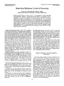
haemodialysis treatment 3 days every week for the previous 17 years. She presented to the emergency eye clinic with a sudden onset bilateral reduction of vision occurring soon after waking in the morning. One day previously she had noticed visual obscurations in her left eye also after waking in the morning, which she described as temporary episodes of blurred vision lasting ∼2 min. She had attended a session for haemodialysis the day prior to when these visual symptoms started. She denied any headache. On examination her Snellen visual acuities were found to be 6/5 in the right eye and 6/9.5 in her left eye. Fundoscopy revealed bilateral swollen discs, more so in the left eye than the right (Figure 1). Visual field testing showed an inferior altitudinal defect in both eyes (Figure 2). An MRI scan of the brain was performed to rule out any space-occupying lesion or visual pathway pathology and was found to be normal. She had also recently been diagnosed with episodes of hypotension associated with dizziness in the mornings. Two weeks prior, she was found to be anaemic with a haemoglobin of 7.5 mmol/L and subsequently received a blood transfusion of 1 unit. The patient was managed conservatively and monitored for the first few weeks and her anaemia improved to 9.5 mmol/L. One month following her initial presentation she was admitted to hospital with a post-operative foot infection and was treated with intravenous clindamycin. Following this, her vision appeared to progressively decline and was measured at hand movements in her right eye and counting fingers in the left. On this occasion, the right optic disc was found to be swollen, consistent with NAAION. Her usual blood pressure had been measured at 110–120/80 and on this occasion had been found to be lower than her norm at 90/60 mmHg. She was placed on erythropoietin and Midodrine therapy to control her anaemia and hypotension. Her subsequent BP had improved and was measured in clinic at 101/61. Approximately 6 weeks later, her
© The Author 2014. Published by Oxford University Press on behalf of ERA-EDTA. All rights reserved. For permissions, please email:
[email protected].
388
S. Bansal et al.
Fig. 1. Fundus photographs right and left eye showing bilateral optic disc swelling.
Fig. 2. Humphrey’s visual fields of the right and left eyes respectively showing bilateral inferior altitudinal loss.
visual acuity improved in both eyes to 6/9 in both eyes; however, her visual field defect remained unchanged.
Case 2 The second case is a 49-year-old male with chronic renal failure for the previous 11 years for which he was receiving thrice weekly haemodialysis. He had a longstanding history of persistent hypotension which fluctuated ∼100/70 mmHg, peripheral vascular disease, polyneuropathy, a previous episode of ischaemic bowel disease and had on one occasion suffered a cardiac arrest during a period of dialysis. The patient’s visual symptoms started 4 days before he was seen in the emergency eye clinic. He describes these symptoms as ‘seeing blank with a few spots of clear vision’ which lasted ∼1 h then progressed to complete visual loss. Two weeks prior to this he had fractured his right femur for which he was managed conservatively. He had also been found to have extremely low blood pressure by his GP in the few days preceding his loss if vision, measuring as low as 70/50 mmHg. In the eye clinic his visual acuities were no perception of light in both eyes. Fundoscopy showed bilateral pallid disc
swelling (Figure 3). He denied any headache, temple tenderness, jaw claudication, weight loss or proximal myalgia. His C-reactive protein was slightly elevated at 30 mg/L; however, this could be explained by his recent fracture and chronic renal failure. Despite the pallid disc swelling, a diagnosis of temporal arteritis was thought to be unlikely given his age and the absence of symptoms to support it. In addition there was no evidence of any intraocular inflammation or co-existent retinal pathology. His optic discs had a ‘crowded’ appearance which would have been consistent with discs ‘at risk’ of developing ischaemic optic neuropathy. This coupled with their swollen appearance and history of hypotension supported the diagnosis of anterior ischaemic optic neuropathy (AION) was made, and he was given pulsed iv 1 g methylprednisolone per day for 3 days followed by oral prednisolone 1 mg/kg per day in an attempt to hasten any opportunity for recovery of the disc swelling. Attempts were made to control his blood pressure (which improved to his previous ‘norm’ of ∼100/70 mmHg). His vision improved to 6/9.5 in the left eye; however perception of light remained only in the right eye. The patient was left with an inferior altitudinal and superonasal visual field defect on confrontational analysis in the left eye.
Non-arteritic ischaemic optic neuropathy
389
Fig. 3. Fundus photographs of right and left eyes showing bilateral optic disc swelling as well as pallor (more marked in the right eye than the left).
Table 1. A chronological summary of cases reported of NAAION as a result of dialysis-induced hypotension First author
Journal
Year
N
Unilateral/bilateral
Other features
1986
1
Bilateral
First report
Michaelson Haider Connolly
Am Journal of kidney diseases Br J Ophthalmol Eye Am J Ophthalmol
1989 1993 1994
1 1 1
Bilateral Unilateral Unilateral
Patient also had optic disc drusen
Sabeel Jackson
Nephrol Dial Transp Am J Ophthalmol
1998 1999
1 1
Bilateral Bilateral
Basri
Saudi Journal Kid Disease
2002
1
Bilateral
Cuxart Tay Nieto
Nefrologia Neuroophthalmology Indian J Ophthalmol
2005 2009 2010
1 3 2
Prat Bartlett Buthaina
Surv Ophthal BMJ Case Reports Oman J Ophthal
2011 2011 2013
1 1 3
Bilateral 2/3 bilateral Bilateral (one within weeks and one simultaneous) Unilateral Bilateral within weeks Bilateral simultaneous
Servilla
Discussion AION is a commonly occurring acute optic neuropathy in the elderly population, caused by infarction of the short posterior ciliary artery which supplies the optic nerve head with resultant ischaemia, oedema and a compartment syndrome leading to further vascular compromise. It may be subdivided broadly into two types: arteritic or non-arteritic according to the underlying pathology. Whilst arteritic AION is associated with giant cell arteritis, the mechanisms underlying the non-arteritic form are in many ways less well understood. Vasculopathic risk factors have been identified including raised blood pressure, cholesterol, poorly controlled diabetes and smoking [2]. An anatomically ‘crowded’ or small optic disc is believed to be ‘at risk’ or prone to such ischaemic events. Hayreh investigated the correlation between nocturnal dips in arterial blood pressure and predisposition to NAAION, postulating that hypotension may be the ‘final insult in a multifactorial situation’ [2]. This is further supported by the observation that patients with visual loss due to NAAION frequently present with symptoms on waking. The combination of such factors is thought to lead to reduced blood flow in the short posterior ciliary arteries and thus reduced blood supply to the optic nerve head and consequent ischaemia.
Reversal of hypotension resulted in partial visual recovery Occurred during dialysis Ambulatory blood pressure recorded as low as 91/ 41 mmHg No improvement with pulsed intravenous methylprednisolone Two given steroids and one improved vision No improvement in vision with oral steroids
AION occurring as a result of haemodialysis-associated hypotension was first reported by Groggel in 1986 [4]. Since then, a further 17 other episodes have been reported, mostly in the form of isolated case reports (Table 1) [5–17]. Although there is yet much to be understood about the underlying mechanisms of NAAION and the exact role of blood pressure, the reported cases do share some common features. Firstly, all cases were associated with extremely low blood pressure during the time period of visual loss. In one case, bilateral visual loss occurred during the haemodialysis session [8]. Jackson et al. studied ambulatory blood pressure in a patient undergoing peritoneal dialysis who suffered bilateral NAAION and recorded diastolic readings as low as 41 mmHg [9]. The paper made the point that although blood pressure readings taken in a clinic setting had been normal, 24 h ambulatory readings showed significant dipping predisposing the patient to risk of NAAION. Both of our patients had persistently low blood pressure recordings, which were as low as 70/50 mmHg in Case 2. One of our patients was also found to be significantly anaemic, a finding also commonly seen in renal failure patients and may be an additional risk factor for NAAION [6, 10, 12]. Another common finding in these cases is the bilateral occurrence of the optic nerve insult. Data from the ION decompression trial show that second eye involvement in
390
NAAION was seen in only 14.7% of patients over a 5-year follow-up period [18]. Out of 20 cases reported (including ours) 17 suffered bilateral loss of vision, a highly unusual finding in other forms of NAAION in which the visual loss tends to be unilateral. In most cases of these hypotensioninduced episodes the second eye became involved either simultaneously or within weeks of the first eye. Generally, NAION is not associated with pallid disc swelling but with a hyperaemic disc swelling. The exception to this is the NAION caused by acute hypotension. Pallid swelling, as in Case 2, is a described but not often remembered feature of dialysis-related NAION [19]. In our second patient, we used steroids with a significant improvement. The rationale for the use of steroids is that they result in faster resolution of disc oedema, reduced compression of optic nerve head capillaries and thus improved perfusion and some recovery of surviving axons [20]. The benefits of this are disputed and there is no definite evidence that final visual outcome is improved. There is also no proven role of surgical decompression of the optic nerve— as shown by the Cochrane database systematic review [21]. In patients undergoing haemodialysis, the question remains as to whether alternative therapies to raise blood pressure can improve visual outcomes. Connolly reported visual recovery after aggressive attempts to raise blood pressure in three hypotensives with AION, one of which was receiving dialysis [7]. In our experience, the visual acuity in Case 1 did gradually improve, albeit with some residual visual field defect. They received blood pressure stabilization therapy with an alpha 1 adrenergic pressor agent; however, it is difficult to establish if this had a direct causative effect. Finally, as the search for alternative management strategies turns to the option of neuroprotection, it has been shown that intramuscular injections of memantine (NMDA receptor antagonist) in the rabbit prevented ganglion cell loss in experimentally induced ischaemic optic neuropathy [22]. The role of neuroprotection however in clinical practice with respect to ischaemic optic neuropathy is yet to be defined.
Conclusion Patients undergoing haemodialysis often have co-existent vasculopathic risk factors. Hypotension is a frequent observation and may provide the ‘final insult’ required to diminish flow to the optic nerve head resulting in often bilateral simultaneous visual loss. Although there are as yet no universally acknowledged treatment modalities for this condition, physicians must firstly be aware that severe visual loss may be a complication of hypotension in haemodialysis patients. Recognition of early signs in the form of transient visual symptoms, especially on waking or during haemodialysis sessions is of importance and a prompt referral to an ophthalmologist may be advisable. Control of blood pressure with avoidance of sudden ‘dipping’ is important and 24 h ambulatory blood pressure monitoring may be required to identify patients with this tendency. Anaemia is an additional risk factor in ischaemic optic neuropathy and must be monitored and controlled. Conflict of interest statement. None declared. (See related articles by Bodaghi et al. The eye: a window on kidney diseases. Clin Kidney J 2014; 7: 337–338 and by Wong et al.
S. Bansal et al. Exudative detachment as a masquerader in hypoalbuminaemic patients. Clin Kidney J 2014; 7: 406–410)
References 1. Sulowiczi W, Radziszewski A. Pathogenesis and treatment of dialysis hypotension. Kidney Int 2006; 70: S36–S39 2. Newman NJ. The ischaemic optic neuropathy decompression trial. Arch Ophthalmol 2007; 125: 1568–1570 3. Hayreh SS, Zimmerman MB, Podhaisky P. Nocturnal arterial hypotension and its role in optic nerve head and ocular ischaemic disorders. Am J Ophthalmol 1994; 117: 603–624 4. Servilla KS, Groggel GC. Anterior ischaemic optic neuropathy as a complication of hemodialysis. Am J Kidney Dis 1986; 8: 61–63 5. Michaelson C, Behrens M, Odel J. Bilateral anterior ischaemic optic neuropathy associated with optic disc drusen and systemic hypotension. Br J Ophthalmol 1989; 73: 762–764 6. Haider S, Astbury NJ, Hamilton DV. Optic neuropathy in uraemic patients on dialysis. Eye 1993; 7: 148–151 7. Connolly SE, Gordon KB, Horton JC. Salvage of vision after hypotension-induced ischaemic optic neuropathy. Am J Ophthalmol 1994; 117: 235–242 8. Sabeel A, Al-Hazaa S, Alfurayh O et al. The dialysed patient who turned blind during a haemodialysis session. Nephrol Dial Transplant 1998; 13: 2957–2958 9. Jackson T, Farmer C, Kingswood C et al. Hypotensive ischemic optic neuropathy and peritoneal dialysis. Am J Ophthalmol 1999; 128; 109–111 10. Basile C, Addabbo G, Montanaro A. Anterior ischaemic optic neuropathy and dialysis: role of hypotension and anaemia. J Nephrol 2001; 14: 420–423 11. Basri BA, Shaheen FA. Visual loss in uraemic patients on dialysis: a case report and review of literature. Saudi J Kidney Dis Transplant 2002; 13: 45–49 12. Cuxart M, Matas M, Picazo M et al. Acute bilateral visual loss in a haemodialysed patient. Nefrologia 2005; 25: 703–705 13. Tay E, Andreou P, Graham E et al. Anterior ischaemic optic neuropathy (AION) associated with post dialysis hypotension. Neuroophthalmologt 2009; 33: 168–173 14. Nieto J, Zapata MA. Bilateral anterior ischaemic optic neuropathy in patients on dialysis: a report of 2 cases. Indian J Nephrol 2010; 20: 48–50 15. Prat NM, Sanchez-Dalmau BF, Faroozan R. Not just for men. Surv Ophthalmol 2011; 56: 173–177 16. Bartlett S, Cai A, Cairns H. Non-arteritic ischaemic optic neuropathy after first return to haemodialysis. BMJ Case Rep 2011. bcr0420114072. doi: 10.1136/bcr.04.2011.4072 17. Buthaina S. Anterior ischaemic optic neuropathy and dialysis: effect of hypotension. Oman J Ophthalmol 2013; 6: 64–65 18. Newman NJ, Scherer R, Langenberg P et al. The fellow eye in NAAION: report from the ischaemic optic neuropathy decompression trial follow up study. Am J Ophthalmol 2002; 134: 317–328 19. Liu G, Volpe N, Galetta S. Neuro-Ophthalmology: Diagnosis and Management, 2nd edn. Saunders Elsevier, 2010, pp. 163 20. Hayreh SS, Zimmerman BM. Non arteritic anterior ischaemic optic neuropathy: role of systemic corticosteroid therapy. Graefes Arch Clin Exp Ophthalmol 2008; 246: 1029–1046 21. Dickersin K, Manheimer E, Li T. Surgery for nonarteritic anterior ischaemic optic neuropathy. Cochrane Database Syst Rev 2012; 1: CD001538 22. Kim TW, Kim DM, Park KH et al. Neuroprotection effects of memantine in rabbit model of optic nerve ischaemia. Korean J Ophthalmol 2003; 16: 1–7 Received for publication: 6.12.13; Accepted in revised form: 25.3.14