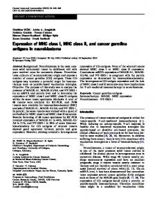
The Journal of Immunology
Two Structurally Related Rat Ly49 Receptors with Opposing Functions (Ly49 Stimulatory Receptor 5 and Ly49 Inhibitory Receptor 5) Recognize Nonclassical MHC Class Ib-Encoded Target Ligands1 Christian Naper,*† Ke-Zheng Dai,* Lise Kveberg,‡ Bent Rolstad,‡ Ere´ne C. Niemi,† John T. Vaage,2* and James C. Ryan† The Ly49 family of lectin-like receptors in rodents includes both stimulatory and inhibitory members. Although NK alloreactivity in mice is regulated primarily by inhibitory Ly49 receptors, in rats activating Ly49 receptors are equally important. Previous studies have suggested that activating rat Ly49 receptors are triggered by polymorphic ligands encoded within the nonclassical class Ib region of the rat MHC, RT1-CE/N/M, while inhibitory Ly49 receptors bind to widely expressed classical class Ia molecules encoded from the RT1-A region. To further investigate rat Ly49-mediated regulation of NK alloreactivity, we report in this study the identification and characterization of two novel paired Ly49 receptors that we have termed Ly49 inhibitory receptor 5 (Ly49i5) and Ly49 stimulatory receptor 5 (Ly49s5). Using a new mAb (mAb Fly5), we showed that Ly49i5 is an inhibitory receptor that recognizes ligands encoded within the class Ib region of the u and l haplotypes, while the structurally related Ly49s5 is an activating receptor that recognizes class Ib ligands of the u haplotype. Ly49s5 is functionally expressed in the high NK-alloresponder PVG strain, but not in the low alloresponder BN strain, in which it is a pseudogene. Ly49s5 is hence not responsible for the striking anti-u NK alloresponse previously described in BN rats (haplotype n), which results from repeated alloimmunizations with u haplotype cells. The present studies support the notion of a complex regulation of rat NK alloreactivity by activating and inhibitory Ly49 members, which may be highly homologous in the extracellular region and bind similar class Ib-encoded target ligands. The Journal of Immunology, 2005, 174: 2702–2711.
N
atural killer lymphocytes are capable of killing neoplastic, virally infected, or allogeneic target cells without prior sensitization. Although early studies suggested that NK cells were not MHC restricted, more recent studies have shown that NK cells possess a myriad of MHC-binding receptors that control NK cell effector functions. In the presence of self MHC class I ligands, target cells are protected from NK cytolysis, consistent with the missing self hypothesis; i.e., that target cells are killed if they fail to express a full array of self class I ligands (1). Inhibitory Ly49 receptors were the first class I-binding receptors to be characterized in mouse NK cells (2). In rats, functional genetic studies predicted the existence of activating NK receptors directed against nonclassical class Ib MHC alleles, in addition to inhibitory receptors for classical class Ia ligands (3, 4). Mapping of the NK alloresponder locus nka to the Ly49 part of the rat NK gene com-
*Institute of Immunology, Rikshospitalet University Hospital, University of Oslo, Rikshospitalet, Oslo, Norway; †Veterans Affairs Medical Center, Northern California Institute for Research and Education, and University of California, San Francisco, CA 94121; and ‡Department of Anatomy, Institute of Basic Medical Sciences, University of Oslo, Oslo, Norway Received for publication October 29, 2004. Accepted for publication December 14, 2004. The costs of publication of this article were defrayed in part by the payment of page charges. This article must therefore be hereby marked advertisement in accordance with 18 U.S.C. Section 1734 solely to indicate this fact. 1 This work was supported by grants from the Norwegian Cancer Society and the Research Council of Norway and Mjåland’s Fund for Cancer Research. K.-Z.D. is a fellow of the Norwegian Cancer Society. 2
Address correspondence and reprint requests to Dr. John T. Vaage, Institute of Immunology, Rikshospitalet University Hospital, NO-0027 Oslo, Norway. E-mail address:
[email protected] Copyright © 2005 by The American Association of Immunologists, Inc.
plex (NKC),3 suggested that Ly49 alloreceptors were involved (5). Rat NK cells can mount strong alloresponses in vivo that bear superficial resemblance to adaptive immune reactions. Repeated alloimmunizations of BN strain rats with fully MHC-mismatched u haplotype cells result in a striking i.p. accumulation of alloreactive NK cells (6 – 8). These NK cells display a much more potent anti-u cytolytic activity than those recruited to the peritoneal cavity by the cytokine IL-2 alone (9). Subsequent studies in mice have identified Ly49 receptors that activate, rather than inhibit, NK cell functions (10). These include Ly49D and Ly49H (11, 12). Although Ly49D can recognize one class Ia allele (H-2Dd) of mouse MHC (13, 14), Ly49H fails to recognize any known mouse MHC molecule, but rather recognizes a class I-like homologue encoded within the genome of mouse CMV (mCMV) (15, 16). Although the Ly49H expression by mouse NK cells is clearly associated with enhanced survival after mCMV challenge, the physiologic roles of activating NK receptors for MHC class I have not been firmly established. Indeed, several strains of recombinant inbred mice have no activating Ly49 receptors, yet appear to be healthy (17). It has been speculated that, in mice, the activating Ly49 receptors predominantly screen for specific pathogens and that their recognition of allogeneic MHC is a chance evolutionary vestige (18). Indeed, in vivo studies in mice have shown that overriding effects of inhibitory receptors largely
3 Abbreviations used in this paper: NKC, NK gene complex; CHO, Chinese hamster ovary; cRPMI, complete RPMI; HCV, hepatitis C virus; KIR, killer cell Ig-like receptor; Ly49i, Ly49 inhibitory receptor; Ly49s, Ly49 stimulatory receptor; mCMV, murine CMV; ORF, open reading frame.
0022-1767/05/$02.00
The Journal of Immunology
2703
overshadow NK-activating effects mediated by MHC-binding receptors (19). In contrast, studies in rats have provided ample evidence for stimulatory NK allorecognition (3, 20 –22). These studies have suggested that rat NK cells express a potent array of activating receptors for several distinct target cell allodeterminants encoded within the class Ib region of the rat MHC, termed RT1CE/N/M (3). The RT1-CE/N/M region is located telomeric to the class II/III regions, in the same position as H2-D,L,Q/T/M in mouse, and it has recently been fully mapped and sequenced in the BN rat strain (23, 24). The whole region spans roughly 2 Mb and harbors several clusters of class I genes that are separated by framework genes conserved between mouse, rat, and human. The first or the most centromeric class Ib cluster, termed the RT1-CE cluster, harbors 16 class I genes in the BN strain, of which 13 may be functional (24). Analysis of an RT1-CE-deleted mutant rat strain (LEW.1LM1), which has lost ⬃100 kb of chromosomal DNA from this cluster, indicated that it encodes target ligands triggering alloreactive rat NK cells (3, 25). This contention has received support from studies showing that target cells transfected with selected RT1-CE genes, i.e., RT1-Cl (LW2) and RT1-Eu, are rendered more sensitive to allospecific NK lysis (7, 26). Previously, we have identified a variety of MHC-binding Ly49 receptors, including Ly49 inhibitory receptor 2 (Ly49i2), which inhibits NK killing of targets expressing the class Ia MHC ligand; RT1-A1c (27, 28); as well as the activating Ly49 stimulatory receptor 3 (Ly49s3) (29). Unlike the prototypical activating mouse Ly49D receptor from mice, which is specific for a solitary class Ia allele, the rat Ly49s3 receptor recognizes a broad array of nonclassical MHC-encoded determinants from rat strains of the c, av1, lv1, l, and n, but not of the u or b haplotypes (29) (our unpublished data). These data support the previous genetic data predicting that activating rat Ly49 receptors bind public class Ib epitopes that may be shared between selected RT1 haplotypes (4). It is likely that the Ly49s3 ligand(s) is encoded from the first class Ib cluster, i.e., RT1-CE. To more fully analyze the phenomenon of NK allorecognition in rats, we have used a series of gene discovery techniques, including homology screening and complementation cloning, to identify Ly49i5 and Ly49s5, novel paired Ly49 receptors with opposing NK cell signaling functions and overlapping MHC class Ib ligand repertoires in rats.
Materials and Methods Animals Four to 6-wk-old female BALB/c mice and BALB/c nu/nu mice were from Simonsen Laboratories and were reared at the Veterans Affairs Medical Center under conventional conditions in accordance with institutional guidelines. The rat strains used and their MHC haplotypes are listed in Table I. The following strains were reared at the Institute of Basic Medical Sciences: PVG, PVG.R23, PVG.1N, PVG.1L, and PVG.1LV1. AO, DA, PVG.1AV1, PVG.1U, and PVG.R8 rats were obtained from Harlan; WAG rats from Harlan Netherlands; and F344, LEW, and BN rats from Møllegaard. Rats were regularly screened for common pathogens and housed in compliance with guidelines set by the Experimental Animal Board under the Ministry of Agriculture of Norway. They were sacrificed at 8 –16 wk of age.
Cloning of Ly49i5 cDNA Ly49i5 was cloned using the ZAP-Express system (Stratagene) and a previously described cDNA library from KLRH1⫹ NK cells (30), except that the primary cDNA was ligated into the ZAP-Express vector instead of into pMET7. Complexity of the primary phage library comprised ⬎2 ⫻ 106 clones. Ten plates, each containing 50,000 clones, were screened with a mixture of 32P-labeled cDNAs, rat Ly49i2 (28), rat Ly49s2 (formerly Ly49.12), and rat Ly49s1 (formerly Ly49.29) (5), and mouse Ly49H (12). Hybridization of nitrocellulose filters was performed overnight at 42°C in 6⫻ SSC, 5⫻ Denhart solution, 0.5% SDS, 200 g/ml sonicated salmon sperm DNA, and 50% formamide. The filters were washed twice with 2⫻ SSC, 0.1% SDS and twice with 0.5 ⫻ SSC, 0.1% SDS, and films were developed after 3 days of exposure at ⫺80°C. Cross-hybridizing plaques were subjected to additional rounds of screening, and the resultant clonal phages were converted into pBK-CMV phagemids by in vivo excision, according to the manufacturer’s instructions (Stratagene), and sequenced on both strands. One Ly49i5 clone was obtained that was fully sequenced on both strands.
Transfection of cells Cell lines were routinely grown in complete RPMI (cRPMI; RPMI 1640 supplemented with 10% FCS, 5 ⫻ 10⫺5 M 2-ME, L-glutamine, and penicillin/streptomycin). Chinese hamster ovary (CHO) cells were from the American Type Culture Collection, and were transfected by electroporation in cRPMI in 2-mm cuvettes at 7.5 ⫻ 106 cells/ml with 20 g of plasmid using a BTX Electroporator at 120 mV, 850 F, and 129 ⍀. After plating in 96-well plates at 103 cells/well in selection medium (cRPMI supplemented with 1 mg/ml G418), clones surviving selection were examined for receptor expression by flow cytometry. The 293T and DT381 cells (293T cells stably transfected with a DAP12 fusion protein tagged with an extracellular FLAG epitope (FLAG/DAP12)) were a gift from L. Lanier (University of California, San Francisco, CA) and were transiently transfected using Fugene transfection reagent according to the manufacturer’s instructions (Roche).
Table I. MHC constitution of the rat strains used RT1 Regions Strain
RT1 (Rat MHC) Haplotype
A
B/D
CE/N/M
PVG PVG.1LV1 PVG.1L PVG.1AV1 PVG.1U PVG.R8 PVG.R23 PVG.1N BN DA F344 LEW AO WAG
c lv1 l av1 u r8 r23 n n av1 lvl l u u
c l l a u a u n n a l l u u
c l l a u u a n n a l l u u
c lv1 l av1 u u av1 n n av1 lv1 l u u
a
Recombination point.
2a 2a
Abbreviation
(c-c-c) (l-l-lv1) (l-l-l) (a-a-av1) (u-u-u) (a-u-u) (u-a-av1) (n-n-n) (n-n-n) (a-a-av1) (l-l-lv1) (l-l-l) (u-u-u) (u-u-u)
Non-MHC Background
PVG PVG PVG PVG PVG PVG PVG PVG BN DA F344 LEW AO WAG
2704
MHC CLASS Ib RECOGNITION BY TWO PAIRED RAT Ly49 RECEPTORS
Production of the mAb Fly5 A cDNA construct was engineered in the EMCV-SR␣ vector to express Ly49i5 as a fusion protein tagged with an extracellular FLAG epitope (Ly49i5/FLAG). After stable transfection and selection in cRPMI with G418, CHO cells that stained with the anti-FLAG mAb M2 were sorted by flow cytometry and expanded. A total of 107 transfected cells was injected i.p. into 6-wk-old female BALB/c mice. After three biweekly immunizations, mice were boosted i.p. 72 h before fusion of splenocytes with SP2/0 myeloma cells using Hybrimax PEG/DMSO solution (Sigma-Aldrich) and DMEM according to standard procedures, as previously described (29). Following metabolic selection in DMEM supplemented with 10% FCS, 1⫻ hypoxanthine/aminopterine/thymidine, and antibiotics, hybridoma supernatants were screened by flow cytometry for the presence of anti-Ly49i5 reactivity against 293T cells transiently transfected with Ly49i5/FLAG and not with other FLAG-tagged constructs, and one hybridoma (Fly5) was selected. The mAb Fly5 was produced in bulk as malignant ascites in pristane-primed BALB/c nu/nu mice, purified by solvent-accessible surface precipitation or protein G affinity purification, and dialyzed against PBS. The mAb Fly5 was determined to be of isotype IgG1 by IsoStrip Ab typing (Roche).
Primary cells, flow cytometry, and cytotoxicity assay Peritoneal cells from alloimmunized BN rats were retrieved as described (7). A total of 107 mononuclear splenocytes from PVG.1U rats, obtained by Lymphoprep centrifugation, was injected i.p. once weekly for 4 wk. The peritoneal cells were retrieved 3– 4 days after the last immunization by injection of 30 ml of PBS i.p. and aspiration after gentle massage of the abdomen, followed by separation by Lymphoprep centrifugation. Fly5-enriched NK cell cultures were obtained by a modification of a previously described method (31), by positive selection of mononuclear splenocytes with streptavidin-coated M280 magnetic Dynabeads (Dynal Biotech) preincubated with mAb Fly5-biotin. Selected Fly5⫹ cells were cultured in cRPMI supplemented with 1 mM Na pyruvate and rat or human rIL-2. The cells were used after 1–2 wk in culture, yielding a purity of Fly5⫹ cells that was routinely above 95% (data not shown). The generation of Con A-activated lymphoblast target cells and 4-h 51Cr release assays was performed, as previously described (3). In mAb-blocking experiments, 3–5 g of purified mAb Fly5 or the isotype-matched negative control mAb TIB96 (mouse IgG1, directed against mouse IgD b allotype; American Type Culture Collection) was added to effectors 20 min before the addition of target cells. Spontaneous release was usually between 5 and 15% of the total cpm in the cells, and the SE for each data point was ⬍5%. The results are presented as median values from triplicates for each E:T cell ratio. A total of 50 l of cells (2–10 ⫻ 106 cells/ml) was incubated with unconjugated or conjugated primary mAb for 30 min on ice, washed, and, if appropriate, incubated with F(ab⬘)2 of FITC-conjugated secondary reagent or streptavidin-RPE/-RPE-Cy5. For multicolor flow cytometry, mononuclear splenocytes and peritoneal cells were depleted of Ig⫹ cells with sheep anti-rat Ig-coated M450 Dynabeads or nylon wool, before labeling with a combination of directly conjugated mAbs: FITC-conjugated anti-NKR-P1A/B (mAb 3.2.3), anti-Ly49s3 (mAb DAR13 (29)), antiLy49i2 (mAb STOK2 (31)), or anti-KLRH1 (mAb STOK9 (30)); PE-conjugated anti-CD3 (mAb G4.18) or anti-NKR-P1A/B (mab 10/78; both from BD Pharmingen); and biotinylated mAb Fly5 (mouse IgG1, biotinylated according to standard procedures). Cells were analyzed on a FACScan or a FACSCalibur (BD Biosciences) or sorted using a FACStar (BD Biosciences).
Cloning of Ly49s5 cDNA Ly49s5 cDNA was cloned by a variation of the complementation cloning approach previously described for Ly49s3 (29). In short, a monolayer of DT381 cells was transiently transfected with the cDNA expression library referred to above in the pMET7 vector (30). After 2 days, cells were stained with mAb Fly5, washed, and panned on petri dishes precoated with rabbit anti-mouse IgG (Valeant Pharmaceuticals/Cappel). Adherent cells were lysed in Hirt solution (0.6% SDS, 10 mM EDTA), and NaCl was added to a final concentration of 1 M. The reaction mixture was precipitated overnight at 4°C and centrifuged at 15,000 ⫻ g for 30 min. The primary cDNA sublibrary, obtained from the Hirt supernatant, was amplified in ElectroMAX DH10B Escherichia coli (Invitrogen Life Technologies) and purified on Qiagen Tip-500 columns. In two consecutive rounds of immunoenrichment, FLAG⫹ Fly5⫹ double-positive transfectants were obtained by FACS sorting after staining with FITC-conjugated anti-FLAG M2 mAb and biotinylated Fly5/PE-conjugated streptavidin. DT381 cells transiently transfected with the tertiary sublibrary were 10 –20% FLAG⫹ Fly5⫹ double positive. Four Ly49s5 clones, which induced Fly5 and FLAG
staining on DT381 cells, were obtained by screening of 480 individual bacterial colonies by dot blotting with a Ly49i5 cDNA probe. A total of 50 l of bacterial growth from each clone was blotted onto nitrocellulose filters, lysed, and DNA denatured using 0.4 M NaOH for 10 min. The filters were neutralized for 10 min in 6⫻ SSC, 0.5 M Tris-HCl (pH 7), washed for 10 min with 6⫻ SSC, baked at 80°C for 2 h, and probed with a 32P-labeled full-length Ly49i5 probe at 42°C for 1 h in 6⫻ SSC, 5⫻ Denhart solution, 0.5% SDS, 200 g/ml sonicated salmon sperm DNA, and 50% formamide. Filters were washed with 0.1% SSC, 1% SDS, and exposed to films.
RT-PCR Expression levels of Ly49i5 and Ly49s5 were determined by RT-PCR in FACS-sorted NK subsets, as indicated. A total of 106 cells was lysed in 250 l of TRIREAGENT (Sigma-Aldrich) and supplemented with 5 g of carrier tRNA. RT-PCR were performed from total RNA, according to the manufacturer’s recommendations (Sigma-Aldrich). The following upper and lower primers, respectively, were used: Ly49i5 (PVG and BN), 5⬘TTCCCAGCAATACTCTTCTGA-3⬘ and 5⬘-GACCTCATCTCTCTAT TCATG-3⬘; Ly49s5 (PVG), 5⬘-CCGGGAAAGGTCAACTCT-3⬘ and 5⬘GAAGCGCATTGCTGGAAG-3⬘. Primers for rat CD45 were included as a control. Typically, PCR were performed on a GeneAmp PCR thermocycler (Applied Biosystems) using hot start for 3 min at 96°C. Pyrococcus woesi DNA polymerase was added at 80°C before running for 35 cycles at 95°C, 20 s; 54°C, 30 s; and 72°C, 30 s. PCR products were resolved by agarose gel electrophoresis (2% Tris acetate EDTA buffer). The coding region of the Ly49i5 and Ly49s5 alleles in BN strain rats was isolated by RT-PCR with the following upper and lower primers: Ly49i5, 5⬘-TCG CAT CGG GAT AGA GAC-3⬘ and 5⬘-CCT GTG GAC CTC ATC TCT CT-3⬘; Ly49s5, 5⬘-AGA CAC AGA AAA CAC TCA AT-3⬘ and 5⬘-ATC GAG TTC TCC ATG TGG TC-3⬘. PCR conditions were as above, except that the extension time was 90 s at 72°C. The PCR products were cloned into a plasmid vector (pCR2.1-TOPO, TOPO-TA cloning system; Invitrogen Life Technologies) and transfected into TOP10 E. coli by electroporation. Single bacterial colonies were selected by blue/ white screening, followed by PCR with vector-specific primers. Five positive clones of the BN alleles of Ly49i5 and Ly49s5 (a pseudogene) were fully sequenced on both strands.
Results Molecular cloning of Ly49i5 cDNA by sequence homology screening Previous studies have suggested that NK cells from the high NK alloresponder PVG rat strain express a broad panel of Ly49 receptors (5, 28, 29). To expand our knowledge of rat Ly49 receptors, we performed homology screening using a cDNA library generated from a PVG NK cell subset (KLRH1⫹) with heightened allogeneic reactivity (30). Hybridization was performed at low stringency using a mixture of broadly divergent Ly49 genes from the rat and mouse. These included our previously published rat genes Ly49i2, Ly49s1, and Ly49s2, and the mouse NK receptor for mCMV-infected cells, Ly49H. Among other receptors, we identified a novel inhibitory Ly49 molecule that we have termed Ly49i5. This receptor is ⬃80% identical with Ly49i2 at the amino acid level (90% at the cDNA level in the open reading frame (ORF)) (Fig. 1A and data not shown). Identity is somewhat lower for Ly49s3, at the amino acid level being 70% (but is more homologous at the cDNA level; 86% in the ORF). In the extracellular region corresponding to the stalk (exon 4) and the lectin-like domain (exons 5–7), Ly49i5 and Ly49i2 or Ly49s3 are 76 and 75% identical, respectively. Hence, Ly49i5 and Ly49s3 differ mostly in the intracellular and transmembrane parts (only 56% amino acid identity for exons 2 and 3), reflecting their different signaling properties. Similar to Ly49i2, Ly49i5 has an ITIM motif (ITIM; VTYTTV, motif-specific amino acids underlined) intracellularly, which predicts inhibitory function (32). Similarity between these two receptors is striking both in the intracellular (exon 2, 100% amino acid identity) and transmembrane (exon 3, 93% identity) regions. Ly49i5 lacks a charged transmembrane amino acid (R) characteristic of activating Ly49 receptors such as Ly49s3 (Fig.
The Journal of Immunology
2705 mAb Fly5 staining of NK and NKT cells: marked coexpression with the Ly49i2 and Ly49s3 receptors in distinct subsets of NK cells
FIGURE 1. The Ly49i5 and Ly49s5 receptors are highly related in the ligand-binding extracellular region, but diverge in the transmembrane and intracellular parts. A, Although Ly49i5 contains an intracellular ITIM (VTYTTV), Ly49s5 possesses the DAP12-associating transmembrane residue, arginine. B, A dendrogram shows that Ly49i5 and Ly49s5 have a high degree of homology to each other, as well as to the Ly49i2 and Ly49s3 receptors previously cloned from highly alloreactive PVG NK cells (P stands for PVG). Weaker homology was observed to the F344 strain Ly49 receptors Ly49i1, Ly49s1, and Ly49s2 (F stands for F344). The BN Ly49i5 allele (B stands for BN) was identical with the PVG allele in the ORF. C, The mAb Fly5 (mouse IgG1) specifically reacts with DT381 cells transiently transfected with the Ly49i5 and Ly49s5 cDNA, but not with mocktransfected control (CTR.) cells. Accession numbers of novel cDNA sequences: Ly49i5 (PVG), AY649836 (protein id ⫽ AAV74510); Ly49s5 (PVG), AY649832 (protein identification ⫽ AAV74506); Ly49i5 (BN), AY846264.
As shown in Fig. 2A, mAb Fly5 stained a small, but distinct population (2–3%) of mononuclear splenocytes from PVG strain rats, while reactivity with mesenteric lymph node cells and thymocytes was close to background, suggesting that it does not react with the vast majority of T and B lymphocytes. To further evaluate its reactivity pattern, we analyzed freshly isolated splenocytes by three-color flow cytometry. As shown in Fig. 2B, Fly5 reacted with subsets of CD3⫺NKR-P1A/Bbright NK cells and CD3⫹NKR-P1A/ Bdim T cells (hereafter referred to as NKT cells), but not with the major population of CD3⫹NKR-P1A/B⫺ T cells in PVG.1L and LEW strain rats. This reaction pattern mirrored previous results for Ly49i2 (31), as well as for the distantly related KLRH1 inhibitory receptor (30). As can be deduced from Table II, mAb Fly5 reacted with a minor subset of NK cells in all rat strains tested, but there were strain-specific differences in the number of Fly5⫹ cells. The percentage of mAb Fly5⫹ NK cells in PVG, F344, and LEW was higher than that seen in the other strains. Among CD3⫹ NKRP1A/Bdim NKT cells, the Fly5⫹ subset was larger in DA, F344, LEW, and BN strain rats. One interesting observation was that while the Fly5-staining pattern was homogeneous among NK cells from most strains tested, including in the LEW strain, it was markedly heterogeneous in NK cells from MHC congenic strains on the PVG background, as shown in Fig. 2B for PVG.1L. The PVG.1L
1A). Nevertheless, Ly49i5 is much more homologous to both Ly49i2 and Ly49s3 than with the previously published receptors Ly49i1, Ly49s1, and Ly49s2, which have all been isolated from the low NK alloresponder F344 rat strain (Fig. 1B).
Generation of mAb Fly5 by immunization with a Ly49i5/FLAG chimeric protein To functionally characterize the Ly49i5 receptor, we generated a mAb against Ly49i5. CHO cells were stably transfected with a FLAG-tagged Ly49i5 expression construct (Ly49i5/FLAG). These transfectants expressed the Ly49i5/FLAG fusion protein on their surface, as determined by flow cytometry using the anti-FLAG mAb M2 (data not shown). Following immunization of BALB/c mice and fusion with SP2/0 myeloma cells, culture supernatants were screened for specific staining of 293T cells transiently transfected with the Ly49i5/FLAG construct, and the hybridoma Fly5 (mouse IgG1) was isolated. As shown in Fig. 1C, mAb Fly5stained DT381 cells (293T cells stably transfected with FLAG/ DAP12) transiently transfected with the Ly49i5 cDNA, and not untransfected DT381 control cells. mAb Fly5 also reacted with primary cells, staining a significant proportion of IL-2-activated PVG strain NK cells (data not shown).
FIGURE 2. mAb Fly5 stains subsets of rat NK cells and NKT cells. A, mAb Fly5 stains a small, but distinct subset of freshly isolated mononuclear cells from the spleen, but not from mesenteric lymph nodes or the thymus of PVG rats. B, Fly5 staining is restricted to NKR-P1A/Bbright NK cells and NKR-P1A/Bdim NKT cells, but not to NKR-P1A/B⫺ conventional T cells. Note the heterogeneous Fly5-staining pattern of NK cells from PVG.1L, but not from the MHC identical LEW rat strain. C, A major proportion of Ly49i2⫹, Ly49s3⫹, and KLRH1⫹ PVG NK cells costains with mAb Fly5.
2706
MHC CLASS Ib RECOGNITION BY TWO PAIRED RAT Ly49 RECEPTORS
Table II. The mAb Fly5 stains subsets of NK and NKT cells from all rat strains testeda % Fly5⫹ Cells
Strain
RT1 Haplotype
NK Cells (CD3⫺ NKR-P1A/Bbright)
NKT Cells (CD3⫹ NKR-P1A/Bdim)
PVG DA F344 LEW BN AO WAG
c av1 lv1 l n u u
16 6.4 11 13 6.4 4.5b 4.2b
13 22 31 33 37 11 10
a Mononuclear spleen cells, depleted of Ig⫹ cells, were analyzed by three-color flow cytometry for the percentage of Fly5⫹ cells. Values represent medians of two to seven observations. b In AO and WAG rats was a population of Fly5dim cells that could not be clearly discriminated from the negative cells, and these cells were not included in the calculation.
‘ and LEW strains express the same rat MHC haplotype (l), so the phenotypical differences between these two strains could not be ascribed to an MHC selection effect during NK maturation. Rather, these data point to strain-specific differences in NKC gene content. For NKT cells, mAb Fly5 staining was homogeneous for all strains tested, including PVG.1L (Fig. 2B and data not shown). Patterns of costaining of mAb Fly5 with other NKC-encoded molecules were determined by three-color flow cytometry. Among ex vivo isolated splenic NK cells, a significant number of Fly5⫹ cells coexpressed Ly49i2, Ly49s3, and the distantly related marker KLRH1 (Fig. 2C). Ly49i2, Ly49s3, and KLRH1 were all markedly overrepresented in Fly5⫹ cells (⬃3– 4⫻). Hence, while only ⬃7% of the NKR-P1A/Bbright splenocytes were Ly49i2⫹, ⬃28% of the Fly5⫹ cells coexpressed Ly49i2, and there was a comparable overrepresentation of Fly5⫹ cells in the Ly49s3⫹ and KLRH1⫹ subsets. It was not possible to extend this analysis to other pertinent rat strains (BN, F344, LEW, DA) because both the anti-Ly49i2 and anti-KLRH1 mAb fail to react with cells from these strains (30, 31). Similar findings, however, were obtained in the high NK alloresponder AO strain (data not shown), suggesting that expression of Ly49 receptors may not be evenly distributed among NK cells. Rather, it appears to concentrate to certain rat NK subsets. ⫹
Proportion and phenotype of Fly5 NK cells are influenced by rat MHC haplotype In rodents, the surface density of Ly49 receptors is typically downregulated in the presence of their cognate MHC ligands, and rel-
ative cell numbers may also be reduced (33, 34). In the rat, such MHC-modulating effects have been observed both for the activating Ly49s3 receptor, and for the inhibitory Ly49i2 receptor (27, 29, 31). To glean clues as to potential Ly49i5 ligands, we examined the MHC influence on the mAb Fly5 reactivity in a set of congenic and intra-MHC recombinant strains on the PVG background (refer to Table I). These data reveal that the proportion of Fly5⫹ NK and NKT cells is influenced by the MHC haplotype of the host strain. Notably, the Fly5⫹ NK cell subset is decreased from 16% of PVG NK cells to 4 – 4.5% of NK cells in MHC congenic PVG rats that are haplotype u at the RT1-CE/N/M class Ib region (i.e., in PVG.1U and PVG.R8). It should be noted that calculation of Fly5⫹ NK cells in these two strains was based on detection of Fly5 bright cells only, as there was no clear distinction between Fly5 dim and negative cells; see below. By contrast, the number of Fly5⫹ NK cells was largely unaffected in PVG rats congenic for MHC haplotypes av1, r23, l, lv1, or n (Table III). When examined more closely, however, these expression data provided additional information. The MHC constitution of the host strain affected not only the number of Fly5⫹ NK cells, but also the intensity of surface staining. As shown in Fig. 3, each PVG congenic rat strain expressed a Fly5bright and Fly5dim positive population. The expression levels of both the Fly5bright and Fly5dim NK cells were similar to those seen in wild-type PVG (c-c-c) in haplotypes a-a-av1, u-a-av1, l-l-lv1, and n-n-n rats, while they were down-regulated in PVG.1U (u-u-u) and PVG.R8 (a-u-u). These results suggest that the mAb Fly5-reactive receptors on both the Fly5bright and Fly5dim NK cells might selectively recognize class Ib ligands encoded within the RT1-CE/N/M region of haplotype u. Analysis of NK cells from haplotypes l-l-l and l-l-lv1, being disparate in the RT1-CE/N/M region only, was particularly intriguing. The Fly5dim population, but not the Fly5bright population, was selectively down-regulated in PVG.1L (l-l-l), but not in PVG.1LV1 (l-l-lv1), suggesting that a class Ib-encoded structure unique to PVG.1L (RT1-CE/N/Ml) might serve as a ligand for a Fly5-reactive receptor. Together these data not only suggested possible MHC ligands for Fly5-binding receptors, but also that mAb Fly5 might be public for more than one NK receptor in PVG strain rats. Expression cloning of Ly49s5 Initial attempts to block Ly49i5 function on IL-2-activated PVG NK cells using mAb Fly5 failed to reveal the expected functional effect, i.e., increased allospecific natural killing as a result of blockade of Ly49i5. Instead, we observed a reduction of lysis of u haplotype target cells (data not shown). Considering the biphasic staining pattern of PVG NK cells by mAb Fly5, we speculated that
Table III. MHC influence on the number of Fly5⫹ NK cells is determined by the class Ib RT1-CE/N/M regiona % Fly5⫹ Cells
Strain
PVG PVG.1AV1 PVG.R23 PVG.1U PVG.R8 PVG.1L PVG.1LV1 PVG.1N
RT1 Haplotype
c-c-c a-a-av1 u-a-av1 u- u-u a- u-u l-l-l l-l-lvl n-n-n
NK Cells (CD3⫺ NKR-P1A/Bbright)
NKT Cells (CD3⫹NKR-P1A/Bdim)
n
16 (14–17) 15 (15–17) 18 (17–20) 4.5b (4.3–5.4) 4.0b (3.7–4.4) 19 (18–21) 19 (18–20) 13 (11–14)
13 (12–13) 10 (8.2–10) 10 (8.7–11) 10 (9.2–11) 8.5 (8.2–9.2) 11 (10–13) 12 (11–14) 15 (14–18)
7 3 7 5 4 6 8 9
a Mononuclear spleen cells, depleted of Ig⫹ cells, were analyzed by three-color flow cytometry for the percentage of Fly5⫹ cells. Values represent medians and observed ranges. b In PVG.IU and PVG.R8 rats was a population of Fly5dim cells that could not be clearly discriminated from the negative cells, and these cells were not included in the calculation.
The Journal of Immunology
2707 population contained primarily Ly49s5 mRNA species (Fig. 4). Although these results were clear-cut for PVG.1AV1 and PVG.1U rats, the Fly5bright NK cells from PVG.1L had appreciable amounts of both Ly49i5 and Ly49s5, although a trend toward more Ly49i5 transcript was observed. The Fly5dim NK cells mainly expressed the Ly49s5 transcript (Fig. 4). Recognition of class Ib ligands encoded from the u and l rat MHC haplotypes by Ly49i5 and Ly49s5
FIGURE 3. Identification of Fly5bright and Fly5dim NK subpopulations that are differentially regulated by the host MHC. Freshly isolated CD3⫺ NKR-P1A/Bbright splenic NK cells from the PVG.1AV1 (RT1-Aa-B/Da-CE/ N/M av1 or a-a-av1), PVG.R23 (u-a-av1), PVG.R8 (a-u-u), PVG.1U (u-uu), PVG.1L (l-l-l), PVG.1LV1 (l-l-lv1), PVG (c-c-c), and PVG.1N (n-n-n) rats were analyzed by three-color flow cytometry for reactivity with mAb Fly5. Careful analysis of mAb Fly5 staining demonstrates distinct Fly5bright and Fly5dim populations that appear to be differentially regulated by host MHC haplotype. Both populations exhibit less staining in host strains expressing the u haplotype in the RT1-CE/N/M class Ib region (top row, last two panels).
The RT-PCR data in Fig. 4 combined with the data presented in Fig. 3 suggested that Ly49i5 (Fly5bright) and Ly49s5 (Fly5dim) both recognized a structure encoded by the nonclassical RT1-CE/ N/M region of rat haplotype u. In addition, we predicted that an RT1-CE/N/M-encoded structure from haplotype l specifically binds and down-regulates Ly49s5 on the Fly5dim NK cell population in PVG.1L strain rats. To investigate these possibilities, we functionally analyzed purified Fly5⫹ NK effectors from several of the MHC congenic and intra-MHC recombinant PVG strains. Fly5⫹ NK cells from the PVG.R23 strain (haplotype u-a-av1) efficiently lysed lymphoblast targets expressing the u haplotype in the nonclassical RT1-CE/N/M region, i.e., PVG.1U (u-u-u) and PVG.R8 (a-u-u), while they spared targets expressing the av1 haplotype in the same region, i.e., PVG.1AV1 (a-a-av1) and PVG.R23 (u-a-av1; Fig. 5A, first four panels). Killing of PVG.1U (u-u-u) and PVG.R8 (a-u-u) targets was markedly reduced by addition of blocking quantities of mAb Fly5, suggesting that this anti-u NK cytolytic response was triggered by the Ly49s5 receptor (Fig. 5A, first two panels). In contrast, Fly5⫹ NK cells from PVG.R8 rats (a-u-u) failed to lyse RT1-CE/N/M-matched PVG.1U and PVG.R8
mAb Fly5 might recognize an activating receptor in addition to Ly49i5. Among the Ly49 receptors identified by our homology screen and by other methods, only Ly49i5 was recognized by the mAb Fly5 (data not shown). We therefore used a variation of the complementation cloning approach previously used for Ly49s3 (29), which is based on the detection of an N-terminal FLAG epitope on DT381 cells (293T cells stably transfected with a FLAG/DAP12 chimeric molecule). The FLAG/DAP12 fusion protein is only stabilized on the cell surface in the presence of a paired DAP12-coupled activating receptor, and its presence can be inferred by staining with the antiFLAG mAb M2. To select for Ly49i5-related receptors that couple to DAP12, we transfected DT381 cells with the cDNA library referred to above (30), and performed positive selection with both the Fly5 and anti-FLAG M2 mAb. Using this system, we expression cloned Ly49s5, a novel receptor that stabilized FLAG/DAP12 on the surface of DT381 cells (data not shown) and stained with mAb Fly5 (Fig. 1C). The Ly49i5 and Ly49s5 receptors are highly homologous in the extracellular region (90% amino acid identity in the stalk and lectin-like domain; 95% identity in the lectin-like domain). In the cytoplasmic region, Ly49s5 lacks a cytoplasmic ITIM seen in Ly49i5, but it contains the DAP12-associating transmembrane residue, arginine, which is lacking in Ly49i5 (Fig. 1A). Preferential expression of Ly49i5 and Ly49s5 in the respective Fly5bright and Fly5dim NK subsets The demonstration of two paired Ly49i5 and Ly49s5 receptors led to the obvious question as to the identities of the Fly5 Ag in the Fly5bright and the Fly5dim NK populations. To examine the Ly49 receptors in these subsets, we sorted the bright and dim NK populations from MHC congenic PVG rat strains and assessed their Ly49i5 and Ly49s5 mRNA expression using receptor-specific oligonucleotides to prime the PCR. With oligonucleotides described in Materials and Methods, we were able to show that the Fly5bright population contained primarily Ly49i5 mRNA, while the Fly5dim
FIGURE 4. Preferential expression of the Ly49s5 and Ly49i5 receptors in the respective Fly5dim and Fly5bright NK subsets. RT-PCR was performed on sorted CD3⫺Fly5bright and CD3⫺Fly5dim NK cells from the indicated MHC congenic PVG rat strains.
2708
MHC CLASS Ib RECOGNITION BY TWO PAIRED RAT Ly49 RECEPTORS
FIGURE 5. Identification of class Ib ligands for the Ly49s5 and Ly49i5 receptors in the u and l haplotypes. Natural killing of purified Fly5⫹ NK cells from PVG.R23 (A) and PVG.R8 (B) against Con A lymphoblast target cells in the presence (f) and absence (䡺) of blocking quantities (5 g/well) of mAb Fly5. The Con A blasts were from the PVG.1U (RT1-Au-B/Du-CE/N/Mu or u-u-u), PVG.R8 (a-u-u), PVG.R23 (u-a-av1), PVG.1AV1 (a-a-av1), PVG.1L (l-l-l), and PVG.1LV1 (l-l-lv1) strains. These data suggest that mAb Fly5 primarily blocks an activating receptor (Ly49s5) for a uencoded class Ib ligand in PVG.R23 NK cells, and an inhibitory receptor (Ly49i5) directed against class Ib ligands from the u and l haplotypes in PVG.R8 NK cells (for details, see text).
targets, but killing was augmented by the addition of mAb Fly5 (Fig. 5B, first two panels). Minimal effects were seen on the killing of PVG.R23 (u-a-av1) and PVG.1AV1 (a-a-av1) targets (Fig. 5B, middle two panels). These data suggested that Ly49i5 functions as an inhibitory self receptor for a u haplotype-encoded RT1-CE molecule in PVG.R8 NK cells. It could be noted that one reason the inhibitory effects of Ly49i5 were so easily discernible using the IL-2-activated NK effectors from PVG.R8 was that these cells were Fly5bright (data not shown), and hence, preferentially expressed Ly49i5 (see above). Staining of the Fly5dim splenocytes from PVG.R8 (see Fig. 3, third panel) was apparently insufficient for positive selection using our enrichment techniques (for details, refer to Materials and Methods). Data presented in Fig. 3 showed that the Fly5dim, but not the Fly5bright subset undergoes ligand-induced down-regulation of expression in l, but not in lv1 haplotype rats, which differ only in the RT1-CE/N/M class Ib region. These data suggest that the l haplotype contains a ligand for Ly49s5 (on the Fly5dim population), but not for Ly49i5 (Fly5bright population). Unfortunately, the Fly5⫹ effectors from PVG.R23 (Fig. 5A, fifth panel) or other strains available for study (data not shown) were not informative concerning Ly49s5/haplotype l functional interactions, as they failed to kill l targets. It is possible that the l targets were protected against allospecific natural killing by some uncharacterized inhibitory receptor that counteracted Ly49s5-dependent alloactivation. In contrast, NK cells from PVG.R8 effectors, which were preferentially Fly5bright (mainly expressed Ly49i5), failed to lyse l targets, while they efficiently killed the lv1 targets (Fig. 5B, last two panels). This suggested that an l-encoded class Ib ligand protected against natural killing by the Fly5⫹ NK cells. In accordance with this notion, lysis of l targets was induced by the addition of mAb Fly5, while killing of lv1 targets was unaffected (Fig. 5B, last two panels). Taken together, these cytotoxicity data indicate that Ly49i5 recognizes a RT1-CE/N/M-encoded class Ib ligand from haplotypes u and l, while Ly49s5 minimally recognizes an RT1-CE/N/M-encoded ligand from haplotype u.
Ly49s5 is nonfunctional in BN strain rats Our experiments showed that Ly49s5 from PVG rats recognizes a class Ib ligand encoded by the u MHC haplotype, but the exact molecular ligand for Ly49s5 remains unknown. Previous data from Petersson et al. (7) suggested that the class Ib allele, RT1-Eu, codes for an activating ligand for NK cells from BN strain rats. They showed that BN rats (haplotype n) immunized with allogeneic (haplotype u) cells increased the number of NK cells in the peritoneum, and that these NK cells demonstrated enhanced lysis of targets transfected with RT1-Eu. It was presumed that the presence of the RT1-Eu ligand in the peritoneum resulted in a selective recruitment or expansion of the subset of NK cells expressing an activating receptor for RT1-Eu. These data suggested that the RT1-Eu molecule might be a candidate ligand for Ly49s5. As in PVG strain rats, a subset of splenic NK cells from BN rats stained with the mAb Fly5, indicating that these cells express the BN allele of Ly49i5 and/or Ly49s5 (Table II). In agreement with previous studies (6), there were essentially no NK cells in the peritoneal cavity of nonimmunized BN rats, but many appeared as a result of repeated immunizations i.p. with u haplotype cells (splenocytes from PVG.1U rats; Fig. 6A). Only a very small proportion (1.7%) of these peritoneal NK cells, however, stained with mAb Fly5, a percentage that is lower than that in the spleens of the same rats (5.7%; Fig. 6B). These data suggested that i.p. immunization of BN rats with haplotype u cells does not select for mAb Fly5⫹ cells, but selects for other NK cell subsets. Accordingly, sequence analysis of the Ly49s5 allele obtained by RT-PCR from BN NK cells indicated that Ly49s5 is a pseudogene in the BN strain. It contains a stop codon in exon 5 (in the first part of the lectin-like domain), and this stop codon is also present in the BN genomic sequence available at National Center for Biotechnology Information (Fig. 7 and data not shown). The Ly49i5 allele in BN, in contrast, was identical in its ORF with the corresponding PVG allele, as indicated at the translated protein level in the dendrogram in Fig. 1B. There were two nucleotide differences in the 3⬘-untranslated region (data not shown). That the Fly5⫹ NK cell
The Journal of Immunology
FIGURE 6. Fly5⫹ NK cells from BN strain rats express the Ly49i5 receptor for a u-encoded class Ib ligand; failure to be recruited to the peritoneal cavity of BN rats (n haplotype) following repeated u alloimmunizations. A, Peritoneal exudate cells from immunized and nonimmunized control BN rats were analyzed for the presence of NK cells, NKT cells, and T cells. B, Fly5 staining of peritoneal and splenic NK cells from an immunized BN rat. Only 1.7% of the CD3⫺NKR-P1A/Bbright peritoneal NK cells were Fly5⫹, which was less than among splenic NK cells from the same animal (5.2%). C, Detection of Ly49i5 mRNA in Fly5⫹, but not Fly5⫺ BN peritoneal NK cells by RT-PCR.
subset in BN rats indeed expresses Ly49i5 was confirmed by RTPCR (Fig. 6C). These data do not preclude RT1-Eu as a Ly49s5 ligand in the PVG strain, but they exclude any pertinence of Ly49s5 in the BN rat experiments previously described (7). Furthermore, they suggest that an uncharacterized anti-u-triggering BN receptor is involved.
Discussion In the last decade, it has become clear that NK cells are restricted by target cell MHC class I molecules (17). The NK cell receptors
FIGURE 7. The Ly49s5 allele in BN strain rats is a pseudogene. The BN sequence (accession AY846265) is given below its functional counterpart from the PVG strain (upper line; accession AY649832 (protein id ⫽ AAV74506)). The ORF starts in exon 2 and ends in exon 7, the gaps mark the exon borders. Translated BN and PVG amino acid sequences are given below and above their corresponding nucleotide sequences, respectively. Hyphens mark identity. The Ly49s5 gene from BN contains a stop codon in exon 5 (the first lectin domain) indicated by an asterisk in the amino acid sequence, and predicting a nonfunctional truncated protein.
2709 involved in this recognition include the rodent Ly49 receptors, the human KIR, and the CD94/NKG2 receptors in humans and rodents (35). Most of these receptors are specific for classical MHC class Ia molecules. The physiological functions of the nonclassical class Ib molecules often remain unknown. Class Ib molecules typically have a limited tissue distribution and low surface expression and do not in general function as restriction elements for conventional T cells (26). Some class Ib molecules, such as the nonpolymorphic molecules Qa-1 in mice and HLA-E in humans, bind the paired activating and inhibitory CD94/NKG2 lectin-like receptors (36). The MIC-A and MIC-B molecules serve as targets for the activating innate immune receptor NKG2D (37), whereas HLA-G is recognized by the conserved KIR2DL4 receptor in humans. In addition, Qa-2 molecules have been implicated as inhibitory ligands for mouse NK cells (38). Early studies in rats clearly implicated nonclassical class Ib molecules in the triggering of NK cell allogeneic responses. Studies of the LEW.1LM1 mutant rat strain, containing a 100-kb chromosomal deletion within the first cluster of class Ib genes, the RT1-CE cluster (23), suggested that PVG strain NK cells express triggering alloreceptors for ligands encoded within this deletion (3), as well as inhibitory receptors for the same set of class Ib molecules (25). The present study shows that both Ly49s5 and Ly49i5 recognize ligands encoded within the class Ib RT1-CE/N/M region of the u and l haplotypes, and is therefore in good accordance with our previous genetic studies. Although we have not yet been able to fine map the genes for the specific Ly49i5/Ly49s5 ligands within the nonclassical RT1-CE/N/M region, the lm1 data strongly suggest that some NK cell allogeneic ligands reside within the first RT1-CE cluster. It could be noted that the content of class I RT1-CE genes varies remarkably between different haplotypes (39), and it can hence prove difficult to generalize findings obtained in a single rat haplotype. The rat MHC for the BN haplotype has recently been fully sequenced and the genes mapped (24). This study shows that the entire nonclassical region in BN rats is comprised of ⬎45 class I genes, located in clusters that are separated by conserved framework genes. A more detailed analysis shows that the first and most centromeric class Ib cluster, the RT1-CE cluster, encompasses 16 genes encoded between the framework genes Bat1 and Pouf5f1 in the BN rat, with 13 of them predicted to be functional. The orthologous region in the mouse genome contains the H2-D, L, and Q (Qa2) loci, but not Qa1, which is in the second cluster. The corresponding human region encodes the HLA-B, HLA-C,
2710
MHC CLASS Ib RECOGNITION BY TWO PAIRED RAT Ly49 RECEPTORS
MIC-A, and MIC-B molecules (24). The MIC genes are absent in the mouse and rat genomes. We are currently in the process of cloning and characterizing rat RT1-CE molecules from the various rat strains to further characterize them as potential NK cell receptor ligands. The Ly49i5 and Ly49s5 receptors are highly similar in the extracellular region, and functional studies suggest that they recognize ligands encoded from the nonclassical class I region of the u and/or l MHC haplotypes. This is the first pair of activating and inhibitory Ly49 receptors that has been functionally characterized in the rat, although the Ly49s1 and Ly49i1 receptors derived from the F344 rat strain constitute another set of highly homologous receptors at the structural level (5). Analysis of the recently available rat genomic sequence in the BN strain has revealed a surprisingly large rat Ly49 gene cluster predicted to contain ⬎30 genes, considerably more than in mice (40 – 42). Most of the rat Ly49 genes can be subdivided into three major blocks based on sequence homology, and this subdivision corresponds with their respective physical positions within the NKC (42). The Ly49i5 and Ly49s5 genes are located adjacent to each other in block II, while Ly49i1 and Ly49s1 cluster together in block III. Such related pairs of Ly49 receptors may have evolved by unequal crossing over and/or by gene conversion events. Pairs of NK receptors have previously been described both in the human and in the mouse system (43– 45). The human inhibitory KIR2DL1 and activating KIR2DS1 receptors recognize HLA-C alleles, while CD94/NKG2A (inhibitory) and CD94/NKG2C (activating) recognize HLA-E. In the mouse, pairs of Ly49 receptors such as Ly49A/P, Ly49O/D, and Ly49G/W have been identified, and their MHC ligands have been characterized (46, 47). In two previous analyses of the available BN genomic sequence (40, 41), the Ly49i5 sequence predicts a pseudogene. However, as we have shown in this study, the BN allele of Ly49i5 is complete and is functionally expressed in NK cells (Figs. 1 and 6). The previously described faulty categorization of Ly49i5 as a pseudogene was most likely caused by gaps and an incomplete genomic sequence. The Ly49s5 cDNA sequence presented in this study, in contrast, is in accordance with the genomic assembly and confirms that Ly49s5 is a pseudogene in BN rats. The Ly49s3 gene, which encodes an important NK alloactivating receptor in the PVG strain NK cells (29), is lacking altogether in the BN strain. It is likely that the functional absence of several NK stimulatory Ly49 receptors may contribute to the limited allorepertoire observed for BN NK cells (9). Why should such opposing NK receptors be present in the same rat? If present on the same cell, such coexpression could possibly enable a more sophisticated regulation of cellular activation against a common ligand. It could also render target cells insensitive to NK cells expressing the activating receptor, because an inhibitory receptor might negate signals from an activating receptor (4, 48). Our studies using the PCR suggest that in PVG rats, most of the Ly49i5 and Ly49s5 receptors are expressed on separate NK cell subpopulations (Fig. 4). Greater insights into the physiologic functions of paired activating and inhibitory receptors in rat NK cells await our identification of the molecular ligands for Ly49s5 and Ly49i5. Although these receptors recognize shared MHC haplotypes, the exact ligands recognized by Ly49i5 and Ly49s5 may be structurally distinct. Moreover, the ligands for Ly49i5 and Ly49s5 may be expressed on different cell types and might be differentially regulated during states of inflammation and disease, allowing for the selective activation of NK cells despite the presence of inhibitory receptors. Insights into innate immune receptor evolution can be gleaned from comparisons of NK cell functions in primates and rodents.
Allele-specific inhibitory NK functions are mediated primarily by KIR in primates and lectin-like Ly49 receptors in rodents (34, 35). Humans only have a solitary Ly49 pseudogene, and rodents express a limited number of functionally uncharacterized KIR genes on the X chromosome (49, 50). Recently, it was demonstrated that the resistance to mCMV is mediated by the activating Ly49H receptor, which directly recognizes the virally encoded protein m157 on the surface of infected target cells (15, 16). Other data suggest that innate immune antiviral effects may be modulated by inhibitory innate immune receptors. Clearance of hepatitis C virus (HCV) in humans is associated with constitutive weak inhibitory KIR-HLA interactions, suggesting that HCV is more readily cleared in individuals whose NK cells have less tonic inhibition, and thus have a lower threshold for innate immune activation during HCV infection (51). Thus, it appears that innate receptors can alter host immune responses through the direct recognition of pathogens and through indirect mechanisms on host responsiveness. Similarities between rodent and primate NK receptor systems are obvious. Still, some have speculated that innate immune receptors may serve distinct functions in even closely related species. Significant differences between the genomic organization of chimpanzee and human KIR clusters have manifested themselves within the relatively short evolutionary time span (4 million years) that separates humans and chimpanzees. The extreme diversity in innate immune receptor use has led some to speculate that innate immune receptors are subjected to powerful evolutionary pressures as new species occupy specific environmental niches (52). This hypothesis presupposes that different species might use these receptor systems in similar, yet distinct roles, in host defense against divergent pathogens. A direct comparison of the mouse and rat MHC genomic structure suggests that the nonclassical RT1-CE/ N/M region of the rat MHC contains genomic features that strongly support independent evolution following the speciation of mouse and rat (24), and the same is true for the Ly49 family of receptors in these two closely related species (40 – 42). Although the physiological functions of RT1-CE molecules in rats have not been clearly defined, we anticipate that they might serve to regulate innate immune responses to environmental agents through interactions with activating and inhibitory Ly49 receptors.
Disclosures The authors have no financial conflict of interest.
References 1. Ljunggren, H.-G., and K. Ka¨rre. 1990. In search of the ‘missing self’: MHC molecules and NK cell recognition. Immunol. Today 11:237. 2. Karlhofer, F. M., R. K. Ribaudo, and W. M. Yokoyama. 1992. MHC class I alloantigen specificity of Ly-49⫹ IL-2-activated natural killer cells. Nature 358:66. 3. Vaage, J. T., C. Naper, G. Løvik, D. Lambracht, A. Rehm, H. J. Hedrich, K. Wonigeit, and B. Rolstad. 1994. Control of rat natural killer cell-mediated allorecognition by a major histocompatibility complex region encoding nonclassical class I antigens. J. Exp. Med. 180:641. 4. Naper, C., B. Rolstad, K. Wonigeit, G. W. Butcher, and J. T. Vaage. 1996. Genes in two MHC class I regions control recognition of a single rat NK cell allodeterminant. Int. Immunol. 8:1779. 5. Dissen, E., J. C. Ryan, W. E. Seaman, and S. Fossum. 1996. An autosomal dominant locus, Nka, mapping to the Ly-49 region of a rat natural killer (NK) gene complex, controls NK cell lysis of allogeneic lymphocytes. J. Exp. Med. 183:2197. 6. Ericsson, P.-O., J. Hansson, M. Dohlsten, H.-O. Sjo¨gren, J. C. Hiserodt, and G. Hedlund. 1992. In vivo induced allo-reactive natural killer cells. J. Immunol. 149:1504. 7. Petersson, E., R. Holmdahl, G. W. Butcher, and G. Hedlund. 1999. Activation and selection of NK cells via recognition of an allogeneic, non-classical MHC class I molecule, RT1-E. Eur. J. Immunol. 29:3663. 8. Petersson, E., and G. Hedlund. 1999. Proliferation and differentiation of alloselective NK cells after alloimmunization— evidence for an adaptive NK response. Cell. Immunol. 197:10.
The Journal of Immunology 9. Løvik, G., J. T. Vaage, C. Naper, H. B. Benestad, and B. Rolstad. 1995. Recruitment of alloreactive natural killer cells to the rat peritoneum by a transfected cell line secreting rat recombinant interleukin-2. J. Immunol. Methods 179:59. 10. Smith, K. M., J. Wu, A. B. H. Bakker, J. H. Phillips, and L. L. Lanier. 1998. Ly-49D and Ly-49H associate with mouse DAP12 and form activating receptors. J. Immunol. 161:7. 11. Smith, H. R. C., F. M. Karlhofer, and W. M. Yokoyama. 1994. Ly-49 multigene family expressed by IL-2-activated NK cells. J. Immunol. 153:1068. 12. Brennan, J., D. Mager, W. Jefferies, and F. Takei. 1994. Expression of different members of the Ly-49 gene family defines distinct natural killer cell subsets and cell adhesion properties. J. Exp. Med. 180:2287. 13. Nakamura, M. C., P. A. Linnemeyer, E. C. Niemi, L. H. Mason, J. R. Ortaldo, J. C. Ryan, and W. E. Seaman. 1999. Mouse Ly-49D recognizes H-2Dd and activates natural killer cell cytotoxicity. J. Exp. Med. 189:493. 14. George, T. C., L. H. Mason, J. R. Ortaldo, V. Kumar, and M. Bennett. 1999. Positive recognition of MHC class I molecules by the Ly49D receptor of murine NK cells. J. Immunol. 162:2035. 15. Arase, H., E. S. Mocarski, A. E. Campbell, A. B. Hill, and L. L. Lanier. 2002. Direct recognition of cytomegalovirus by activating and inhibitory NK cell receptors. Science 296:1323. 16. Smith, H. R. C., J. W. Heusel, I. K. Mehta, S. Kim, B. G. Dorner, O. V. Naidenko, K. Iizuka, H. Furukawa, D. L. Beckman, J. T. Pingel, et al. 2002. Recognition of a virus-encoded ligand by a natural killer cell activation receptor. Proc. Natl. Acad. Sci. USA 99:8826. 17. Yokoyama, W. M., S. Kim, and A. R. French. 2004. The dynamic life of natural killer cells. Annu. Rev. Immunol. 22:405. 18. Ryan, J. C., C. Naper, S. Hayashi, and M. R. Daws. 2001. Physiologic functions of activating natural killer (NK) complex-encoded receptors on NK cells. Immunol. Rev. 181:126. 19. George, T. C., J. R. Ortaldo, S. Lemieux, V. Kumar, and M. Bennett. 1999. Tolerance and alloreactivity of the Ly49D subset of murine NK cells. J. Immunol. 163:1859. 20. Rolstad, B., and W. L. Ford. 1983. The rapid elimination of allogeneic lymphocytes: relationship to established mechanisms of immunity and to lymphocyte traffic. Immunol. Rev. 73:87. 21. Vaage, J. T., E. Dissen, A. Ager, S. Fossum, and B. Rolstad. 1991. Allospecific recognition of hemic cells in vitro by natural killer cells from athymic rats: evidence that allodeterminants coded for by single major histocompatibility complex haplotypes are recognized. Eur. J. Immunol. 21:2167. 22. Fossum, S., A. Ager, and B. Rolstad. 1987. Specific inhibition of natural killer (NK) activity against different alloantigens. Immunogenetics 26:329. 23. Ioannidu, S., L. Walter, R. Dressel, and E. Gu¨nther. 2001. Physical map and expression profile of genes of the telomeric class I gene region of the rat MHC. J. Immunol. 166:3957. 24. Hurt, P., L. Walter, R. Sudbrak, S. Klages, I. Mu¨ller, T. Shiina, H. Inoko, H. Lehrach, E. Gu¨nther, R. Reinhardt, and H. Himmelbauer. 2004. The genomic sequence and comparative analysis of the rat major histocompatibility complex. Genome Res. 14:631. 25. Naper, C., J. T. Vaage, D. Lambracht, G. Løvik, G. W. Butcher, K. Wonigeit, and B. Rolstad. 1995. Alloreactive natural killer cells in the rat: complex genetics of major histocompatibility complex control. Eur. J. Immunol. 25:1249. 26. Rolstad, B., J. T. Vaage, C. Naper, D. Lambracht, K. Wonigeit, E. Joly, and G. W. Butcher. 1997. Positive and negative MHC class I recognition by rat NK cells. Immunol. Rev. 155:91. 27. Naper, C., J. C. Ryan, R. Kirsch, G. W. Butcher, B. Rolstad, and J. T. Vaage. 1999. Genes in two major histocompatibility complex class I regions control selection, phenotype, and function of a rat Ly-49 natural killer cell subset. Eur. J. Immunol. 29:2046. 28. Naper, C., S. Hayashi, E. Joly, G. W. Butcher, B. Rolstad, J. T. Vaage, and J. C. Ryan. 2002. Ly49i2 is an inhibitory rat natural killer cell receptor for an MHC class 1a molecule (RTZ-A1c). Eur. J. Immunol. 32:2031. 29. Naper, C., S. Hayashi, L. Kveberg, E. C. Niemi, L. L. Lanier, J. T. Vaage, and J. C. Ryan. 2002. Ly49s3 is a promiscuous activating rat NK cell receptor for nonclassical MHC class I-encoded target ligands. J. Immunol. 169:22. 30. Naper, C., S. Hayashi, G. Løvik, L. Kveberg, E. C. Niemi, B. Rolstad, E. Dissen, J. C. Ryan, and J. T. Vaage. 2002. Characterization of a novel killer cell lectinlike receptor (KLRH1) expressed by alloreactive rat NK cells. J. Immunol. 168:5147.
2711 31. Naper, C., J. C. Ryan, M. C. Nakamura, D. Lambracht, B. Rolstad, and J. T. Vaage. 1998. Identification of an inhibitory MHC receptor on alloreactive rat natural killer cells. J. Immunol. 160:219. 32. Nakamura, M. C., E. C. Niemi, M. J. Fisher, L. D. Shultz, W. E. Seaman, and J. C. Ryan. 1997. Mouse Ly-49A interrupts early signaling events in natural killer cell cytotoxicity and functionally associates with the SHP-1 tyrosine phosphatase. J. Exp. Med. 185:673. 33. Olsson, M. Y., K. Ka¨rre, and C. L. Sentman. 1995. Altered phenotype and function of natural killer cells expressing the major histocompatibility complex receptor Ly-49 in mice transgenic for its ligand. Proc. Natl. Acad. Sci. USA 92:1649. 34. Raulet, D. H., R. E. Vance, and C. W. McMahon. 2001. Regulation of the natural killer cell receptor repertoire. Annu. Rev. Immunol. 19:291. 35. Lanier, L. L. 1998. NK cell receptors. Annu. Rev. Immunol. 16:359. 36. Lo´pez-Botet, M., T. Bello´n, M. Llano, F. Navarro, P. Garcia, and M. de Miguel. 2000. Paired inhibitory and triggering NK cell receptors for HLA class I molecules. Hum. Immunol. 61:7. 37. Cerwenka, A., A. B. H. Bakker, T. McClanahan, J. Wagner, J. Wu, J. H. Phillips, and L. L. Lanier. 2000. Retinoic acid early inducible genes define a ligand family for the activating NKG2D receptor in mice. Immunity 12:721. 38. Chiang, E. Y., M. Henson, and I. Stroynowski. 2002. The nonclassical major histocompatibility complex molecule Qa-2 protects tumor cells from NK celland lymphokine-activated killer cell-mediated cytolysis. J. Immunol. 168:2200. 39. Roos, C., and L. Walter. 2005. Considerable haplotypic diversity in the RT1-CE class I gene region of the rat major histocompatibility complex. Immunogenetics. In press. 40. Wilhelm, B. T., and D. L. Mager. 2004. Rapid expansion of the Ly49 gene cluster in rat. Genomics 84:218. 41. Hao, L., and M. Nei. 2004. Genomic organization and evolutionary analysis of Ly49 genes encoding the rodent natural killer cell receptors: rapid evolution by repeated gene duplication. Immunogenetics 56:343. 42. Nylenna, Ø., C. Naper, J. T. Vaage, P. Y. Woon, D. Gauguier, E. Dissen, J. C. Ryan, and S. Fossum. 2005. The genes and gene organization of the Ly49 region of the rat natural killer cell gene complex. Eur. J. Immunol. 35:261. 43. Biassoni, R., C. Cantoni, M. Falco, S. Verdiani, C. Bottino, M. Vitale, R. Conte, A. Poggi, A. Moretta, and L. Moretta. 1996. The human leukocyte antigen (HLA)-C-specific “activatory” or “inhibitory” natural killer cell receptors display highly homologous extracellular domains but differ in their transmembrane and intracytoplasmic portions. J. Exp. Med. 183:645. 44. Braud, V. M., D. S. Allan, C. A. O’Callaghan, K. So¨derstro¨m, A. D’Andrea, G. S. Ogg, S. Lazetic, N. T. Young, J. I. Bell, J. H. Phillips, et al. 1998. HLA-E binds to natural killer cell receptors CD94/NKG2A, B and C. Nature 391:795. 45. Borrego, F., M. Ulbrecht, E. H. Weiss, J. E. Coligan, and A. G. Brooks. 1998. Recognition of human histocompatibility leukocyte antigen (HLA)-E complexed with HLA class I signal sequence-derived peptides by CD94/NKG2 confers protection from natural killer cell-mediated lysis. J. Exp. Med. 187:813. 46. Anderson, S. K., J. R. Ortaldo, and D. W. McVicar. 2001. The ever-expanding Ly49 gene family: repertoire and signaling. Immunol. Rev. 181:79. 47. Kane, K. P., E. T. Silver, and B. Hazes. 2001. Specificity and function of activating Ly-49 receptors. Immunol. Rev. 181:104. 48. Ba¨ckman-Petersson, E., G. W. Butcher, and G. Hedlund. 2000. Self-MHC class Ia (RT1-An) protects cells co-expressing the activatory allogeneic MHC class Ib molecule (RT1-Eu) from NK lysis. Int. Immunol. 12:843. 49. Westgaard, I. H., S. F. Berg, S. Orstavik, S. Fossum, and E. Dissen. 1998. Identification of a human member of the Ly-49 multigene family. Eur. J. Immunol. 28:1839. ¨ . Slettedal, J. C. Ryan, 50. Hoelsbrekken, S. E., O 兾 . Nylenna, P. C. Saether, I. O S. Fossum, and E. Dissen. 2003. Molecular cloning of a killer cell Ig-like receptor in the mouse and rat. J. Immunol. 170:2259. 51. Khakoo, S. I., C. L. Thio, M. P. Martin, C. R. Brooks, X. Gao, J. Astemborski, J. Cheng, J. J. Goedert, D. Vlahov, M. Hilgartner, et al. 2004. HLA and NK cell inhibitory receptor genes in resolving hepatitis C virus infection. Science 305:872. 52. Khakoo, S. I., R. Rajalingam, B. P. Shum, K. Weidenbach, L. Flodin, D. G. Muir, F. Canavez, S. L. Cooper, N. M. Valiante, L. L. Lanier, and P. Parham. 2000. Rapid evolution of NK cell receptor systems demonstrated by comparison of chimpanzees and humans. Immunity 12:687.