Dec 3, 1986 - Zhong-Ru Gan and William W. Wells$. From the Department ..... S-S-R. E. + H+ + RSSR d E. + RSH. (1). SH. H. S-S-R. S. /. / '. E. FfE. + RSH. (2).
THEJOURNAL OF BIOLOGICAL CHEMISTRY 01987 by The American Society of Biological Chemists, Inc.
Vol. 262, No. 14, Issue of May 15, pp. 67044707 1987 Printed in d..S.A.
Identification and Reactivity of the Catalytic Site of Pig Liver Thioltransferase* (Received for publication, December 3, 1986)
Zhong-Ru Ganand William W. Wells$ From the Department of Biochemistry, Michigan State University, East Lansing, Michigan 48824
The active site cysteineof pig liver thioltransferase was identified as C Y S ~The ~ . kinetics of the reaction between Cys2' of the reduced enzyme and iodoacetic acid as a function of pH revealed that the active site sulfhydryl group had a pK,, of 2.5. Incubation of reduced enzyme with [l-'4CJcystineprevented the inactivation of the enzyme by iodoacetic acid at pH 6.5, andno stable protein-cysteine disulfide was found when the enzyme was separated from excess [1-14C] cystine, suggesting an intramolecular disulfide formation. The results suggested a reaction mechanism for thioltransferase. The thiolated CysZ2first initiatesa nucleophilic attack on a disulfide substrate, resulting in the formation of an unstable mixed disulfide between CysZ2 and the substrate. Subsequently, the sulfhydryl group at CysZ6is deprotonated as a result of micro-environmental changes within the active sitedomain, releasing the mixed disulfide and forming an intramolecular disulfide bond. Reduced glutathione, the second substrate, reduces the intramolecular disulfide forming a transient mixed disulfide which is then further reduced by glutathione to regenerate the reduced enzyme and form oxidized glutathione. The rate-limiting step for a typical reaction between a disulfide and reduced glutathione is proposed to be the reduction of the intramolecular disulfide formof the enzyme byreduced glutathione.
yet been established which of these dithiol pairs areinvolved in the catalytic events. In this article,we report that the active centerof pig liver thioltransferase islocated at cysteine 22. The sulfhydryl group of this cysteine has a pK, of 2.5. The reduced enzyme was oxidized by [l-'4C]cystine toform an intramoleculardisulfide enzyme which was resistant to iodoacetic acid inactivation. These observations may suggestapossible mechanism for thioltransferase catalysis. EXPERIMENTALPROCEDURES
Materiak;-Iod~[l-'~C]acetic acid with a specific activity of6.25 mCi/mmol was from ICN Radiobiochemicals. DL-[l-"C]Cystine with a specific activity of74 mCi/mmol was from Research Products International C o p . Reduced glutathione, dithiothreitol, L-cystine, iodoacetic acid, and glutathione reductase were purchased from Sigma. Sephadex (3-25 and a Mono Q HR 5/5 FPLC column were from Pharmacia. A reversed-phase C18 HPLC' column (Micro Pak, 0.4 X 30 cm) was from Varian. S-Sulfocysteine was prepared by the method of Segel and Johnson (13), and theproduct was recrystallized three times from aqueous ethanol. All the other reagents are either HPLC or analytical grade. Thioltransferase Activity Assay-The enzyme was assayed as describedpreviously (8).Briefly, the reaction mixture (500pl) contained 0.5 mM GSH, 1.2 units of glutathione reductase, 2.5 mM S-sulfocysteine, 0.35 mM NADPH, 0.137 M sodium phosphate buffer, pH 7.5, and the enzyme to be assayed. The reaction proceeded a t 30 "C, and thioltransferase activity was measured spectrophotometrically a t 340 nm. Preparation of Reduced Pig Liver Thioltransferase-The homogeneous enzyme was purified by a method described previously (9). The reduced enzyme was obtained by incubation with 20 mM dithiothreitol Cytosolic thioltransferase functions as a thiol-disulfide ox- for 20 min at room temperature in 0.1 M sodium phosphate buffer, pH 7.0. Subsequent desalting was performed on a Sephadex G-25 gel idoreductase which catalyzes the reduction of low molecular filtration column (1 X 45 cm) which was equilibrated and eluted with weight disulfides and some protein disulfides by reduced glu- water. The protein was collected through a pipettetip intoa test tube tathione (1-7). The reduced enzyme is very sensitive to al- and concentrated by lyophilization. To protect the enzyme from kylation reagents; however, preincubation of thioltransferase oxidation, the following storage conditions were used. The enzyme with its disulfide substratecanprotectthe enzyme from was kept at a high concentration (13-20 mg/ml), aliquots of 0.1 ml inactivation (8, 9). Accordingly, it has been postulated that of the reduced enzyme were stored in a freezer, and each aliquot was thawed only once. Under the conditions described above, 99% of the the active center of thioltransferase is cysteine-dependent(8, enzyme was typically found in the reduced form, as demonstrated by 9). A similar protein from Escherichia coli, thioredoxin, has 2 iodoacetic acid inactivation. cysteine residues located near the N terminus at positions Oxidation of the ReducedEnzyme-The reduced enzymewas - C y ~ ~ ~ - G l y - P r o - Cwhich y s ~ ~ -participate in the active site of treated with [l-'4C]cystine at an enzyme concentration of 0.42 mM of the reactivity and a cystine concentration of 0.67 mM at room temperature for 5 the enzyme (10).Furthermore, kinetic studies of the sulfhydryl groups toward iodoacetic acid and iodoacet- min in the presence of 0.1 M sodium phosphate buffer, pH 6.5. The oxidized enzyme was separated from excess cystine on a Sephadex Gamide as a function of pH revealed that thepK, of Cys3' was 25 column (1X 45 cm) which was equilibrated and eluted with water. 6.7, whereas that of C Y Swas ~ ~ 9.0 (11).In a companion paper The protein was lyophilized. (12), we have shown that pig liver thioltransferase contains 2 Carboxymethylution-Protein was carboxymethylated with iodocysteine pairs with sequences of -CysZ2-Pro-Phe-Cysz5- and acetic acid as described by Gracy (14). Excess iodoacetic acid was removed by Sephadex G-25 chromatography as described above. - C y ~ ~ ~ - I l e - G l y - G l y - C yrespectively. s~~-, However, it has not Tryptic and ChymotrypticCleavage of the Protein and Peptide * This work was supported in part by United States Public Health Purification-The protein cleavage and the peptide purification were Service Grant AM 32930. The costs of publication of this article were carried out under the same conditions described in a companion paper defrayed in part by the payment of page charges. This article must (12). therefore be hereby marked "advertisement" in accordance with 18 The abbreviation used is: HPLC, high performance liquid chroU.S.C. Section 1734 solelyto indicate this fact. matography. $ To whom correspondence should be addressed.
6704
PigThioltransferase Liver
of Site Active The
Sequencing of the Active Center Peptide-The methods applied in the sequencing of the peptide were described in a companion paper (12). Protein Assay-Protein was assayed by the method of Lowry et al. (15) with bovine serum albumin as a standard.
6705
of reduced thioltransferase and iodoacetic acid are used, the reaction should follow second orderreaction kinetics described as follows: k=
RESULTS
1 t([TT-O] - [TT-CM])
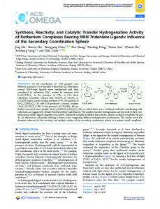
Identification of the Active Site Cysteine of Pig Liver Thiol- where k is the apparent rate constant,t is the time, [TT-0] is transferase-Preliminary results have shown that more than the concentrationof reduced enzyme at time 0, and [TT-CM] 90% of thioltransferase activity was lost after 20 min incu- is the concentration of carboxymethylated enzyme at time t. bation of 0.12 mM reduced enzyme with 0.24 mM iodoacetic The term [TT-O] - [TT-CM] should be a function of the acid at a pH of 6.5 and at room temperature (data notshown). thioltransferase activity. Equal concentrations (0.12 mM)of To identify which amino acid residue(s) react(s) with iodoa- reduced enzyme and iodoacetic acid were incubated at room cetic acid, thereduced enzyme (0.12 mM) was incubated with temperature in the presenceof 0.1 sodium citrate buffer, pH i~do[l-'~C]acetic acid (0.24 mM) inthepresence of 0.1 M 6.0. A plot of l/thioltransferase activity against time gave a sodium phosphate, p H 6.5, at room temperature. After20 min straight line with a rate constant of 0.96 mM" min" and a incubation, cold iodoacetic acid was added t o give a concen- half-time of 8.7 min (Fig. 1).Since it isknown that thiols are form (16),thereactionrate of tration of 5 mM, and the excess iodoacetic acid was removed alkylatedintheirthiolate alkylation should be strongly pH-dependent. The rate conon a Sephadex G-25 column. Subsequent carboxymethylation, tryptic digestion, andpeptidepurification were performed stants of the reactionbetween reduced enzymeand iodoacetic under the same condition used in the companion paper (12). acid were determined over a pH range of 1.4 to 6.6. A plot of The results of radioactivity counting of the tryptic peptides the apparent rate constant against pH isgiven in Fig. 2. The showed that only peptide T 3 was labeled by i~do[l-'~C]acetic apparent rate constants between pH 5 and 6.6 were not pHacidwithmore than background radioactivity. Peptide T 3 dependent, whereas they increased between pH 5 and 3, and was then subjected to automated Edman degradation and the decreased below pH 3. The rate constant was near 0 at pH total radioactivity of each residue was counted. The results of 14C was given inTable I indicatedthatincorporation sharply increased at residue 9 which corresponds to cysteine 22 of the protein. The total counts/min of the residues recovered before residue 9 were at the background level. The radioactivity observed with the residues following residue 9 was the result of carry-over, a common phenomenon which occurs in automated Edman degradation of peptides. These resultsstrongly suggest thattheactivesiteis located at cysteine 22 of the protein. To further confirm this conclusion, the i~do[l-'~C]aceticacid-labeledenzyme was digested by chymotrypsin, and the peptides were separated as described (12). It was found that peptideC3, containing only 1cysteine, Cys", and terminating a t Phe24 (12), was the only peptide 0 5 IO 1 5 2 0 2 5 labeled by [l-'4C]iodoacetic acid. Time (rnin) Kinetics of the Reaction between CysZ2and Iodoacetic AcidFIG. 1. Rate plot of the reaction between equal concentraSince alkylation of CysZ2will destroy catalytic activityof the enzyme, thioltransferase activity measurements should be a tions (0.12 mM) of reduced pig liver thioltransferase and f k c t i o n of the concentrationof reduced enzyme in an alkyl- iodoacetic acid. The reactions were carried out at room temperature in the presence of 0.1 M sodium citrate buffer, pH 6.0. Thioltransferation reaction of the enzyme. If only Cys2*reacts with iodo- ase activity was determined at various times after at least a 1000-fold acetic acid at pH 6.5 as seenabove, and equal concentrations dilution. [ TT-SH]represents the reduced enzyme concentration cal-
L
culated from the remaining thioltransferase activity.
TABLEI Identification of the active site amino acid of pig liver thioltransferase Peptide T3 (15 nmol) was subjected to automated Edman degradation. Each degradation product was transferred to a scintillation vial with methanol. Total counts/min of each residue were counted. Residue Amino No.
Yield acid
cpm X IO-', nmol total
cpm X per
lo-*,
nmol
Val 9.3 Val 9.2 Val 8.7 Phe 11 Ile 6.4 7.9 LYS Pro 2.1 Thr" R8 R9 CYS 1.0 R10 Pro 0.8 R11 P he 0.7 R12 Cys 0.4 R13 Arg 0.2 The yield was not determined. R1 R2 R3 R4 R5 R6 R7
2.3 2.3 2.2 2.1 2.5 2.2 2.6 1.8 401 189 78 14 8.5
0.2 0.2 0.3 0.2 0.4 0.3 1.2 410 236 111 35 42
7.-
-
3 -
E
= -E
2 -
a a
y"
1 -
PH
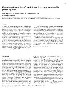
FIG. 2. pH dependence of second order apparent rate constants of the reactions between equal concentrations (0.12 mM) of reducedpig liver thioltransferase and iodoacetic acid. The kappwere determined by the methods described in Fig. 1. Blank thioltransferase activity was not affected when the pHdecreased over the indicated range.
Site Active The
6706
of
Pig Thioltransferase Liver
1.4. Since the degree of hydrogen ion dissociation of a sulfhydryl group should decrease as the pH decreases, an increase of the k value at pH 5 to 3is probably not due to theincreasing protonation of the sulfhydryl group, but instead to effects on the alkylating reagent. Since iodoacetic acid has apK, of 3.12 (17), the degree of carboxyl protonation of the reagent will increase and this decrease in negative charge may facilitate the reactivity of iodoacetic acid with the sulfhydryl group of the enzyme. This is in accord with the finding of Kallis and Holmgren (11)that the apparent rateconstant of iodoacetamide with no negative charge was 20-fold higher than that of iodoacetic acid at pH7.2, in similar studies with thioredoxin. However, it is possible that the low pH may cause a conformational change of the protein whereby iodoacetic acid could come into closer proximity to the sulfhydryl group, resulting in the increased reaction rate. The sharp decrease of the apparent rate constantsbelow pH 3may be caused by protonation of the active site sulfhydryl group in agreement with an estimatedpK, of 2.5 for the Cys22sulfhydryl group (see below). Theinterpretation of the above kinetics is based on the assumption that the alkylation reaction of the enzyme is a function of the catalytic activity, and only one sulfhydryl group of the enzyme reacts with iodoacetic acid under the conditions used. If this is true, the degree of iodoacetic acid incorporation into the enzyme should be proportional to the apparent rate constants at different pH. To investigate this hypothesis further, reduced pig liver thioltransferase (0.12 mM)was alkylated by [l-'4C]iodoacetic acid (0.15 mM) in the presence of0.1 M sodium citrate buffer, at different pH. Excess cold iodoacetic acid was added to the mixture after 3 min incubation at room temperature, and the enzyme was separated from excess iodoacetic acid by a Sephadex G-25 column. The radioactivity incorporated into the enzyme is given in Table 11. The incorporation pattern is in good agreement with the apparent rate constant curve shown in Fig. 2. The protein labeled at different pH was subjected to chymotryptic digestion and the chymotryptic peptides were purified by reversed-phase HPLC. It was found that only peptide C3 was labeled, suggesting that cysteine 22 was the only amino acid residue of the protein which reacted with iodoacetic acid under the conditions used. These studies indicate that the enzyme Cys22has a pK, of approximately 2.5. Disulfide Protection o/ the Enzyme from lodoacetate Inuctiuation-Previous results showed thatthe disulfide substrates (RSSR) of the enzyme can protect the enzyme from inactivation by iodoacetic acid (8,9), but the mechanism was not clear. According to the results described above, i.e. only one sulfhydryl group reacted with iodoacetic acid, three mech-
S-
/
E
S-S-R
+ H+ + RSSR & E
\
/ \
SH
+ RSH
(1)
SH
S-
2E
/ \
+ 2 H+ + RSSR *
(2)
SH
+ 2 RSH
HS-E--S-S-E-SH
S-
/
\
RSH
+ H' + RSSR &
E
SH
(3)
S-S-R
S
/
E
\
SH
\I
S
To test these mechanisms, reduced pig liver thioltransferase (0.42 mM) was incubated with [l-14C]cystine (0.67 mM) at room temperature for 5 min in the presence of 0.1 M sodium phosphate buffer, pH 6.6. The subsequent Sephadex G-25 chromatography was performed to separate the enzyme from excess [l-'4C]cystine, and the thioltransferase activity and the radioactivity of the fractions were measured. Consistent with previous results (8, 9), thioltransferase was totally inactivated by iodoacetic acid before incubation with [I-'4c]cystine, whereas the enzyme, preincubated with [l-"Clcystine, retained its full enzymatic activity and insensitivity to iodoacetic acid treatment, but was unlabeled with l4c(Fig. 31, ruling out mechanism 1.Another possibility for the protection mechanism is the formation of a dimeric species of the enzyme (Equation 2). In this mechanism, the enzyme would be protected against iodoacetic acid treatment and would not be radioactively labeled.However,when the[l-'4Clc~stinetreated enzyme was run on sodium dodecyl sulfate-polyacrylamide gel electrophoresis under nonreducing conditions in the presence of excess iodoacetic acid, no shift in the molecular weight of the enzyme on the gel (data not shown) was observed, ruling out mechanism 2. However, the results obtained in this study fully support the mechanism described in reaction 3, namely the formation of an intramolecular d i d fide bond. DISCUSSION
TABLEI1 Incorporation of [l -14C/iodoaceticacid into pig liver thioltransferase at different p H Reduced enzyme (120 PM) wasincubated with [l-14CC]iodoacetic acid (150 WM) in 0.1 M sodium citrate buffer at different pH. After 3 min incubation at room temperature,excess cold iodoacetic acid was added to give a final concentration of 5 mM, and the mixtures were immediately loaded onto Sephadex G-25 columns to desalt. Total counts/min and protein content of protein fractions of Sephadex G25 chromatography were determined. The molecular weight of the enzyme used in the calculation of moles of carboxymethyl groups incorporation/mol of enzyme was 11,500. pH
2.10.4
anisms can be proposed
3.0
5.0 4.0
6.0
Moles carboxymethyl groups 0.04 0.10 0.49 0.37 0.21 0.22 incorporated per mol of enzyme
The alkylation of a thiol, as well as the reduction of a disulfide by thiols, requires a thiol anion which can initiate a nucleophilic attack (16). The ionization of a thiol is strongly influenced by neighboring group effects. The pK, values of cysteine thiols maybe determined by their pH-dependent reactivity with an appropriate disulfide (16).The present work demonstrated that only CysZ2of pig liver thioltransferase reacted with iodoacetic acid in a physiological pH range. From the studies reported here, the sulfhydryl group of cysteine 22 has a pK, of approximately 2.5. It has been reported that the reaction rate of disulfide reduction by a thiol is electrostatically dependent (16). For example, positively charged disulfides react with thiol more rapidly than those with negative charge. Thus, compared with low molecular weightthiols, the ionization status of protein thiols are more strongly affected by their microenvironment. An unusual feature of the primary
The Active Site of Pig Liver Thioltransferase
6707
structure of pig liver thioltransferase is that there are no negatively charged amino acid residues from the N terminus to residue 29, whereas 5 basic amino acids were found. In particular, 3positivelycharged amino acidresidues,Lys”, and Lys“, surround Cys2’. While the primary structure of the protein may facilitate the low pK,, of CysZ2,the threedimensional structure of the proteinmay also play an important role in the ionizationof the sulfhydryl group of Cys”. Several proteins have been reported to catalyze sulfhydryldisulfide interchange (18-21). The active site of thioredoxin has been well characterized with a sequence of -Trp-Cys3’Gly-Pro-Cy~~~-Lys-, which undergoes a thiol-disulfide interchange in the presenceof the NADPH-dependent thioredoxin reductase system (11).The sulfhydryl group of cysteine 32 has a pK, of 6.7, which can effectively react with disulfides at Frwtion No. (1.1 ml a physiological pH. The location of the reactive thiol proFIG. 3. Interaction between [1-”Clcystine and reduced Pig trudes outfrom the surfaceof the protein, making it accessible liver thioltransferase. The solid circles and triangles represent to other disulfide surfaces (22). The similarsequences, -Trp- thioltransferase activity and counts/min of the fractions, respectively. Cys-Gly-His-Cys-Arg- in the protein disulfide isomerase (20) For details, see “Experimental Procedures.” and -Thr-Cys-Pro-Tyr-Cys-Lys- inglutaredoxin(23), have been presumed to be the active site of these enzymes. Howcysteines,analogous to CysZ2 and C y P , are required for ever, the ionization properties of thethiols of these two catalysis. The next steps in the model are involved in the enzymes have not been studied. We report here that the active regeneration of the reduced enzyme by GSH for which 2 mol site of thioltransferase hasa sequence of -Thr-CysZ2-Pro-Phe- of GSH are required (Equations 3 and 4). Since the pK, of CysZ5-Arg-Lys-.The low pK, of the sulfhydryl group of CysZ2 the sulfhydryl group of CysZ2is about 2.5, the initial nucleosuggests that thioltransferase will have a relatively negative philic attack on thedisulfide by the enzyme is probably very E,, although these measurements have not yet been made. A fast at a physiological pH. Reaction 2 is also assumed to model for the action of the enzyme for a typical thiol-disulfide proceed rapidly because of the rapid deprotonation of CysZ5 oxidoreduction is proposed as follows: in the transient intermediate complex and by its close proxSS-S-R imity to the initial mixed disulfide. Our previous work showed that pig liver thioltransferase had an optimum pH of 8.5 to 9, / / + H+ + RSSR d E + RSH (1) andthioltransferaseactivity E was stronglydependenton \ \ [GSH] (9). Whether the proposed Reactions 3 and 4 are rateSH limiting for the catalytic mechanism of thioltransferase remains to be determined. S S-S-R E
/ \
/ ’
+ RSH
FfE SH
\I
S
S
S-S-G
+ GSH d E
lE ’
\
S
/ (3)
\
E
\
+ GSH Ft E SH
SH
S-
S-S-G
/
(2)
/ \
+ H+ + GSSG
(4)
SH
In themodel above, RSSR andE represent disulfide substrate and enzyme, respectively. The sulfhydryl group of CysZ2initiates a nucleophilic attack on the disulfide, resulting in an enzyme-substrate mixed disulfide complex. The mixed disulfide will be rapidly reduced by an intramolecular rearrangement in which deprotonated CysZ5,presumably in close juxtaposition, cleaves the mixeddisulfidebond, creatingthe intramolecular enzyme disulfide bond. Although theoretically possible, it is unlikely that either Cys7*or Cyssz would take part in this intramolecular disulfide formation since they are likely to be aconsiderable distance from theactive site. Furthermore, in E. coli thioredoxin or glutaredoxin, only 2
Acknowledgments-Wewish to thank Dr. Young Moo Lee and Melanie Markel for valuable assistance in active site peptide sequencing. REFERENCES 1. Racker, E. (1955) J. Biol. Chem. 217,867-874 2. Axelsson, K., Eriksson, S., and Mannervik, B. (1978) Biochemistry 17, 2978-2984 3. Mannervik, B., and Axelsson, K. (1980)Biochem. J . 190,125-130 4. Axelsson, K., and Mannervik, B. (1983) FEES Lett. 152, 114-118 5. Mannervik, B., Axelsson, K., Sundewall, A.-C., and Holmgren, A. (1983) Biochem. J . 213. 519-523 6. Hatakeyama, M., Tanimoto, Y., and Mizoguchi, T. (1984) J . Biochem. (Tokyo) 95,1811-1818 7. Ziegler, D. M. (1985) Annu. Reu. Biochem. 54,305-329 8. Gan, Z., and Wells, W. W. (1986) J. Biol. Chem. 261, 996-1001 9. Gan, Z.-R., and Wells, W. W. (1987) Anal. Biochem., in press 10. Holmgren, A. (1968) Eur. J. Biochem. 6,475-484 11. Kallis, G.-B., and Holmgren, A. (1980) J. Biol. Chem. 255, 10261-10265 12. Gan, 2.-R., and Wells, W. W. (1987) J. Biol. Chem. 262, 6699-6703 13. Segel, J. H., and Johnson, M. J. (1963) Anal. Biochem. 5, 330-337 14. Gracy, J. H. (1977) Methods Enzymol. 47,195-204 15. Lowry, 0. H., Rosebrough, N. J., Farr, A. L., and Randall, R. J. (1951) J. Biol. Chem. 193,265-275 16. Creighton, T.E. (1978) Prog. Biophys. Mol. Biol. 33,231-297 17. Weast, R. C. (ed) (1972) in Handbook of Chemistry and Physics, 53rd. Ed. p. D-120, CRC Press, Cleveland, OH 18. Holmgren, A. (1979) J. Biol. Chem. 254, 9627-9632 19. Carmichael, D. F., Keefe, M., Pace, M., and Dixon, J. E. (1979) J . Biol. Chem. 254,8386-8390 20. Edman, J. C., Ellis, L., Blacher, R. W., Roth, R. A,, and Rutter, W. J. (1985) Nature 317, 267-270 21. Luthman, M., and Holmgren, A. (1982) J. Biol. Chem. 257,6686-6690 22. Holmgren, A., Sderberg, B.-O., Eklund, H., andBranden, C.-I. (1975) Proc. Natl. Acad. Sci. U. S. A. 7 2 , 2305-2309 23. Klintrot, L.-M., Hoijg, J.-O., Jornvall, H., Holmgren, A., and Luthman, M. (1984) Eur. J. Biochem. 144,417-423