The EMBO Journal Vol.17 No.2 pp.575–589, 1998
Identifying the right stop: determining how the surveillance complex recognizes and degrades an aberrant mRNA
Maria J.Ruiz-Echevarrı´a1, Carlos I.Gonza´lez1 and Stuart W.Peltz1,2,3 1Department of Molecular Genetics and Microbiology, Robert Wood Johnson Medical School-UMDNJ and 2Cancer Institute of New Jersey, 675 Hoes Lane, Piscataway, NJ 08854, USA 3Corresponding author e-mail:
[email protected]
The nonsense-mediated mRNA decay (NMD) pathway functions by checking whether translation termination has occurred prematurely and subsequently degrading the aberrant mRNAs. In Saccharomyces cerevisiae, it has been proposed that a surveillance complex scans 39 of the premature termination codon and searches for the downstream element (DSE), whose recognition by the complex identifies the transcript as aberrant and promotes its rapid decay. The results presented here suggest that translation termination is important for assembly of the surveillance complex. Neither the activity of the initiation ternary complex after premature translation termination has occurred nor the elongation phase of translation are essential for the activity of the NMD pathway. Once assembled, the surveillance complex is active for searching and recognizing a DSE for ~200 nt 39 of the stop codon. We have also identified a stabilizer sequence (STE) in the GCN4 leader region that inactivates the NMD pathway. Inactivation of the NMD pathway, as a consequence of either the DSE being too far from a stop codon or the presence of the STE, can be circumvented by inserting sequences containing a new translation initiation/termination cycle immediately 59 of the DSE. Further, the results indicate that the STE functions in the context of the GCN4 transcript to inactivate the NMD pathway. Keywords: cis-acting stability elements/cis-acting instability elements/mRNA decay/nonsense mutations/ translation termination
Introduction Recent studies have demonstrated that cells have evolved elaborate mechanisms to rid themselves of aberrant proteins and transcripts that can dominantly interfere with their normal functioning (reviewed in Jacobson and Peltz, 1996; Ruiz-Echevarria et al., 1996; Gottesman et al., 1997; Suzuki et al., 1997; Weng et al., 1997). Such pathways can function either as regulators of gene expression or as sensors for inappropriate polypeptide synthesis. The nonsense-mediated mRNA decay pathway (NMD) is an example of such a pathway (reviewed in Maquat, 1995; Caponigro and Parker, 1996; Jacobson and Peltz, 1996; Ruiz-Echevarria et al., 1996; Weng et al., 1997). The © Oxford University Press
NMD pathway has been observed in all eukaryotic systems examined so far, and appears to have evolved as a surveillance mechanism to ensure that transcripts containing premature nonsense codons are degraded rapidly, thus preventing synthesis of incomplete and potentially deleterious proteins. Interestingly, the clinical manifestation and severity of several human genetic diseases that are a consequence of nonsense mutations can increase under conditions in which the nonsense-containing transcript is stabilized (Dietz et al., 1993, 1995; Hall and Thein, 1994). The fact that premature translation termination affects the stability of mRNAs, combined with numerous other observations coupling protein synthesis and mRNA turnover, establishes a strong relationship between the two processes (reviewed in Jacobson and Peltz, 1996). Our studies focus on understanding this relationship. The NMD pathway has been investigated extensively in the yeast Saccharomyces cerevisiae (reviewed in Caponigro and Parker, 1996; Jacobson and Peltz, 1996; Ruiz-Echevarria et al., 1996; Weng et al., 1997). Based on these studies, a model for the mechanism of the NMD pathway has been proposed (reviewed in Caponigro and Parker, 1996; Jacobson and Peltz, 1996; Ruiz-Echevarria et al., 1996; Weng et al., 1997). Either concurrently or immediately after export of the mRNA to the cytoplasm, ribosomes become associated with the transcript and begin translation. The presence of a nonsense mutation causes the ribosome to terminate translation prematurely. Following termination, a complex which might consist of ribosomes, ribosomal subunits and/or associated factors scans 39 of the termination codon and interacts with specific sequences. These sequences have been defined as downstream elements (DSEs) and are required for rapid destabilization of nonsense-containing mRNAs. We propose that this interaction leads to an RNP structure that renders the mRNA susceptible to decapping independently of shortening of the poly(A) tail. The uncapped nonsensecontaining mRNA is then rapidly degraded by the 59→39 Xrn1 exoribonuclease. The key in determining whether premature translation termination has occurred depends upon the recognition of a DSE 39 of a stop codon. Transcripts containing premature termination codons, but lacking a DSE, are not degraded by the NMD pathway (Ruiz-Echevarria and Peltz, 1996). We hypothesize the presence of a surveillance complex acting as a sensor that scans 39 of the termination site and searches for a sequence containing a DSE. This complex should consist of factors that: (i) facilitate scanning 39 of the stop codon; (ii) recognize a sequence containing a DSE; and (iii) transduce a signal to the 59-end of the transcript to promote rapid degradation of the mRNA. Three components essential for the activity of the NMD pathway have been identified. Mutations in the UPF1, 575
M.J.Ruiz-Echevarrı´a, C.I.Gonza´lez and S.W.Peltz
UPF2 and UPF3 genes were shown selectively to stabilize mRNAs containing early nonsense mutations without affecting the decay rate of most wild-type mRNAs (Leeds et al., 1991, 1992; Cui et al., 1995; He and Jacobson, 1995; Lee and Culbertson, 1995). Recent studies suggest that Upf1p, Upf2p and Upf3p interact to form a complex (He and Jacobson, 1995; Weng et al., 1996a; He et al., 1997). Elucidating the NMD pathway requires understanding how the surveillance complex searches for and recognizes the DSE. Therefore, it is necessary to define the sequences that constitute a DSE and establish the rules that govern how the surveillance complex identifies this element to promote NMD. To this end, we recently have developed a system using the leader region of the GCN4 transcript (Ruiz-Echevarria and Peltz, 1996). The Gcn4 protein is a transcriptional activator of amino acid biosynthetic genes and its expression is regulated by a translation control mechanism that monitors the concentration of uncharged tRNAs to sense the amino acid pool sizes (reviewed in Hinnebusch, 1996). This control mechanism operates at the level of translational initiation/termination and is mediated by four upstream open reading frames (uORFs; uORF1–uORF4) that are located in the leader region of the GCN4 mRNA. Following translation of uORF1 or uORF2, a substantial fraction of ribosomes can resume scanning and reinitiate translation at downstream initiation sites. Translation of uORF3 and uORF4, however, prevents reinitiation at downstream translation start sites, precluding synthesis of the Gcn4p (reviewed in Hinnebusch, 1996). We have taken advantage of the unique termination, scanning and reinitiation properties of the GCN4 leader region to develop a system to investigate how the surveillance machinery recognizes a DSE and promotes accelerated decay of aberrant nonsense-containing transcripts (Ruiz-Echevarria and Peltz, 1996). Using this system, we have previously shown that: (i) the GCN4 leader region does not harbor a DSE but can be degraded by this pathway if a DSE is inserted; (ii) the DSE is functional only after a translation initiation and termination cycle has been completed; and (iii) translation reinitiation downstream of a uORF is not sufficient to promote NMD (Ruiz-Echevarria and Peltz, 1996). These results indicate that premature translation termination in a transcript is not sufficient to distinguish it as an aberrant RNA, but requires a DSE 39 of the termination codon. The experiments reported here establish the criteria utilized by the surveillance machinery to assess whether an aberrant termination event has occurred. The results presented suggest that the elongation phase of translation is dispensable for the activation of the NMD pathway. Further, the activity of the eukaryotic translation initiation ternary complex (eIF2–GTP–itRNAMet) does not influence the activity of this pathway. We also demonstrate that following translation termination, the complex can search ~200 nt 39 of a stop codon for a DSE. The DSE is not recognized by the complex when located at a distance .200 nt from the stop codon, presumably because it becomes inactivated or disassembles. Significantly, we have also identified a ‘stabilizer element’ (STE) in the GCN4 leader region that inactivates the NMD pathway when positioned between the termination codon and the DSE. Deletion of the STE from the GCN4 transcript 576
makes this mRNA susceptible to NMD. The STE is a general determinant that blocks the activity of the NMD pathway when inserted into heterologous transcripts. Interestingly, inactivation of the NMD pathway, as a consequence of either the DSE being too far from a stop codon or the presence of the STE, can be reversed by inserting sequences containing a new translation initiation/ termination cycle. This suggests that the surveillance complex can reassemble or be reactivated during a new translation cycle. The consequences of these results for the decay of both wild-type and nonsense-containing transcripts will be discussed.
Results A key to understanding how a nonsense-containing RNA is identified as aberrant and rapidly degraded lies in determining how the DSE is recognized by the surveillance complex. To this end, we have developed a system based on the leader region of the GCN4 transcript. The GCN4 gene, or a construct in which a GCN4–LacZ fusion gene is inserted 39 of the GCN4 leader region, encodes moderately stable mRNAs which are resistant to NMD. However, insertion of a DSE immediately 39 of uORF1 in the GCN4 leader region or in the GCN4–LacZ construct results in rapid decay of the corresponding transcripts through the NMD pathway that is followed by a decrease in β-galactosidase activity (Ruiz-Echevarria and Peltz, 1996). These results demonstrated that the system reflects the activity of the DSE. We have utilized the leader region of the GCN4 mRNA to characterize key parameters that determine how recognition of the DSE by the surveillance complex occurs and how this recognition can be modulated. A translation initiation/termination cycle lacking the elongation phase can promote nonsense-mediated mRNA decay
Recognition of an aberrant transcript by the surveillance complex requires translation termination and subsequent recognition of a DSE. We have shown previously that a translation initiation/elongation/termination cycle is required for the activation of the NMD pathway (RuizEchevarria and Peltz, 1996). This result suggests that either all, or a subset, of the translation cycle is required for the assembly of the complex that recognizes the DSE. Our previous results, however, indicate that Upf1p protein can affect translation termination (Weng et al., 1996a,b). Certain mutations in the Upf1p promote efficient suppression of a nonsense codon while having no effect on the ability of the Upf1p to promote rapid decay of nonsensecontaining transcripts. These results suggest that the complex may assemble at the termination site and that the elongation phase of translation may not be required for the NMD pathway to be functional. To test this possibility, the three hybrid genes shown in Figure 1 were constructed. The translation initiation codons at uORF1, uORF2 and uORF3 of the GCN4 leader were mutated to non-AUG codons in all the constructs. In construct 1A, a segment containing the wild-type uORF1 was inserted in place of the corresponding sequences of uORF4 and immediately followed by a DSE from the PGK1 gene (Figure 1, construct 1A; Peltz et al., 1993). The wild-type uORF1
Identifying the appropriate termination codon
tion were degraded rapidly in a UPF11 strain (half-lives of ø3 min), but were stabilized 4-fold in a upf1∆ strain (constructs 1A and 1B). The untranslated hybrid transcript, lacking an initiation site, was moderately stable and its decay rate did not change in UPF11 or upf1∆ strains (construct 1C). These results indicate that the elongation phase of translation is dispensable for activating the NMD pathway. Only translation initiation and/or termination are required to assemble the surveillance complex and activate the NMD pathway. Similar results have been reported for the triosephosphate isomerase transcript in mammalian cells (Zhang and Maquat, 1997). The distance between the translation termination codon and the DSE is important for the activity of the surveillance complex
Fig. 1. The elongation phase of translation is dispensable for nonsense-mediated mRNA decay. Schematic diagram of the hybrid GCN4–PGK1 constructs. A small segment from the GCN4 leader region containing the uORF1 coding region or the mutant derivatives (indicated by a boxed 1), plus 16 nt 59-flanking and 57 nt 39-flanking sequences (open bars) were inserted in the place of the corresponding sequences of uORF4. The Xs represent point mutations that inactivate the AUG codons of uORF1, uORF2 and uORF3. The shaded region represents the DSE from the PGK1 gene (Peltz et al., 1993). Construct 1A contains the wild-type uORF1 coding sequence. In construct 1B, the second and third codons of uORF1 have been mutated to UAA (underlined). In construct 1C, the AUG codon of uORF1 has been mutated to AAG (indicated by a black circle). The black box represents the 39-UTR of the PGK1 gene (containing the transcription termination and polyadenylation signals). mRNA decay rates of these alleles were determined in either the RY262 (UPF11) or RY262– (upf1∆) strain as described in Materials and methods. The RNA blots for these experiments are shown below the schematic representations of the hybrid alleles.
contains an elongation phase of translation. This construct does not harbor any of the sequences normally found 39 of uORF4 and it only contains a unique AUG codon from uORF1. Construct 1B is identical to construct 1A except that the elongation phase of translation was eliminated by substituting the second and third codons of uORF1 with two consecutive UAA termination codons (Figure 1, construct 1B). As a control for these experiments, translation initiation at uORF1 was inactivated by a point mutation (construct 1C). These three constructs were transformed into isogenic UPF11 and upf1∆ strains harboring the rpb1-1 temperature-sensitive allele of the RNA polymerase II, and mRNA decay rates were determined (see Materials and methods). In these experiments, as in all the following, the abundances of the RNAs were normalized to the abundance of the U3 snRNA (see Materials and methods; data not shown). The results demonstrated that regardless of whether translation elongation occurred, the hybrid transcripts that initiated transla-
The model for degradation of nonsense-containing transcripts suggests that a transcript is recognized as aberrant when the surveillance complex scans 39 of the stop codon and recognizes a DSE. We determined whether there are limitations on the distance that the complex can scan 39 of a termination codon to search for a DSE. For this purpose, a GCN4 leader region was utilized in which the translation initiation codon of uORF1 was functional, but those of uORF2, uORF3 and uORF4 were inactivated by point mutations (Figure 2). A DSE from the PGK1 gene (Peltz et al., 1993) was inserted at 57, 153, 220 and 325 nt from the uORF1 translation termination codon (Figure 2, constructs 2A–2D). The decay rates of the corresponding transcripts were determined in isogenic UPF11 and upf1∆ strains (see Materials and methods) and the results are shown in Figure 2. The hybrid mRNAs in which the DSE is located either 57 or 153 nt from the stop codon of uORF1 were degraded rapidly in a UPF11 strain, with half-lives ,3 min. These transcripts were stabilized 4-fold in a upf1∆ strain (constructs 2A and 2B). The transcript in which the DSE was inserted 220 nt 39 of the stop codon of uORF1 showed a moderate stability in a UPF11 strain, with a half-life of 8 min, and was stabilized to 12 min in a upf1∆ strain (construct 2C). However, the transcript in which the DSE was inserted 325 nt 39 of the stop codon of uORF1 showed a decay rate of 12 min in a UPF11 strain which did not change in a upf1∆ strain (construct 2D). The stabilization of the transcript observed when the DSE is located at a distance .220 nt from the termination codon (constructs 2C and 2D) could be due to: (i) the insertion of a sequence that specifically inactivates the decay pathway; or (ii) the inactivation of the surveillance complex as a consequence of an increased distance between the termination codon and the DSE. To distinguish between these possibilities, we first tested whether the sequence in the GCN4 leader region that delineates between sensitivity and resistance to NMD, defined here as segment c, harbors sequences that inactivate this pathway (Figure 2). Segment c was inserted into construct 2A (Figure 2), yielding construct 2E. Measurement of the decay rate of this mRNA demonstrated that it was sensitive to NMD (construct 2E). This result indicates that segment c does not harbor sequences that inactivate the NMD pathway and suggests that scanning a distance .220 nt renders the surveillance complex unable to recognize the DSE. To test this hypothesis, a region in the GCN4 leader that does not prevent rapid mRNA decay (regions a and 577
M.J.Ruiz-Echevarrı´a, C.I.Gonza´lez and S.W.Peltz
Fig. 2. Effect of the distance between the stop codon and the downstream element on nonsense-mediated mRNA decay. A DSE from the PGK1 gene (shaded region; Peltz et al., 1993) was inserted at different positions from the stop codon of uORF1. Constructs 2A–2D contain the GCN4 leader region up to position 1297, 1393, 1458 and 1565 respectively (where 11 is defined as the transcription start site). The distance between the translation termination codon of uORF1 and the DSE in each of the constructs is indicated. The sequences that encompass segment a, b, c or d are as indicated. In construct 2E, segment c was inserted immediately downstream of segment a. In construct 2F, the region corresponding to segments a and b was duplicated and inserted 59 of the DSE. Other symbols are as indicated in the legend to Figure 1. mRNA decay rates of these alleles were determined in strains RY2621 (UPF11) and RY262– (upf1∆). The RNA blots are shown below the schematic representations of the hybrid GCN4–PGK1 alleles.
b, see constructs 2A and 2B) was duplicated and inserted between the uORF1 stop codon and the DSE (construct 2F). The DSE in this construct is located 312 nt from the termination codon as a consequence of the repeated sequence (construct 2F). This transcript was resistant to NMD, indicating that the increased distance between the termination codon and the DSE led to inactivation of the NMD pathway (construct 2F). These results suggest that the surveillance complex has a limited range (~200 nt) in which it can search for a DSE, after which the complex becomes progressively inactivated. This suggests that the activity of the NMD pathway can be modulated as a function of the distance between the stop codon and the DSE. Alteration of the translation termination or reinitiation processes does not affect the activity of the surveillance complex
Translational control of GCN4 gene expression is mediated by the uORFs present in the mRNA leader and by 578
phosphorylation of the α-subunit of the eukaryotic initiation factor, eIF-2 (reviewed in Hinnebusch, 1993, 1996). The model proposed to explain translational regulation of GCN4 expression suggests that after translation of uORF1, ribosomes resume scanning and must rebind aminoacylated initiator tRNAMet, thought to be delivered to the ribosome in a ternary complex with eIF-2 and GTP, before they can recognize a downstream AUG (Abastado et al., 1991a). Under non-starvation conditions, eIF2–GDP is recycled readily to eIF-2–GTP, leading to high levels of ternary complex formation, allowing reinitiation at uORF2, uORF3 or uORF4. Under conditions of amino acid starvation, phosphorylation of the α-subunit of eIF-2 leads to low levels of ternary complex formation. Therefore, many ribosomes scanning 39 of uORF1 bypass uORF2, uORF3 and uORF4 and become competent to reinitiate translation while scanning the interval between uORF4 and the GCN4 start codon (reviewed in Hinnebusch, 1996). The model for how the surveillance complex identifies a nonsense-containing mRNA suggests that following translation termination, it scans 39 of the stop codon searching for a DSE. The process of translation reinitiation also requires translation termination, scanning and subsequent recognition of a downstream AUG. Based on the similarities between both pathways, we asked whether a relationship exists between the ability of a ribosome to reinitiate translation and the recognition of a DSE following translation termination. We envisaged that it is possible that the ribosome scanning downstream from the stop codon needs to regain its ability to reinitiate translation in order to recognize the DSE; alternatively, it is possible that once the ribosome becomes capable of reinitiating translation it loses its competence to recognize the DSE. To distinguish between these two possibilities, we determined whether recognition of the DSE by the surveillance complex is modulated by the availability of an active ternary complex, since it has been shown to modulate translation reinitiation. The mRNA decay rates of the constructs harboring the uORF1 of the GCN4 gene in which the DSE is located at various distances from the stop codon of uORF1 (see Figure 2, constructs 2A–2D) was determined in cells grown under induced amino acid starvation conditions in the presence of 3-aminotriazole (3-AT; Materials and methods). The results, shown in Table I, demonstrated that the decay rates of these mRNAs in UPF11 cells were equivalent to their decay rates when grown in rich medium in the absence of 3-AT (Table I). The GCN4 fusion transcripts in which the DSE was located 57 and 153 nt from the termination codon were degraded rapidly, with a half-life of ,3 min (Table I, constructs 2A and 2B). Conversely, the transcripts in which the downstream element was located 220 and 325 nt 39 of the termination codon were moderately stable, with half-lives of 8 and 12 min, respectively (Table I, constructs 2C and 2D). Analysis of the mRNA abundance of the HIS4 transcript demonstrated that the starvation conditions used in these experiments led to transcriptional derepression of the HIS4 gene (Table I), yielding a 3-fold increase in HIS4 mRNA abundance and indicating a reduction in the efficiency of translation reinitiation. These results suggest that the activity of the NMD pathway is not modulated by the availability of active ternary complex.
Identifying the appropriate termination codon
Table I. Activation of the NMD pathway under amino acid starvation Constructa
Distance of DSE to stop codon (nt)
mRNA half-life (min)b SD 1 3-ATc SD – 3-AT
Construct 2A Construct 2B Construct 2C Construct 2D HIS4 mRNA relative abundanced
57 153 220 325
,3 ,3 8 12 3.0–3.5
,3 ,3 8 12 1.0
aSchematic representation and details of the constructs are given in Figure 2. bTo measure mRNA decay rates, plasmids were transformed into strain RY260 (UPF11). cTransformants were grown in SD medium containing or lacking 3-AT, as described in Materials and methods. Total mRNA was extracted and subjected to Northern blot analysis. The mRNA decay rates were determined as described in Materials and methods. dThe increase in the relative abundance of the HIS4 mRNA when cells were grown in the presence of 3-AT was approximately the same for each of the four strains. The half-life of the HIS4 mRNA did not change (14 min) under the different growth conditions.
In a second test to determine whether translation reinitiation efficiency affects the activity of the surveillance complex, we took advantage of the distinct termination/ reinitiation efficiencies of the different uORFs in the GCN4 leader region (Mueller and Hinnebusch, 1986). Translation of the uORF1 or uORF2 allows a large fraction of the ribosomes to scan and reinitiate translation efficiently. In contrast, translation of uORF3 and uORF4 leads to very inefficient translation reinitiation. The sequences surrounding the termination codon of the uORFs play a critical role in determining the degree of translation reinitiation following their translation (Miller and Hinnebusch, 1989). We examined the ability of the four GCN4 uORFs to modulate the activity of the surveillance complex to promote NMD. For this purpose, a DSE from the PGK1 gene (Peltz et al., 1993) was inserted 39 of either uORF1, uORF2, uORF3 or uORF4 (Figure 3, constructs 3A–3D). In each of these constructs, only one uORF is active (Figure 3). Measurement of the mRNA decay rates demonstrated that all these transcripts were degraded rapidly in a UPF11 strain, with half-lives of ~3 min, and were stabilized 3- to 4-fold in a upf1∆ strain (Figure 3). These results indicate that the DSE can be recognized even when translation reinitiation is impaired, suggesting that the events that influence translation reinitiation are different from the events that allow the surveillance complex to assemble and search for the DSE. Moreover, these results confirm the observation that the rules governing translation reinitiation are different from the requirements to activate the surveillance complex which leads to NMD (RuizEchevarria and Peltz, 1996; Zhang and Maquat, 1997). Distance-mediated inactivation of the NMD pathway can be reversed by a new translation initiation/termination cycle
The results shown in Figure 2 indicated that when the DSE was inserted 325 nt from the stop codon of uORF1, the mRNA is not susceptible to accelerated decay (Figure
Fig. 3. Effect of the different GCN4 uORFs on nonsense-mediated mRNA decay. The DSE (shaded region) was inserted 39 of the GCN4 uORFs. The functional uORFs are indicated by boxed numbers. Constructs 3A–3D contain the GCN4 leader region up to position 1297, 1393, 1458 and 1498 respectively (where 11 is defined as the transcription start site). Other symbols are as in the legends to Figures 1 and 2. The mRNA decay rates for these alleles were determined in strains RY2621 (UPF11) or RY262– (upf1∆), and the RNA blots are shown below the schematic representations of the hybrid GCN4–PGK1 alleles.
2 or Figure 4A, construct 4A), suggesting that the surveillance complex becomes inactive as a consequence of scanning a long distance from the stop codon. However, as shown in Figure 4A (construct 4B) and previously reported (Ruiz-Echevarria and Peltz, 1996), accelerated mRNA decay can occur when an additional ORF from the PGK1 gene is inserted immediately 59 of the DSE (Figure 4A, construct 4B). Two mechanisms can account for the rapid decay of the transcript containing the PGK1 ORF: (i) the surveillance complex reassembles during the new initiation/termination cycle and becomes competent to recognize the DSE; or (ii) a fraction of the ribosomes fail to initiate at uORF1 and initiate directly at the AUG codon of the PGK1 gene. In an attempt to distinguish between these two possibilities, several constructs were prepared containing multiple initiation/termination sites from the GCN4 leader region, 59 of the PGK1 ORF (Figure 4A, constructs 4C and 4D). Having multiple uORFs makes it unlikely for a ribosome to scan past multiple efficient translation start sites and to initiate directly at the PGK1 ORF (Abastado et al., 1991b). The relevant features of these constructs are shown in Figure 4A (constructs 4C and 4D). Constructs 4C and 4D contain three functional ORFs upstream of the DSE or the miniPGK1 gene, respectively. The mini-PGK1 encodes a short ORF just 59 of a DSE (Peltz et al., 1993). The decay rates of the corresponding transcripts were determined in isogenic UPF11 and upf1∆ strains as described above. The transcript in which the DSE is located 325 nt from the stop codon of the third uORF is moderately stable and 579
M.J.Ruiz-Echevarrı´a, C.I.Gonza´lez and S.W.Peltz
from the HIS4 gene also triggered rapid decay of the transcript (Figure 4B, construct 4E). Further, deletion of the DSE (construct 4F) or introduction of a mutation that removes the stop codon (construct 4G) from construct 4B (Figure 4A) renders this transcript resistant to accelerated decay. These results indicate that the rapid decay of the transcript encoded by construct 4B was dependent on a termination codon 59 of the DSE (compare construct 4B with constructs 4F and 4G). Taken together, these results suggest that the surveillance complex can reassemble during a new translation initiation/termination cycle, regaining the ability to search and recognize a DSE and to promote decay of the aberrant mRNA. Identification of a ‘stabilizer element’ (STE) that inactivates the nonsense-mediated mRNA decay pathway
Fig. 4. Insertion of a translation initiation/termination cycle reactivates nonsense-mediated mRNA decay. (A) The schematic representation at the top of the figure depicts the GCN4–PGK1 hybrid alleles used in these experiments. Functional uORFs are indicated by boxed numbers. Sequences derived from the PGK1 gene are represented by black rectangles except for the DSE which is indicated by a shaded rectangle. In construct 4A, the DSE is inserted immediately 39 of the GCN4 leader. The distance between the stop codon of uORF1 and the DSE is indicated (325 nt). Construct 4B contains a 23 amino acid long ORF from the PGK1 gene (black rectangle) inserted immediately upstream of the DSE. Constructs 4C and 4D contain a duplication of a segment containing uORF1 and uORF2. mRNA decay rates of the corresponding transcripts were determined in strains RY2621 (UPF11) and RY262– (upf1∆). The RNA blots for these alleles are shown below the schematic representation. The blots corresponding to constructs 4C and 4D show an additional upper band (*) corresponding to the endogenous GCN4 transcript. (B) The ‘reactivation’ effect requires the presence of a stop codon and a DSE and is not specific to the PGK1 ORF. The PGK1 sequences (black boxes) in construct 4B were substituted by HIS4 sequences (shaded boxes) in construct 4E. Constructs 4F and 4G are identical to 4B except that construct 4F lacks a DSE and in 4G a point mutation has been introduced that inactivates the termination codon 59 of the DSE. Other symbols are as in the legend to Figure 1.
its decay rate did not change significantly in UPF11 or upf1∆ strains (construct 4C). However, the transcript which contains a uORF from the PGK1 gene in close proximity to the DSE was degraded rapidly in a UPF11 strain (t1/2 5 3 min) and stabilized 4-fold in a upf1∆ strain, demonstrating susceptibility to NMD (construct 4D). These results indicate that insertion of an additional uORF immediately upstream of a DSE reactivates the NMD pathway. Additional results demonstrated that this reactivation was not specific to the PGK1 ORF. Substituting the PGK1 ORF in construct 4B by an ORF derived 580
Translation of the uORF4 from GCN4 prevents ribosomes from reinitiating at downstream translation start sites (reviewed in Hinnebusch, 1996). However, the results presented in Figure 3 demonstrate that translation of uORF4 does not inactivate NMD. Using a computer search described previously (Zhang et al., 1995), a putative DSE in the Gcn4 protein-coding region was identified that could be recognized after translation of uORF3 or uORF4 and consequently activate the NMD pathway. However, the GCN4 transcript was shown previously to be resistant to accelerated decay by this pathway (Ruiz-Echevarria and Peltz, 1996). Therefore, we hypothesized that an STE capable of inactivating the NMD pathway must lie between the termination codon of uORF4 and the DSE located in the Gcn4-coding region. To test this hypothesis, constructs were prepared by inserting different segments of the GCN4 leader sequences between the stop codon of uORF1 or uORF4 and the Gcn4-coding region. The relevant features of these constructs are shown in Figure 5. In constructs 5A and 5B, the DSE from the PGK1 gene was inserted 57 nt 39 of uORF4 or uORF1, respectively. In constructs 5C and 5D, an additional 68 nt sequence from the wildtype GCN4 leader (nt 1498 to 1565) was included 39 of uORF4 or uORF1 respectively. The decay rates of these transcripts were determined in UPF11 and upf1∆ strains as described above. The transcripts lacking the 68 nt sequence were degraded rapidly via the NMD pathway (Figure 5, constructs 5A and 5B). Conversely, the transcripts containing the additional 68 nt sequence, 39 of uORF1 or uORF4, were insensitive to this decay pathway (constructs 5C and 5D). Furthermore, when the 68 nt segment was deleted, the transcript became susceptible to NMD (construct 5E). These results indicate that the 68 nt region from the GCN4 leader region (nt 1498 to 1565) functions as an STE that blocks NMD when located downstream of an ORF. The ability of the STE to function in its original context in the GCN4 mRNA was also analyzed. Two constructs were prepared in which the sequence harboring the STE was deleted from the GCN4 mRNA, and the effect of the deletion on the stability of the GCN4 transcript was measured (Figure 5, constructs 5F and 5G). The relevant features of these constructs are shown in Figure 5C (constructs 5F and 5G). In construct 5F, the STE was deleted from a GCN4 allele in which the AUG codons of uORF1, uORF2 and uORF3 were mutated to non-AUG
Identifying the appropriate termination codon
codons. Thus, ribosomes will initiate translation at the GCN4 uORF4. Measurement of the decay rate of this mRNA demonstrated that it was degraded rapidly in a UPF11 strain, with a half-life of ~5–6 min, and stabilized 3-fold in a upf1∆ strain (Figure 5). As expected, the increase in mRNA half-life in the upf1∆ strain corresponded to an increase in mRNA abundance. The halflife of the wild-type GCN4 transcript was equivalent in both strains. A second test to determine the effect of the STE on the stability of the GCN4 mRNA was also performed. A
construct was prepared in which the STE was deleted from a GCN4 allele in which uORF1 and uORF4 were active, but the AUG codons of uORF2 and uORF3 were inactivated (Figure 5C, construct 5G). The decay rate of this transcript was determined in cells grown under both amino acid starvation and amino acid sufficiency conditions (Materials and methods). The ribosomes scanning downstream of uORF1 efficiently reinitiate translation at uORF4 under non-starvation conditions. Therefore, we predicted that in the absence of the STE, the DSE within the Gcn4 protein-coding region would be recognized, promoting rapid NMD. Under starvation conditions, however, translation reinitiation at uORF4 would be prevented and the transcript would be predicted to be stable in the absence of the STE, since the surveillance complex has to scan a large distance to the DSE and would become inactivated. The results of these experiments demonstrated that the transcript encoded by construct 5G was degraded rapidly when the cells were grown under amino acid sufficiency and stabilized when grown under amino acid starvation conditions. Taken together, these results indicate the STE functions in the context of the GCN4 transcript to prevent the recognition of the GCN4 DSE and therefore to impede degradation of this transcript through the nonsense-mediated mRNA decay pathway. The GCN4 stabilizer element is a transferable sequence that functions in heterologous transcripts
The results described above indicate that we have identified an STE in the GCN4 leader region that is required to prevent the NMD pathway from degrading this transcript. We predicted that this STE should be a transferable element that can promote stability to other nonsensecontaining mRNAs. To test this possibility, the 68 nt STE from the GCN4 leader was inserted in the nonsensecontaining mini-PGK1 allele 39 of the stop codon (Figure 6, construct 6B). Previous results have demonstrated that the mRNA produced by this mini-PGK1 allele is degraded via the NMD pathway (construct 6A; Peltz et al., 1993; Hagan et al., 1995). As a control, a 68 nt segment derived from the PGK1 gene (nt 1791 to 1857) was inserted 39 of the stop codon, in the mini-PGK1 allele (construct 6C). Fig. 5. The GCN4 leader region contains an STE that inactivates the nonsense-mediated mRNA decay pathway. (A) The position of the STE in the wild-type GCN4 mRNA is shown on top. All the sequences upstream of the DSE (shaded region) from the PGK1 gene are derived from the GCN4 leader region. A functional uORF1 plus the 16 nt 59-flanking and 25 nt 39-flanking sequences are indicated by a boxed 1 and a thick open line, respectively. uORF4 is indicated by a boxed 4. Constructs 5A and 5B contain GCN4 sequences 1498 to 1298 immediately following the 25 nt 39 flanking of uORF1 or uORF4, respectively. Construct 5E contains the GCN4 leader region up to position 1498. Constructs 5C and 5D contain the GCN4 leader region up to position 1565. The presence of the GCN4 leader sequences containing the STE (nt 1498 to 1565) is indicated in constructs 5C and 5D, as STE. (B) mRNA decay rates of the corresponding transcripts were determined in strains RY2621 (UPF11) and RY262– (upf1∆). (C) Constructs 5F and 5G are identical to the GCN4 wild-type allele, except for the absence of the STE sequence and the presence of point mutations that inactivate the AUG codons of the indicated ORFs in the GCN4 leader region. Other symbols are as in the legend to Figure 1. Transcript abundances were determined in the indicated strains by RNase protection analysis. The presence of two bands is due to the high AU content of the probe.
581
M.J.Ruiz-Echevarrı´a, C.I.Gonza´lez and S.W.Peltz
Fig. 6. (A) The STE can function in heterologous transcripts when located 59 of the DSE. The STE is represented by an unfilled box containing STE. In construct 6C, the control sequence from the PGK1 gene (nt 791–857) is indicated by a box denoted ‘control’. The sequences of the STE and the control region are specified under the schematics of the corresponding alleles. The decay rates of the transcripts were determined in strains RY2621 (UPF11) and RY262– (upf1∆). The RNA blots for these alleles are shown below the schematic representation. (B) The STE is specific for the inactivation of the NMD pathway. Different segments of the GCN4 leader region were inserted upstream of the 39-UTR region of the MFA2 transcript (unfilled rectangle). The presence in construct 6F of the STE is indicated. Other symbols are as in the legend to Figure 1. The mRNA decay rates of the corresponding transcripts were determined in strain RY262– (upf1∆). The RNA blots for these alleles are shown below the schematic representation.
The decay rates of these transcripts were determined as previously described. The mini-PGK1 mRNA containing the STE was stable and its half-life was comparable in both UPF11 and upf1∆ strains (half-life of 16–18 min; Figure 6, construct 6B). By contrast, the transcript containing the mock sequence was degraded rapidly via the NMD pathway (construct 6C; half-life of ,3 min in a UPF11 strain and 18 min in a upf1∆ strain). These results clearly demonstrate that the 68 nt segment from GCN4 functions as a general modulator of the NMD pathway when positioned between a stop codon and a DSE. We next determined whether the STE could function if it is translated. For this purpose, construct 6D (Figure 6) 582
was prepared in which the STE was inserted in the translated portion of the nonsense-containing mini-PGK1 allele. A translation stop codon and a DSE were inserted immediately 39 of the STE. The decay rate of this transcript was determined as previously described. The transcript was degraded rapidly in a UPF11 strain (half-life of ,3 min) and stabilized 6-fold in a upf1∆ strain. This result demonstrated that the STE did not stabilize the nonsense-containing mini-PGK1 transcript when located within the protein-coding region, indicating that translation of the element inactivated its ability to turn off the NMD pathway. We also determined whether the STE can function if it is located 39 of the DSE. To test this possibility, construct 6E (Figure 6) was prepared in which the STE was inserted in the nonsense-containing mini-PGK1 allele 39 of the DSE. Measurement of the decay rate of this transcript demonstrated that it was degraded rapidly in a UPF11 strain, with a half-life of ~3 min, and was stabilized 6fold in a upf1∆ strain (Figure 6, construct 6E). This result indicated that the STE did not stabilize the nonsensecontaining mini-PGK1 transcript when positioned 39 of the DSE, suggesting that the STE inactivates the NMD pathway prior to recognition of the DSE. We also determined whether the STE could block the decay of transcripts that are degraded through another decay pathway. The rapid turnover of the MFA2 transcript (half-life of 3 min) in the yeast S.cerevisiae is a consequence of an instability determinant in its 39-untranslated region (UTR) (Muhlrad and Parker, 1992). We asked whether the STE could stabilize a hybrid transcript harboring the MFA2 instability determinant. To test this possibility, two constructs were prepared (Figure 6, constructs 6F and 6G). Both constructs contain the GCN4 leader region, in which only uORF4 is functional, fused upstream of the MFA2 39-UTR (constructs 6F and 6G). Construct 6F contains the STE upstream of the MFA2 39-UTR. The decay rates of these transcripts were determined in a upf1∆ strain as described above, ensuring that any alteration in mRNA half-life is not a consequence of the NMD pathway. The results demonstrated that the presence of the STE did not alter the half-life of the transcripts (t1/2 ,3 min; compare constructs 6F and 6G). These results suggest that the STE functions specifically to block activation of the NMD pathway and does not have an effect on alternate mRNA decay pathways. The effect of the STE can be reversed by a new translation initiation/termination cycle
We asked whether the stabilizing effect of the STE can also be reversed by a new initiation/termination cycle. To test this possibility, several hybrid genes were constructed containing or lacking an additional ORF between the STE and the DSE (Figure 7). In construct 7A, uORF1 and uORF4 of the GCN4 leader region were functional and contain the STE followed immediately 39 of it by the DSE. Construct 7B contains a short ORF from the PGK1 gene inserted immediately 39 of the STE but just 59 of the DSE. Measurement of the decay rates of these mRNAs indicated that the transcript containing the PGK1 uORF 59 of the DSE was degraded rapidly by the NMD pathway (Figure 7, construct 7B) while the analogous transcript that lacks the PGK1 ORF was insensitive to the NMD
Identifying the appropriate termination codon
Discussion
Fig. 7. Insertion of a translation initiation/termination cycle eliminates the effect of the STE. The complete GCN4 leader is present in all the constructs. The Xs represent point mutations that inactivate the AUG codons of the uORFs. Sequences derived from the PGK1 gene are shown as black rectangles. In constructs 7A and 7C, the DSE (shaded region) is inserted immediately 39 of the GCN4 leader. Constructs 7B and 7D contain an additional 23 amino acid long ORF from the PGK1 gene (black rectangle) inserted immediately upstream of the DSE. Construct 7E is identical to 7B except that the AUG codon of the PGK1 ORF has been mutated to AAG. Other symbols are as in the legends to Figures 1 and 5. mRNA decay rates of the corresponding transcripts were determined in strains RY2621 (UPF11) and RY262– (upf1∆). The RNA blots for these alleles are shown below the schematic representation.
pathway, being degraded with a 13 min half-life (Figure 7, construct 7A). The same results were obtained when a wild-type GCN4 leader region containing functional uORF1, uORF2, uORF3 and uORF4 was used (constructs 7C and 7D). By including the four functional uORFs from the GCN4 leader, we eliminated the possibility that ribosomes skip all four translation initiation sites and initiate directly at the PGK1 uORF (Figure 7, construct 7D; Abastado et al., 1991b). We wanted to ensure that the reactivation effect was due to the presence of an active PGK1 ORF rather than merely due to an increase in the distance between the STE and the DSE. To test this possibility, the AUG of the PGK1 ORF in construct 7B was inactivated by a point mutation (construct 7E). Measurement of mRNA decay rates demonstrated that this transcript was moderately stable and not susceptible to NMD. Taken together, these results indicate that the surveillance complex reassembles at the new translation initiation/termination cycle and becomes competent to recognize the DSE and promote NMD.
Premature translation termination can produce polypeptides that have deleterious effects on normal cellular functions. Therefore, an elaborate pathway has evolved and been maintained throughout evolution to ensure that RNAs containing premature termination codons are eliminated rapidly from the cell. Our goal has been the characterization of the cis-acting sequences and transacting factors involved in this surveillance pathway. Understanding how a premature termination codon is recognized and subsequently signals accelerated degradation provides a unique window to view how the components of the translation and mRNA turnover machinery communicate to rid the cell of an RNA that encodes a non-functional or deleterious protein. The results presented here establish the criteria necessary for the surveillance complex to evaluate whether premature termination has occurred and subsequently promote degradation of the aberrant mRNA. Using the unique aspects of the GCN4 leader region, we have defined several major requirements for the mechanism that identifies and degrades nonsense-containing transcripts, including: (i) determining the importance of the different phases of protein synthesis on the activity of the surveillance pathway; (ii) defining the range 39 of the termination site that the surveillance complex examines to identify a DSE and promote rapid mRNA decay; (iii) identifying an STE that inactivates the NMD pathway when positioned between the stop codon and the DSE. Of particular significance, our results demonstrate that the STE has a physiological role in maintaining the stability of the GCN4 transcript; and (iv) demonstrating that, once inactivated, the NMD pathway can be reactivated by a new translation initiation/termination cycle. Translation termination is the key event that regulates subsequent steps in translation and mRNA turnover
Previous results demonstrated that the surveillance complex can only recognize a DSE after an initiation/termination cycle has been completed, suggesting that the complex assembles during the translation process (RuizEchevarria and Peltz, 1996). The results presented here demonstrate that the elongation phase of translation is dispensable for the assembly of the complex. The surveillance complex was able to promote accelerated decay of an mRNA harboring an ORF in which the translation initiation site was followed immediately by a termination codon (Figure 1; Zhang and Maquat, 1997). This result indicates that the surveillance complex assembles at the site of either translation initiation or termination. Although it is possible that the complex assembles during 60S subunit joining of the ribosome, recent results suggest that the complex assembles during the process of translation termination. We recently have shown that Upf1p, an integral component of the surveillance complex, can physically interact with the translation termination factors eRF1 and eRF3 (K.Czaplinski, Y.Weng and S.W.Peltz, unpublished results). Consistent with this view, genetic analysis of the Upf1p indicated that it has a role in modulating the translation termination process (Weng et al., 1996a,b). Based on these results, we hypo583
M.J.Ruiz-Echevarrı´a, C.I.Gonza´lez and S.W.Peltz
thesize that assembly of the surveillance complex occurs on the ribosome when complexed with the translation termination machinery. The association of factors at the termination step of translation may be a general mechanism to bring in factors that modulate the process. For example, it has been shown that human eRF1 interacts with the protein phosphatase 2A (PP2A), suggesting that this interaction recruits PP2A into polysomes possibly to act on its targets in the translational machinery (Andjelkovic et al., 1996). Phosphorylation of many components of the eukaryotic protein synthesis machinery has been shown to control translation rates (Hershey, 1989; Clemens, 1996). The results presented here, as well as others, indicate that translation termination may be an important regulatory point in controlling translation and mRNA degradation. For example, it has been reported that a uORF located in the human cytomegalovirus gpUL4 gene represses translation of the downstream cistron by blocking translation termination prior to hydrolysis of the peptidyl-tRNA bond which results in ribosome stalling (Cao and Geballe, 1996). Furthermore, and as described above, events that occur at translation termination in the GCN4 leader region regulate the expression of the GCN4 gene (reviewed in Miller and Hinnebusch, 1989; Hinnebusch, 1996). The activity of the surveillance complex is independent of the factors that regulate translation reinitiation
Several lines of evidence suggest that the NMD pathway is not influenced by the activity of the machinery involved in modulating translation reinitiation. The half-lives of nonsense-containing transcripts, in which the DSE was located at different distances from the termination codon, were not affected when cells were grown under amino acid starvation conditions (Table I). This condition reduces the ability of the translation machinery to reinitiate translation (reviewed in Hinnebusch, 1996). The translation reinitiation efficiency is governed by the availability of eIF-2–GTP–Met-tRNAiMet. As described previously, under amino acid sufficiency, the ternary complex is abundant and can reinitiate translation rapidly. Under amino acid starvation conditions, however, the α-subunit of eIF-2 is phosphorylated, inhibiting the recycling of eIF-2 from its inactive GDP-bound state to the active GTP-bound form (reviewed in Hinnebusch, 1996). Therefore, reinitiation at a downstream ORF occurs efficiently when amino acid pools are sufficient but very inefficiently under amino acid starvation conditions. Reinitiation at the downstream ORF occurs under starvation only when there is a significant distance from the upstream ORF. The half-lives of nonsense-containing transcripts were identical when cells were grown under amino acid sufficiency or amino acid starvation conditions (Table I), and were independent of the position of the DSE with respect to the termination codon (Figure 2 and Table I). These results demonstrate that the level of the ternary complex does not influence the activity of the surveillance complex. Additional results indicate that the activity of the NMD pathway is not affected by events occurring at translation termination but which have been shown to influence translation reinitiation. Sequences surrounding the termination codons of the uORFs in the GCN4 leader region have been shown to modulate their ability to reinitiate 584
translation (Miller and Hinnebusch, 1989). Although translation of uORF3 and uORF4 greatly impair translation reinitiation compared with translation of uORF1 and uORF2, all four uORFs can promote rapid mRNA decay equivalently when a DSE is inserted 39 of the termination site (Figure 3). Moreover, many different 59 uORFs, are able to trigger the decay of nonsense-containing transcripts (Pinto et al., 1992; Cui et al., 1995; Hagan et al., 1995; Oliveira and McCarthy, 1995; Yun and Sherman, 1995), suggesting that any termination event can promote the assembly of the surveillance complex initiating the decay of nonsense-containing transcripts. The surveillance complex remains active to recognize the DSE for a limited distance following the termination event
The results presented here indicate that the DSE must be within a defined range downstream of the stop codon. Insertion of a DSE 220 nt 39 of the uORF1 stop codon reduces the activity of the NMD pathway by 85%, while a DSE 325 nt from the stop codon completely abolishes the activity of this pathway (Figure 2). We propose that components of the surveillance complex are inactivated or stochastically lost as a function of the distance scanned from the stop codon. Interestingly, although the NMD pathway is inactivated, translation reinitiation occurs efficiently when an AUG is located 325 nt from the stop codon of uORF1 (Mueller and Hinnebusch, 1986; RuizEchevarria and Peltz, 1996). This result further supports the idea that a reinitiation-competent ribosome is not sufficient for recognition of a DSE and activation of the NMD pathway. The notion that either the distance of an instability element from a termination codon or events following termination can affect mRNA turnover has been suggested previously. The half-life of the histone transcript is not regulated in response to changes in DNA synthesis when the stability determinant is located at a distance .300 nt from the stop codon (Graves et al., 1987). In addition, several studies indicate that the activity of the AUrich instability elements located in the 39-UTR of many unstable transcripts requires translation of the preceding coding region (Savant-Bhonsale and Cleveland, 1992; Aharon and Schneider, 1993; Winstall et al., 1995). For example, insertion of a strong stem–loop structure between the termination codon and the AU-rich element responsible for promoting decay of the granulocyte–macrophage colony-stimulating factor transcript prevented the rapid decay of this mRNA (Curatola et al., 1995). These results suggest that rapid decay of mRNAs containing AU-rich elements in the 39-UTR requires factors that scan 39 of a termination codon and subsequently interact with these elements. Identification of an STE in the GCN4 leader that inactivates the NMD pathway
We have identified a 68 nt sequence in the GCN4 leader region that inactivates the NMD pathway. This STE is located just 59 of the start site of the Gcn4-coding sequence (position 1498 to 1565 of the GCN4 leader; Miller and Hinnebusch, 1989). We demonstrated that several GCN4 alleles that lacked the STE were sensitive to the NMD pathway (Figure 5C). The presence of the STE can explain
Identifying the appropriate termination codon
why the GCN4 transcript is resistant to NMD despite the presence of DSEs in the Gcn4-coding region (RuizEchevarria and Peltz, 1996). In addition, heterologous nonsense-containing transcripts were stabilized when the STE was inserted between the stop codon and the DSE (Figure 5, constructs 5C and 5D; Figure 6, construct 6B). Furthermore, this stabilization was shown to be attributable to specific sequences within the STE, since inserting diverse sequences between the termination codon and the DSE did not prevent decay of the mRNA (Figure 6, compare constructs 6B and 6C). Why does the GCN4 transcript contain an STE? The Gcn4p is a transcriptional activator of at least 40 different genes encoding amino acid biosynthetic genes (Hinnebusch, 1988). The results presented here demonstrate that the GCN4 mRNA has evolved a sequence element that inactivates the NMD pathway, ensuring the maintenance of an appropriate steady-state level of the GCN4 transcript. A fixed concentration of the GCN4 mRNA can be critical for rapid adjustment to changes in the availability of the amino acid pools in response to sudden alterations in growth conditions. Consistent with this view, deleting the STE from GCN4 alleles renders the transcript sensitive to the NMD pathway. It is conceivable that STEs can be found in other wild-type transcripts, perhaps near the wild-type translation termination codons, to protect these transcripts from degradation by this pathway. The identification of the STE provides an additional mechanism to inactivate the NMD pathway. Previous reports have shown that this pathway can be inactivated when most of the protein-coding region of an mRNA has been translated (Peltz et al., 1993; Hagan et al., 1995). Further analysis is required to determine how these STEs function and whether any similarities exist among them. Sequence analysis of the STE demonstrates that it is an AU-rich sequence, with no apparent secondary structure. Comparison of the STE with the yeast database failed to identify additional regions of the yeast genome with sequence homology that could reflect functional similarities. Ongoing experiments focusing on the specific sequences required for the activity of the STE should provide further insights into the structure and function of this sequence and will aid in the search for functional homologous sequences in other genes. The stability of the long-lived α-globin transcript has been demonstrated to be a consequence of a C-rich STE located in the 39-UTR of the mRNA (Weiss and Liebhaber, 1995). This sequence promotes the assembly of a specific RNP complex which prevents mRNA destabilization (Kiledjian et al., 1995; Wang et al., 1995). Unlike the GCN4 STE, the α-globin STE functions regardless of whether the transcript is translated (Weiss and Liebhaber, 1995). Inactivation of the NMD pathway can be reversed by a second translation initiation/termination cycle
We have shown that the surveillance complex becomes incompetent to promote rapid decay of a nonsense-containing mRNA if the distance between the stop codon and the DSE is .220 nt or if the STE is present between the stop codon and the DSE (Figures 2 and 5). However, the
surveillance complex can re-acquire the ability to promote decay if it encounters a new translation initiation/termination event. Either inserting a short uORF from the PGK1 gene 325 nt 39 of uORF1 or 39 of the STE resulted in rapid decay of the transcript (Figure 4A, constructs 4B and 4D; Figure 7, constructs 7B and 7D). In the absence of the additional uORF, however, these transcripts were not susceptible to this decay pathway. These results indicate that the surveillance complex reassembles during the new translation initiation/termination cycle, probably at the termination step. A model for the mechanism of the NMD pathway
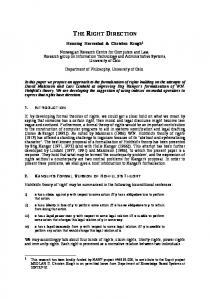
Based on the results described here, we propose a model for how the NMD pathway functions to recognize and degrade a nonsense-containing transcript (Figure 8). A termination codon in the A site of a translating ribosome causes the ribosome to pause. The translation termination factors eRF1 and eRF3 interact at the A site and promote assembly of the surveillance complex (Step 1). The termination factors stimulate peptide hydrolysis and subsequently disassociate from the ribosome. This triggers scanning of the surveillance complex for ~200 nt 39 of the termination codon in search of a DSE (Step 2). An interaction of the complex with the DSE (Step 3) forms an RNP structure in which the mRNA is subsequently decapped, independently of deadenylation, and degraded by a 59→39 exoribonuclease (Step 4). The model described above identifies several potential steps in which the STE may function to inactivate the decay pathway (Figure 8). Since the STE functions only when it is located 59 of the DSE (Figure 5 and data not shown), we suggest that the STE can: (i) prevent the surveillance complex from scanning 39 of the termination codon; (ii) promote disassembly of the surveillance complex from the mRNA; (iii) prevent the complex from being able to recognize the DSE; or (iv) prevent subsequent steps after recognition of the DSE from occurring, such as inhibiting decapping of the transcript. At present, we consider the fourth possibility unlikely. The steps that occur following recognition of the DSE are events that also take place in the degradation of wild-type transcripts (i.e. decapping and 59→39 exonucleolytic degradation). Therefore, it would be anticipated that the STE would also prevent rapid decay of normally unstable wild-type mRNAs. However, a GCN4–MFA2 hybrid transcript was degraded rapidly even in the presence of the STE (Figure 6). The results presented here define a number of key parameters that describe how the surveillance complex searches and recognizes a DSE to promote decay of aberrant transcripts. In addition, we have defined several ways to modulate the activity of the NMD pathway. Understanding the parameters of how the complex identifies a DSE and how the activity of the complex can be modulated is a central issue concerning the functioning and regulation of this pathway. We anticipate that these findings will aid in the understanding of how translation termination affects other processes, including scanning, translation reinitiation and recognition of specific sequences, and how these processes can be modulated. 585
M.J.Ruiz-Echevarrı´a, C.I.Gonza´lez and S.W.Peltz
Fig. 8. A model for the mechanism of the NMD pathway. Step 1: the ribosome pauses when a stop codon occupies the A site. This pause promotes the interaction of the release factors (eRF1 and eRF3) and assembles the surveillance complex. Step 2: following peptide hydrolysis, the termination factors disassociate from the ribosome, triggering scanning of the surveillance complex 39 of the termination codon in search of a DSE. Step 3: the surveillance complex recognizes and interacts with the DSE. Step 4: this interaction promotes an RNP structure that renders the RNA susceptible to decapping. The STE is represented as a rectangle and can exert its effect at several steps in the decay pathway (see text for details).
Materials and methods Yeast strains, growth conditions and transformation procedures The yeast strains used in this study were: RY260 (MATa ura3-52 rpb1-1), RY262 (MATα his4-519 ura3-52 rpb1-1; Nonet et al., 1987) and RY262– (MATα his4-519 ura3-52 upf1::hisG rpb1-1; Hagan et al., 1995), a upf1 deletion strain. RY260 was used to determine mRNA decay rates under starvation conditions. Cells harboring the rpb1-1 allele were grown at 24°C. Yeast media were prepared as described (Rose et al., 1990). Cells were cultured on defined minimal synthetic dextrose (SD) medium lacking uracil to select and maintain the plasmids used in these studies. Cells lacking plasmids were grown non-selectively in YPD media (Ausubel et al., 1992). Starvation conditions were achieved as described before (Hinnebusch and Fink, 1983). Briefly, saturated cultures were diluted 1:50 and grown for 2 h in SD media. After this time, cells were pelleted, washed and resuspended in SD medium lacking histidine and containing 10 mM 3-AT and grown for 6 h. 3-AT is a competitive inhibitor of the HIS3-encoded step of the histidine biosynthetic pathway. Yeast transformations were performed using the lithium acetate protocol (Schiestl and Gietz, 1989). Escherichia coli DH5α strain was used to amplify DNA using standard methods (Sambrook et al., 1989). mRNA decay measurements, RNA preparation and analysis The various GCN4, PGK1 and hybrid GCN4–PGK1 alleles were transformed into strains harboring the temperature-sensitive allele of the RNA polymerase II (rpb1-1) and the mRNA decay rates were determined by Northern analyses as described previously (Peltz et al., 1993; Hagan et al., 1995). The results of these experiments were quantitated using a Bio-Rad model G-250 Molecular Imager or a Bio-Rad model GS-670 Imaging Densitometer. The mRNA abundances were normalized using the U3 RNA (Myslinski et al., 1990). mRNA decay rates of the GCN4derived mRNAs from plasmids p4382 and p4383 were determined by RNase protection assays essentially as described (Sambrook et al., 1989). Probes were generated by in vitro transcription with SP6 or T3 RNA polymerases. Preparation of radioactive probes DNA probes were labeled to high specific activity with [α-32PO4]dCTP. A 0.5 kb EcoRI or a 0.75 kb BamHI–NheI fragment from plasmid p3519 (see below) spanning different parts of the GCN4 leader region was used to monitor the decay of the GCN4–PGK1 hybrid alleles used in
586
this study. A 0.5 kb NheI–HindIII fragment from pUC9PGKH2(3)UAG (Peltz et al., 1993) was used to monitor mRNA decay of the miniPGK1-derived alleles. A 4.0 kb SstI–SphI fragment from pUC18-HIS4 was used to monitor the decay of the HIS4 transcript (Peltz et al., 1992). A 0.5 kb BamHI–HpaI fragment from Bluescript KS-(U3) was used to prepare probes for the U3 small nuclear RNA (Myslinski et al., 1990). For the RNase protection assay, RNA probes were labeled with [α-32PO4]UTP. To monitor decay of GCN4 transcripts, a pGEM-derived plasmid containing the GCN4 leader sequences from 1300 to 1565 was linearized with Asp718 and in vitro transcribed with SP6. To monitor decay of the U3 transcript, a pGEM-derived plasmid containing the U3 gene was cut with SspI and in vitro transcribed with RNA polymerase T3. Plasmid constructions Unless otherwise indicated, all plasmid constructions described below contain a 185 bp DNA fragment derived from the GCN4 gene that contains the signals required for transcription initiation (Hinnebusch, 1984). In addition, they harbor the 39-UTR of the PGK1 gene containing the transcription termination and polyadenylation signals and a tag sequence (Peltz et al., 1993). All the hybrid GCN4–PGK1 constructs contain different regions of the GCN4 leader region followed by a DNA fragment containing the downstream element plus the 39-UTR of the PGK1 gene or a DNA fragment containing the mini-PGK1 gene (Peltz et al., 1993). To prepare the DNA fragment containing the downstream element plus the 39-UTR of the PGK1 gene, plasmid pUC9PGKH2(3)∆1IN1 (Peltz et al., 1993) was cleaved with NheI and HindIII to obtain a 0.65 kb DNA fragment. The fragment containing the mini-PGK1 gene, harboring or lacking the downstream element, was synthesized by PCR using the appropriate oligonucleotides as primers and plasmids pRIPPGKH2(3)∆1 or pRIPPGKH2(3)∆1IN1(1) (Peltz et al., 1993) as templates, respectively. The yeast centromeric plasmid pRIP (Parker and Jacobson, 1990) was used as vector for all the alleles constructed and was cleaved with BamHI and HindIII. To prepare constructs 4C (p4099) and 4D (p4098) in Figure 4A, pRIP was cleaved with BamHI and SphI. In order to facilitate the explanation on the construction of the plasmids, the description of the different plasmids is not shown in the same order as they appear in the Results section. The GCN4 leader regions present in plasmids depicted in Figure 2 were synthesized by PCR using plasmid pM23 (Miller and Hinnebusch, 1989) as a template. Construct 2A (p3518) contains the GCN4 leader region up to position 1297 (taking 11 as the transcription start site). Constructs 2B (p3536),
Identifying the appropriate termination codon 2C (p3537) and 2D (p3519) contain the GCN4 leader region up to positions 1393, 1458 and 1565, respectively. To prepare construct 2E (p3780), a fragment containing the GCN4 sequences 1393 to 1457 plus the PGK1 sequences containing the DSE and 39-UTR was synthesized by PCR, using as a template construct 2C (p3537). This fragment was ligated to a fragment containing the GCN4 leader region up to position 1297 which was synthesized by PCR using plasmid pM23 as a template. To prepare construct 2F (p4174), two PCRs were performed using construct p3536 (construct 2B) as the template. One reaction generated a fragment containing the GCN4 leader region up to position 1393. The second PCR generated a fragment containing GCN4 sequences 1243 to 1393, followed by PGK1 sequences harboring the DSE and 39-UTR. This fragment was ligated downstream of the first PCR fragment described above. In Figure 3, constructs 3B (p4093), 3C (p4095) and 3D (p4159) were made using the same strategy as described above for the construction of plasmids p3536, p3537 and p3519, respectively. However, plasmids p194, p210 (Mueller and Hinnebusch, 1986) and p237 (Grant et al., 1995) were used as templates to generate the GCN4 fragments present in p4093, p4095 and p4159, respectively. In addition, differently from p3519, p4159 contains the GCN4 leader sequences up to position 1498. In Figure 4A, construct 4B (p3493) is as previously described (Figure 3, construct 3 of Ruiz-Echevarria and Peltz, 1996). For constructs 4C (p4099) and 4D (p4098), a 518 nt fragment containing the GCN4 leader sequences up to nucleotide 1335 was synthesized by PCR amplification using plasmid p231 (Figure 3c, construct 2 of Mueller and Hinnebusch, 1986) as template. This fragment was ligated to: (i) a 1094 nt PCR containing the GCN4 sequences from 144 to 1565 followed by PGK1 sequences harboring the DSE and 39-UTR, which was synthesized using p3519 as template (for p4099); or (ii) a 1184 nt PCR fragment containing the GCN4 sequences from 144 to 1565 followed by a mini-PGK1 nonsense-containing allele, synthesized using p3493 as template (for p4098). In these constructs, both copies of uORF1 are preceded by the 186 nt found upstream of this segment and shown to be required for efficient reinitiation after translation of uORF1 (Grant et al., 1995). In Figure 4B, constructs 4E (p3778) and 4F (p3766) were prepared using plasmid pM23 (lacking uORF4; Miller and Hinnebusch, 1989) as the template, generating a 748 bp DNA fragment containing the GCN4 leader region up to position 1565. This PCR reaction was ligated together with: (i) a 796 nt fragment harboring a nonsense-containing mini-HIS4 synthesized using pRIPHIS4∆UAAIN1 (Hagan et al., 1995) as template; or (ii) with a 563 bp DNA fragment harboring the nonsensecontaining mini-PGK1, lacking the DSE (see above), to make p3778 and p3766, respectively. Construct 4G (p3503) has been described previously (Figure 4, construct 2 of Ruiz-Echevarria and Peltz, 1996). In Figure 5, construct 5A (p4170) was prepared by insertion of a sequence harboring a DSE and 39-UTR from the PGK1 gene in the unique EcoRI site of plasmid pM98 (Miller and Hinnebusch, 1989). The resulting plasmid was used as the template in a PCR reaction to generate a fragment that contains the coding sequence of uORF4 followed by its natural 25 39-flanking nucleotides, the GCN4 sequences 1267 to 1298 (normally 39 of uORF1) and PGK1 sequences harboring a DSE and the 39-UTR. A second PCR reaction using p4159 as the template yielded a 600 nt fragment containing the GCN4 leader sequence up to nucleotide 1417 that was ligated upstream of the first one. To prepare construct 5B (p4092), p3518 was used as template in a PCR reaction to synthesize a fragment that contains the coding sequence of uORF1 followed by its 57 39-flanking nucleotides and PGK1 sequences harboring a DSE and the 39-UTR. As described above, a second PCR reaction using p4159 as the template yielded a 600 nt fragment that was ligated upstream of the first one. To prepare construct 5C (p4158), plasmid p237 (Grant et al., 1995) was used as template to generate a 748 bp DNA fragment containing the GCN4 leader region up to position 1565. Construct 5D (p4171) was constructed in the same manner as p4158, except that the template for the PCR reaction was p189 (Grant et al., 1995) containing uORF1 in the position of uORF4. Construct 5E (p4159) is as described above. To prepare construct 5F (p4382), p4159 was used as template in a PCR reaction to synthesize a fragment that contains the GCN4 leader region up to position 1498, in which the AUG codons of uORF1, uORF2 and uORF3 are inactive. A second PCR reaction using p164 (Hinnebusch, 1985) as template yielded a fragment containing the GCN4 sequences from nucleotide 1565 in the leader region to the 39-end of the gene, that was ligated downstream of the first one. Construct 5G (p4383) was constructed in the same manner as p4382, except that the template for the first PCR reaction was uORF4-containing pM23 (Miller and Hinnebusch, 1989), yielding a fragment that contains the GCN4
leader region up to position 1498, in which the AUG codons of uORF2 and uORF3 are inactivated. In Figure 6, construct 6A [pPRIPGK(-AU)∆1UAAIN1] is as already described (Hagan et al., 1993). Construct 6B (p4180) was prepared by insertion of a PCR fragment carrying the GCN4 sequences from 1498 to 1565 in the unique HpaI site of pPRIPGK(-AU)∆1UAAIN1. Construct 6C (p4230) was made as p4180, except that the sequence inserted correspond to PGK1 sequences from 1791 to 1855. Construct 6D (p4385) was prepared by insertion of a PCR fragment carrying the GCN4 sequences from 1498 to 1565 followed by an in-frame UAA termination codon, in the unique NheI site of pPRIPPGKH2(3)∆1IN1(1) (Peltz et al., 1993) and in-frame with the PGK1 sequences. Construct 6E (p4381) was prepared by insertion of a PCR fragment carrying the GCN4 sequences from 1498 to 1565 in the unique BglII site of pPRIPPGKH2(3)∆1IN1(1). In Figure 6B, construct 6F (p4178) was prepared using plasmid p237 (Grant et al., 1995) as the template to generate a 748 bp DNA fragment containing the GCN4 leader region up to position 1565. A second PCR reaction, using as the template pRP455 (Beelman and Parker, 1994), yielded a fragment containing the MFA2 3’-UTR that was ligated downstream of the first one. Construct 6G (p4179) was prepared using the same procedure, except that the fragment synthesized using plasmid p237 (Grant et al., 1995) as the template contains the GCN4 leader sequences up to position 1498. In Figure 7, constructs 7A (p4133) and 7B (p3765) were prepared using uORF4-containing plasmid pM23 (Miller and Hinnebusch, 1989) as the template to generate a 748 bp DNA fragment containing the GCN4 leader region up to position 1565 (containing functional uORF1 and uORF4 and inactive uORF2 and uORF3). This PCR fragment was ligated to: (i) a 0.65 kb DNA fragment carrying a DSE plus the 39-UTR region of the PGK1 gene (described above; construct 7A); or (ii) a DNA fragment harboring the nonsense-containing mini-PGK1 allele (construct 7B). Constructs 7C (p4132) and 7D (p3712) were prepared using the same procedure, except that the PCR reaction used as a template plasmid p164 (Hinnebusch, 1985). To prepare construct 7E (p4341), PCR mutagenesis was performed on p3765, changing the AUG codon of the PGK1 ORF to AAG. In Figure 1, a DNA fragment harboring the uORF1 mutant sequence plus the 16 nt 59-flanking and the 25 nt 39-flanking sequences was obtained by annealing two complementary oligonucleotides (59-AGCTTAAAGATCATTGAAAAATGTAATAATAAACCGATTATATTTTGTTTTTAAAGTA-39; 59-GATCTACTTTAAAAACAAAATATAATCGGTTTATTATTACATTTTTCAATGATCTTTA-39; the mutant uORF1 is underlined). This DNA fragment was inserted in the place of uORF1 in p4092, yielding construct 1B (p4176). To prepare construct 1C (p4186), PCR mutagenesis was performed on p4092, changing the AUG codon of uORF1 to an AAG codon.
Acknowledgements We thank A.G.Hinnebusch and T.Dever for a number of plasmid constructs used for this study. We also thank Kevin Czaplisnki, Jon Dinman, Sam Gundersson, T.Goss Kinzy, T.Thisted and Shuang Zhang for helpful discussions and critical reading of the manuscript. This work was supported by grants from the National Institutes of Health (GM48631) and an American Heart Association Established Investigator Award given to S.W.P. M.J.R.-E. acknowledges the American Heart Association for support during part of this research.
References Abastado,J.P., Miller,P.M., Jackson,B.M. and Hinnebusch,A.G. (1991a) Suppression of ribosomal reinitiation at upstream open reading frames in amino acid-starved cells forms the basis for GCN4 translational control. Mol. Cell. Biol., 11, 486–496. Abastado,J.P., Miller,P.M. Jackson,B.M. and Hinnebusch,A.G. (1991b) A quantitative model for translational control of the GCN4 gene of Saccharomyces cerevisiae. The New Biologist, 3, 511–524. Aharon,T. and Schneider,R.J. (1993) Selective destabilization of shortlived mRNAs with the granulocyte–macrophage colony-stimulating factor AU-rich 39 noncoding region is mediated by a cotranslational mechanism. Mol. Cell. Biol., 13, 1971–1980. Andjelkovic,N., Zolnierowicz,S., Van Hoof,C., Goris,J. and Hemmings, B.A. (1996) The catalytic subunit of protein phosphatase 2A associates with the translation termination factor eRF1. EMBO J., 15, 7156–7167. Ausubel,F.M., Brent,R., Kingston,R.E., Moore,D.D., Seidman,J.G.,
587
M.J.Ruiz-Echevarrı´a, C.I.Gonza´lez and S.W.Peltz Smith,J.A. and Struhl,K. (1992) Current Protocols in Molecular Biology. John Wiley & Sons Inc., New York. Beelman,C.A. and Parker,R. (1994) Differential effects of translational inhibition in cis and in trans on the decay of the unstable yeast MFA2 mRNA. J. Biol. Chem., 269, 9687–9692. Cao,J. and Geballe,A.P. (1996) Inhibition of nascent-peptide release at translation termination. Mol. Cell. Biol., 16, 7109–7114. Caponigro,G. and Parker,R. (1996) Mechanisms and control of mRNA turnover in Saccharomyces cerevisiae. Microbiol. Rev., 60, 233–249. Clemens,M.J. (1996) Protein kinases that phosphorylate eIF2 and eIF2B, and their role in eukaryotic cell translational control. In Hershey,J.W.B., Mathews,M.B. and Sonenberg,N. (eds), Translational Control. Cold Spring Harbor Laboratory Press, Cold Spring Harbor, NY, pp. 139– 172. Curatola,A.M., Nadal,M.S. and Schneider,R. (1995) Rapid degradation of AU-rich element (ARE) mRNAs is activated by ribosome transit and blocked by secondary structure at any position 59 to the ARE. Mol. Cell. Biol., 15, 6331–6340. Cui,Y., Hagan,K.W., Zhang,S. and Peltz,S.W. (1995) Identification and characterization of genes that are required for the accelerated degradation of mRNAs containing a premature translational termination codon. Genes Dev., 9, 423–436. Dietz,H.C., McIntosh,I., Sakai,L.Y., Corson,G.M., Chalberg,S.C., Pyeritz,R.E. and Francomano,C.A. (1993) Four novel FBN1 mutations: significance for mutant transcript level and EGF-like domain calcium binding in the pathogenesis of Marfan syndrome. Genomics, 17, 469–475. Dietz,H.C., Franke,U., Furthmayer,H., Francomano,C.A., De Paepe,A., Devereux,R., Ramirez,F. and Pyeritz,R.E. (1995) The question of heterogeneity in Marfan syndrome. Nature Genet., 9, 228–231. Gottesman,S., Wickner,S. and Maurizi,M.R. (1997) Protein quality control: triage by chaperones and proteases. Genes Dev., 11, 815–823. Grant,C.M., Miller,P.M. and Hinnebusch,A.G. (1995) Sequences 59 of the first upstream open reading frame in GCN4 mRNA are required for efficient translation reinitiation. Nucleic Acid Res., 23, 3980–3988. Graves,R.A., Pandey,N.B., Chodchoy,N. and Marzluff,W.F. (1987) Translation is required for degradation of histone mRNA degradation. Cell, 48, 615–626. Hagan,K.W., Ruiz-Echevarria,M.J., Quan,Y. and Peltz,S.W. (1995) Characterization of cis-acting sequences and decay intermediates involved in nonsense-mediated mRNA turnover. Mol. Cell. Biol., 15, 809–823. Hall,G.W. and Thein,S. (1994) Nonsense codons mutations in the internal exon of the β-globin gene are not associated with a reduction in βmRNA accumulation: a mechanism for the phenotype of dominant βthalassemia. Blood, 83, 2031–2037. He,F. and Jacobson,A. (1995) Identification of a novel component of the nonsense-mediated mRNA decay pathway using an interacting protein screen. Genes Dev., 9, 437–459. He,F., Brown,A.H. and Jacobson,A. (1997) Upf1p, Nmd2p, and Upf3p are interacting components of the yeast nonsense-mediated mRNA decay pathway. Mol. Cell. Biol., 17, 1580–1594. Hershey,J.W.B. (1989) Protein phosphorylation controls translational rates. J. Biol. Chem., 264, 20823–20826. Hinnebusch,A.G. (1984) Evidence for translational regulation of the activator of general amino acid control in yeast. Proc. Natl Acad. Sci. USA, 81, 6442–6446. Hinnebusch,A.G. (1985) A hierarchy of trans-acting factors modulates translation of an activator of amino acid biosynthetic genes in Saccharomyces cerevisiae. Mol. Cell. Biol., 5, 2349–2360. Hinnebusch,A.G. (1988) Mechanisms of gene regulation in the general control of amino acid biosynthesis in Saccharomyces cerevisiae. Microbiol. Rev., 52, 248–273. Hinnebusch,A.G. (1993) Gene-specific translational control of the yeast GNC4 gene by phosphorylation of eukaryotic initiation factor 2. Mol. Microbiol., 10, 215–223. Hinnebusch,A.G. (1996) Translational control of GCN4: gene-specific regulation by phosphorylation of eIF2. In Hershey,J.W.B., Mathews,M.B. and Sonenberg,N. (eds), Translational Control. Cold Spring Harbor Laboratory Press, Cold Spring Harbor, NY, pp. 199– 244. Hinnebusch,A.G. and Fink,G.R. (1983) Repeated DNA sequences upstream from HIS1 also occur at several other co-regulated genes in Saccharomyces cerevisiae. J. Biol. Chem., 258, 5238–5247. Jacobson,A. and Peltz,S.W. (1996) Interrelationship of the pathways of mRNA decay and translation in eukaryotic cells. Annu. Rev. Biochem., 65, 693–739.
588
Kiledjian,M., Wang,X. and Liebhaber,S.A. (1995) Identification of two KH domain proteins in the alpha-globin mRNP stability complex. EMBO J., 14, 4357–4364. Lee,B.S. and Culbertson,M.R. (1995) Identification of an additional gene required for eukaryotic nonsense mRNA turnover. Proc. Natl Acad. Sci. USA, 92, 10354–10358. Leeds,P., Peltz,S.W., Jacobson,A. and Culbertson,M.R. (1991) The product of the yeast UPF1 gene is required for rapid turnover of mRNAs containing a premature translational termination codon. Genes Dev., 5, 2303–2314. Leeds,P., Wood,J.M., Lee,B.-S. and Culbertson,M.R. (1992) Gene products that promote mRNA turnover in Saccharomyces cerevisiae. Mol. Cell. Biol., 12, 2165–2177. Maquat,L.E. (1995) When cells stop making sense: effects of nonsense codons on RNA metabolism in vertebrate cells. RNA, 1, 453–465. Miller,P.F. and Hinnebusch,A.G. (1989) Sequences that surround the stop codons of upstream open reading frames in GCN4 mRNA determine their distinct functions in translational control. Genes Dev., 3, 1217–1225. Mueller,P.P. and Hinnebusch,A.G. (1986) Multiple upstream AUG codons mediate translational control of GCN4. Cell, 45, 201–207. Mulhrad,D. and Parker,R. (1992) Mutations affecting stability and deadenylation of the yeast MFA2 transcript. Genes Dev., 6, 2100–2111. Myslinski,E., Segault,V. and Branlant,C. (1990) An intron for the genes of the U3 small nucleolar RNAs of the yeast Saccharomyces cerevisiae. Science, 247, 1213–1216. Nonet,M., Scafe,C., Sexton,J. and Young,R. (1987) Eucaryotic RNA polymerase conditional mutant that rapidly ceases mRNA synthesis. Mol. Cell. Biol., 7, 1602–1611. Oliveira,C.C. and McCarthy,J.E.G. (1995) The relationship between eukaryotic translation and mRNA stability. A short upstream reading frame strongly inhibits translational initiation and greatly accelerates mRNA degradation in the yeast Saccharomyces cerevisiae. J. Biol. Chem., 270, 8936–8943. Parker,R. and Jacobson,A. (1990) Translation and a forty-two nucleotide segment within the coding region of the mRNA encoded by the MATα1 gene are involved in promoting rapid mRNA decay in yeast. Proc. Natl Acad. Sci. USA, 87, 2780–2784. Peltz,S.W., Brown,A.H. and Jacobson,A. (1993) mRNA destabilization triggered by premature translational termination depends on at least three cis-acting sequence elements and one trans-acting factor. Genes Dev., 7, 1737–1754. Pinto,I., Na,J.G., Sherman,F. and Hampsey,M. (1992) cis- and transacting suppressors of a translational initiation defect at the cyc1 locus of Saccharomyces cerevisiae. Genetics, 132, 97–112. Rose,M.D., Winston,F. and Hieter,P. (1990) Methods in Yeast Genetics. Cold Spring Harbor Laboratory Press, Cold Spring Harbor, NY. Ruiz-Echevarria,M.J. and Peltz,S.W. (1996) Utilizing the GCN4 leader region to investigate the role of the sequence determinants in nonsensemediated mRNA decay. EMBO J., 15, 2810–2819. Ruiz-Echevarria,M.J., Czaplinski,K. and Peltz,S.W. (1996) Making sense of nonsense in yeast. Trends Biochem. Sci., 21, 433–438. Sambrook,J., Fritsch,E.F. and Maniatis,T. (1989) Molecular Cloning: A Laboratory Manual. Cold Spring Harbor Laboratory Press, Cold Spring Harbor, NY. Savant-Bhonsale,S. and Cleveland,D.W. (1992) Evidence for instability of mRNAs containing AUUUA motifs mediated through translationdependent assembly of a .20S degradation complex. Genes Dev., 6, 1927–1939. Schiestl,R.H. and Gietz,R.D. (1989) High efficiency transformation of intact yeast cells using single stranded nucleic acid as a carrier. Curr. Genet., 16, 339–346. Suzuki,C.K., Rep,M., van Dijl,J.M., Suda,K., Grivell,L.A. and Schatz,G. (1997) ATP-dependent proteases that also chaperone protein biogenesis. Trends Biochem. Sci., 22, 118–123. Wang,X., Kiledjian,M., Weiss,I.M. and Liebhaber,S.A. (1995) Detection and characterization of a 39 untranslated region ribonucleoprotein complex associated with human alpha-globin mRNA stability. Mol. Cell. Biol., 15, 1769–1777. Weiss,I.M. and Liebhaber,S.A. (1995) Erythroid cell-specific mRNA stability elements in the alpha 2-globin 39 nontranslated region. Mol. Cell. Biol., 15, 2457–2465. Weng,Y., Czaplinski,K. and Peltz,S.W. (1996a) Identification and characterization of mutations in the UPF1 gene that affect nonsense suppression and the formation of the Upf protein complex but not mRNA turnover. Mol. Cell. Biol., 16, 5491–5506.
Identifying the appropriate termination codon Weng,Y., Czaplinski,K. and Peltz,S.W. (1996b) Genetic and biochemical characterization of the mutations in the ATPase and helicase regions of the Upf1 protein. Mol. Cell. Biol., 16, 5447–5490. Weng,Y., Ruiz-Echevarria,M.J., Zhang,S., Cui,Y. Czaplinski,K., Dinman,J. and Peltz,S.W. (1997) Characterization of the nonsensemediated decay pathway and its effect in modulating translation termination and programmed frameshifting. Modern Cell Biol., 17, 241–263. Winstall,E., Gamache,M. and Raymond,V. (1995) Rapid mRNA degradation mediated by the c-fos 39 AU-rich element and that mediated by the granulocyte–macrophage colony-stimulating factor 39 AU-rich element occur through similar polysome-associated mechanisms. Mol. Cell. Biol., 15, 3796–3804. Yun,D.-F. and Sherman,F. (1995) Initiation of translation can occur only in a restricted region of the CYC1 mRNA of Saccharomyces cerevisiae. Mol. Cell. Biol., 15, 1021–1033. Zhang,J. and Maquat,L.E. (1997) Evidence that translation reinitiation abrogates nonsense-mediated mRNA decay in mammalian cells. EMBO J., 16, 826–833. Zhang,S., Ruiz-Echevarria,M.J., Quan,Y. and Peltz,S.W. (1995) Identification and characterization of a sequence motif involved in nonsense-mediated mRNA decay. Mol. Cell. Biol., 15, 2231–2244. Received July 2, 1997; revised October 30, 1997; accepted November 11, 1997
589