International Journal of
Molecular Sciences Article
Immune-Response Patterns and Next Generation Sequencing Diagnostics for the Detection of Mycoses in Patients with Septic Shock—Results of a Combined Clinical and Experimental Investigation Sebastian O. Decker 1 , Annette Sigl 1 , Christian Grumaz 2 , Philip Stevens 2,3 , Yevhen Vainshtein 2 , Stefan Zimmermann 4 , Markus A. Weigand 1 , Stefan Hofer 5 , Kai Sohn 2,† and Thorsten Brenner 1, *,† 1
2
3 4 5
* †
Department of Anesthesiology, Heidelberg University Hospital, 110, Im Neuenheimer Feld, D-69120 Heidelberg, Germany;
[email protected] (S.O.D.);
[email protected] (A.S.);
[email protected] (M.A.W.) Fraunhofer IGB, 12, Nobelstraße, D-70569 Stuttgart, Germany;
[email protected] (C.G.);
[email protected] (P.S.);
[email protected] (Y.V.);
[email protected] (K.S.) Noscendo GmbH, 9, Meitnerstraße, D-70563 Stuttgart, Germany Department of Infectious Diseases, Medical Microbiology and Hygiene, Heidelberg University Hospital, 324, Im Neuenheimer Feld, D-69120 Heidelberg, Germany;
[email protected] Department of Anesthesiology, Westpfalzklinikum, 1, Hellmut-Hartert-Straß, D-67655 Kaiserslautern, Germany;
[email protected] Correspondence:
[email protected]; Tel.: +49-(0)-6221-5639418 Thorsten Brenner and Kai Sohn share senior authorship.
Received: 14 July 2017; Accepted: 14 August 2017; Published: 18 August 2017
Abstract: Fungi are of increasing importance in sepsis. However, culture-based diagnostic procedures are associated with relevant weaknesses. Therefore, culture- and next-generation sequencing (NGS)-based fungal findings as well as corresponding plasma levels of β-D-glucan, interferon gamma (INF-γ), tumor necrosis factor alpha (TNF-α), interleukin (IL)-2, -4, -6, -10, -17A, and mid-regional proadrenomedullin (MR-proADM) were evaluated in 50 septic patients at six consecutive time points within 28 days after sepsis onset. Furthermore, immune-response patterns during infections with Candida spp. were studied in a reconstituted human epithelium model. In total, 22% (n = 11) of patients suffered from a fungal infection. An NGS-based diagnostic approach appeared to be suitable for the identification of fungal pathogens in patients suffering from fungemia as well as in patients with negative blood cultures. Moreover, MR-proADM and IL-17A in plasma proved suitable for the identification of patients with a fungal infection. Using RNA-seq., adrenomedullin (ADM) was shown to be a target gene which is upregulated early after an epithelial infection with Candida spp. In summary, an NGS-based diagnostic approach was able to close the diagnostic gap of routinely used culture-based diagnostic procedures, which can be further facilitated by plasmatic measurements of MR-proADM and IL-17A. In addition, ADM was identified as an early target gene in response to epithelial infections with Candida spp. Keywords: mid-regional proadrenomedullin; interleukin-17A; β-D-glucan; next-generation sequencing; mycoses; sepsis; septic shock
1. Introduction Sepsis, defined as a life-threatening organ dysfunction caused by a dysregulated host response to infection [1], is most frequently a result of a bacterial infection [2–4], whereas those of a fungal Int. J. Mol. Sci. 2017, 18, 1796; doi:10.3390/ijms18081796
www.mdpi.com/journal/ijms
Int. J. Mol. Sci. 2017, 18, 1796
2 of 32
or viral nature are less common [5–7]. Accordingly, fungemia is only present in 3% of unselected sepsis cases [5,8,9]. Nevertheless, fungi are one of the most isolated species recovered from abdominal foci in peritonitis [10,11] and numerous patients develop fungal colonization during their hospital stay [5]. Although the group of infected patients appears to be small, the number of severe fungal infections is escalating due to an increase in the number of immunocompromised patients, a more aggressive surgical therapy in older patients with relevant co-morbidities, and an increasing number of oncologic diseases [5,8,12,13]. Within this context, three fungal species seem to be of most relevance in Europe: Candida albicans (C. albicans), Candida glabrata (C. glabrata), and Aspergillus fumigatus [14,15]. Sepsis-associated mortality in patients suffering from an invasive fungal infection is known to be high, with up to 42% of mortalities observed due to Candida spp. and even higher numbers in response to Aspergillus spp. infections [16–18]. Especially in patients suffering from fungemia, diagnostic weaknesses may substantially contribute to this alarming mortality. Only a small proportion of affected patients show positive blood cultures, and fungal growth on culture media is known to be very slow. Accordingly, several studies have shown that invasive fungal infections are the most frequently missed diagnoses in critically ill patients [19–21]. Consequently the administration of life-saving antifungal therapy may either be completely absent, or initiated with a minimum delay of 2–3 days [22]. Indeed, such a delay is known to be associated with an increased mortality [23,24]. On the other hand, molecular biological methods for the detection of fungal isolates (e.g., polymerase chain reaction (PCR)-based methods) have already been shown to be of value (saving time, good sensitivity, and specificity) [25]. However, up to now there are still no commercial assays available [26]. The aims of this observational, prospective clinical study were therefore to (1) assess the incidence, risk factors and relevance with regard to the outcome of fungal infections in patients with septic shock; (2) evaluate the additional benefit of a next-generation sequencing (NGS)-based diagnostic procedure; and (3) assess the host response upon fungal infection by investigating the diagnostic value of β-D-glucan (BG), galactomannan (GM), interferon gamma (INF-γ), tumor necrosis factor alpha (TNF-α), interleukins (IL)-2, -4, -6, -10, -17A, and mid-regional proadrenomedullin (MR-proADM) in septic patients as well as by analyzing the overall host response by expression profiling of Candida spp.-infected epithelia in vitro. 2. Results 2.1. Patient’s Characteristics In total, 50 patients with septic shock were included in the presented investigation. Patients’ characteristics are presented in Table 1. The underlying septic focus was the abdomen (n = 43; 86%), followed by the lung (n = 6; 12%), and the urogenital tract (n = 1; 2%). The overall 28 and 90 day mortality rates were 22% (n = 11) and 34% (n = 17), respectively. The median length of intensive care unit (ICU) and hospital stay was 20 and 44 days, respectively.
Int. J. Mol. Sci. 2017, 18, 1796
3 of 32
Table 1. Patient’s characteristics (n = 50).
Parameter
Gender male Age BMI Postoperative peritonitis initial operation Kidney Liver Pancreas GIT VAS Others ≥48 h after hospital admission NYHA 0-I Diabetes mellitus Arterial hypertension Coronary heart disease Chronic obstructive lung disease Renal insufficiency Renal replacement therapy Liver cirrhosis Oncological disease APACHE II # SOFA # SAPS II # Candida colonization Candida infection Candidemia Aspergillus spp. Candida-Score Ventilation duration ICU length of stay Hospital length of stay Tracheotomy Anastomosis leakage Fascia dehiscence 90 day mortality 28 day mortality
Unit
All Patients (n = 50)
Without Fungal Isolates (n = 17)
With Fungal Isolates (n = 33)
p for Patients without Fungal Isolates vs. Patients with Fungal Isolates
(years) (kg/m2 )
38 (76) 66 (61–75) 27.2 (24.4–30.9) 31
11 (64.7) 71 (64–80) 27.2 (25.7–34.9) 9 (52.9)
27 (81.8) 66 (59–74) 26.9 (23.1–30.9) 22 (66.7)
0.160 0.117 0.401 0.206
2 (4) 11 (22) 2 (10) 38 (76) 3 (6) 12 (24) 25 (50) 41 (82) 17 (34) 34 (68) 8 (16) 10 (20) 7 (14) 15 (30) 13 (26) 33 (66) 30 (28–35) 11 (10–14) 65 (49–75) 22 (44) 10 (20) 3 (6) 1 (3) 4 (4–4) 145.5 (67.3–450) 19.5 (12–44) 44 (23.3–68.5) 14 (28) 24 (48) 12 (24) 17 (34) 11 (22)
0 (0) 1 (2.1) 1 (5.9) 14 (82.4) 2 (11.8) 3 (17.6) 7 (41.2) 13 (76.4) 5 (29.4) 12 (70.6) 5 (29.4) 5 (29.4) 1 (5.9) 2 (11.8) 3 (17.6) 11 (64.7) 32 (28–36) 11 (10–14) 72 (48–75) 0 (0) 0 (0) 0 (0) 0 (0) 4 (4–4) 89 (46–145) 12 (3–17) 24 (12–40) 2 (11.8) 7 (41.2) 2 (11.8) 8 (47.1) 7 (41.2)
2 (6.1) 10 (30.3) 1 (3.0) 24 (72.7) 1 (3.0) 9 (27.3) 18 (54.5) 28 (84.8) 12 (36.3) 22 (66.7) 3 (9.1) 5 (15.2) 6 (18.2) 13 (39.4) 10 (30.3) 22 (66.7) 30 (28–34) 11 (10–14) 65 (51–72) 22 (66.7) 10 (30.3) 3 (9.1) 1 (3.0) 4 (4–4) 181 (77–682) 24 (15–46) 51 (39–78) 12 (36.3) 17 (51.5) 10 (30.3) 9 (27.3) 4 (12.1)
0.431 0.047 * 0.569 0.350 0.264 0.350 0.276 0.358 0.434 0.520 0.076 0.204 0.231 0.041 * 0.270 0.566 0.491 0.959 0.467 — — — — 0.080 0.015 * 0.002 ** 0.007 ** 0.063 0.347 0.134 0.175 0.025 *
(h) (days) (days)
Data are presented either as number (with the corresponding percentage value) or as median (with accompanying quartiles (Q1–Q3). Legend: BMI = Body mass index, GIT = gastro intestinal tract, VAS = vascular artery surgery, NYHA = New York Heart Association score, APACHE II = Acute Physiology Health Evaluation score, SAPS II = Simplified Acute Physiology Score, SOFA = Sequential Organ Failure Assessment score; # calculated at sepsis onset. Concerning symbolism and higher orders of significance: * p < 0.05, ** p < 0.01, — : not calculated.
2.2. Fungal Pathogens and Infection Sites Culture-Based Microbiological Diagnostics As assessed by culture-based microbiological diagnostics, fungal pathogens were present in 33 patients (66.0%), whereas 17 patients (34.0%) revealed negative fungal cultures (Figure 1). Fungal isolates were found in single or multiple locations in 25 (75.8%), or 8 (24.2%) patients respectively, and were located at the following sites: respiratory tract (n = 17; 51.5%), abdominal site (n = 21; 63.6%) and blood culture (n = 3; 9.1%) (double-naming feasible, Figure 1). Characteristics of patients with or without fungal pathogens are presented in Table 1. Patients with fungal pathogens more frequently underwent liver surgery prior to study inclusion and the need for renal replacement therapy was shown to be significantly increased. Concerning further markers for morbidity, fungal-positive patients revealed a significantly prolonged duration of mechanical ventilation, and the need for tracheostomy tended to be increased. Moreover, the length of both ICU and hospital stay was significantly prolonged in patients with fungal pathogens. Surprisingly, 28 day mortality was significantly increased in patients without fungal pathogens, whereas 90 day mortality was shown to be comparable.
underwent liver surgery prior to study inclusion and the need for renal replacement therapy was shown to be significantly increased. Concerning further markers for morbidity, fungal-positive patients revealed a significantly prolonged duration of mechanical ventilation, and the need for tracheostomy tended to be increased. Moreover, the length of both ICU and hospital stay was significantly prolonged in patients with fungal pathogens. Surprisingly, 28 day mortality was Int. J. Mol. Sci. 2017, 18, 1796 significantly increased in patients without fungal pathogens, whereas 90 day mortality was shown4 of 32 to be comparable.
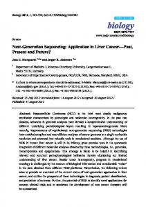
Figure 1. Identification of fungal pathogens in patients with septic shock (n = 50).
Figure 1. Identification of fungal pathogens in patients with septic shock (n = 50).
Based on the group definitions as described in the methods section, fungal colonization and fungal infection was found in 22 (44.0%) and 11in (22.0%) patients,section, respectively (Figure 1). In colonized Based on the group definitions as described the methods fungal colonization and fungal patients, 8 (16.0%) participants exclusively revealed Candida spp. in respiratory secretions (5×patients, C. infection was found in 22 (44.0%) and 11 (22.0%) patients, respectively (Figure 1). In colonized albicans, 1× C. albicans and C. glabrata, 2× C. albicans and C. spp.), whereas in 6 (12.0%) patients Candida 8 (16.0%) participants exclusively revealed Candida spp. in respiratory secretions (5× C. albicans, spp. could only be cultured from drainage fluids (3× C. albicans, 2× C. glabrata, 1× C. albicans and C. 1× C. albicans and C. glabrata, 2× C. albicans and C. spp.), whereas in 6 (12.0%) patients Candida spp. glabrata). In contrast, 8 (16.0%) patients were colonized at both sides (4× C. albicans, 1× C. albicans and could only be cultured from drainage fluids (3× C. albicans, 2× C. glabrata, 1× C. albicans and C. spp., 3× C. albicans and C. glabrata). In infected patients, fungemia was found in 3 (6.0%) patients C. glabrata). In contrast, 8 (16.0%) patients colonized at swabs both sides × C.inalbicans, C. albicans (2× C. albicans, 1× C. glabrata) and positivewere abdominal wound were (4 found 7 (14.0%)1× patients and C. spp., 3 × C. albicans and C. glabrata). In infected patients, fungemia was found in 3 (6.0%) patients (4× C. albicans, 1× C. glabrata, 1× C. krusei, 1× C. albicans and C. glabrata). Moreover, in one (2.0%) patient (2× C. albicans, 1 × C. glabrata) and positive abdominal wound swabs were found in 7 (14.0%) patients Aspergillus fumigatus was isolated in respiratory tract secretions. Detailed characteristics of patients without a fungal infection (colonized patients as C. well as patients any fungal isolates),in patients (4× C. albicans, 1× C. glabrata, 1× C. krusei, 1× albicans andwithout C. glabrata). Moreover, one (2.0%) with a fungal colonization, andisolated patients in with a fungal infection are presented in Tables 2 and 3. of patient Aspergillus fumigatus was respiratory tract secretions. Detailed characteristics Concerning factors, liver surgery prior patients to study as inclusion well aswithout liver cirrhosis couldisolates), be patients without risk a fungal infection (colonized well as as patients any fungal observed more frequently in patients with a fungal infection. Moreover, the duration of ICU stay and patients with a fungal colonization, and patients with a fungal infection are presented in Tables 2 and 3.
Concerning risk factors, liver surgery prior to study inclusion as well as liver cirrhosis could be observed more frequently in patients with a fungal infection. Moreover, the duration of ICU stay and mechanical ventilation was significantly prolonged and the need for tracheotomy was significantly increased in patients suffering from a fungal infection (Table 3). Although morbidity was shown to be increased, mortality at 28 and 90 days did not differ significantly between patients with and without a fungal infection. Table 2. Characteristics of patients with a fungal colonization or a fungal infection. Parameter Male Age BMI Postoperative peritonitis Initial operation Kidney Liver Pancreas GIT VAS Others
Unit
Fungal Colonization (n = 22)
Fungal Infection (n = 11)
p for Patients with Fungal Colonization vs. Patients with Fungal Infection
(years) (kg/m2 )
17 (77.3) 66 (61–74) 25.3 (21.6–30.8) 14 (63.6)
10 (90.1) 65 (58–74) 27.4 (26–30.5) 8 (72.7)
0.329 0.355 0.925 0.454
1 (4.5) 3 (13.6) 1 (4.5) 16 (72.7) 0 (0) 5 (22.7)
1 (9.1) 7 (63.6) 0 (0) 8 (72.7) 1 (9.1) 4 (36.4)
0.563 0.006 ** 0.667 0.653 0.333 0.333
Int. J. Mol. Sci. 2017, 18, 1796
5 of 32
Table 2. Cont.
Parameter
≥48 h after hospital admission NYHA 0-I Diabetes mellitus Arterial hypertension Coronary heart disease Chronic obstructive lung disease Renal insufficiency Renal replacement therapy Liver cirrhosis Oncological disease APACHE II # SOFA # SAPS II # Candida-Score Ventilation duration Tracheotomy Fascia dehiscence Anastomosis leakage ICU length of stay Hospital length of stay 90 day mortality 28 day mortality
Unit
(h)
(days) (days)
Fungal Colonization (n = 22)
Fungal Infection (n = 11)
p for Patients with Fungal Colonization vs. Patients with Fungal Infection
15 (68.2) 17 (77.3) 8 (36.4) 15 (68.2) 2 (9.1) 5 (22.7) 5 (22.7) 8 (36.4) 3 (13.6) 14 (63.6) 30 (29–34) 11 (10–13) 61 (44–72) 4 (4–4) 148.5 (74–239.3) 2 (9.1) 3 (13.6) 11 (50) 21 (13.5–43.5) 50 (34.5–68.5) 4 (18.2) 3 (13.6)
3 (27.3) 11 (100) 4 (36.4) 7 (63.6) 1 (9.1) 0 (0) 1 (9.1) 5 (45.5) 7 (63.6) 8 (72.7) 29 (28–33) 14 (11–15) 68 (57–77) 4 (4–4) 600 (424.5–944) 8 (72.7) 7 (63.6) 6 (54.5) 38 (25.5–64) 53 (47.5–88) 5 (45.5) 1 (9.1)
0.031 * 0.111 0.653 0.546 0.748 0.111 0.329 0.446 0.006 ** 0.454 0.396 0.044 * 0.336 1.0 0.040 * 0.002 ** 0.006 ** 0.549 0.082 0.418 0.120 0.593
Data are presented either as number (with the corresponding percentage value) or as median (with accompanying quartiles (Q1–Q3). Legend: BMI = Body mass index, GIT = gastro intestinal tract, VAS = vascular artery surgery, NYHA = New York Heart Association score, APACHE II = Acute Physiology Health Evaluation score, SAPS II = Simplified Acute Physiology Score, SOFA = Sequential Organ Failure Assessment score, # calculated at sepsis onset. Concerning symbolism and higher orders of significance: * p < 0.05, ** p < 0.01.
Table 3. Characteristics of patients with or without a fungal infection. Parameter Gender male Age BMI Postoperative peritonitis initial operation Kidney Liver Pancreas GIT VAS Others ≥48 h after hospital admission NYHA 0-I Diabetes mellitus Arterial hypertension Coronary heart disease Chronic obstructive lung disease Renal insufficiency Renal replacement therapy Liver cirrhosis Oncological disease APACHE II # SOFA # SAPS II # Candida-Score Candida colonization Candida infection Candidemia Aspergillus spp. Ventilation duration ICU length of stay Hospital length of stay Tracheotomy Anastomosis leakage Fascia dehiscence 90 day mortality 28 day mortality
Unit
Without Fungal Infection (n = 39)
Fungal Infection (n = 11)
p for Patients without Fungal Infection vs. Patients with Fungal Infection
(years) (kg/m2 )
28 (71.8) 66 (63–76) 27.2 (23.2–33.2) 23 (58.9)
10 (90.1) 65 (58–74) 27.4 (26–30.5) 8 (72.7)
0.184 0.460 0.582 0.322
1 (2.6) 4 (10.3) 2 (5.1) 30 (76.9) 2 (5.1) 8 (20.5) 22 (56.4) 30 (76.9) 13 (33.3) 27 (69.2) 7 (17.9) 10 (25.6) 6 (15.4) 10 (25.6) 6 (15.4) 25 (64.1) 31 (28–36) 11 (10–13) 62 (47–74) 4 (4–4) 22 (56.4) 0 (0%) 0 (0%) 0 (0%) 120 (60–200) 16 (10–28.5) 40 (21.5–63) 4 (10.3) 18 (46.2) 13 (33.3) 12 (30.8) 10 (25.6)
1 (9.1) 7 (63.6) 0 (0) 8 (72.7) 1 (9.1) 4 (36.4) 3 (27.3) 11 (100) 4 (36.4) 7 (63.6) 1 (9.1) 0 (0) 1 (9.1) 5 (45.5) 7 (63.6) 8 (72.7) 29 (28–33) 14 (11–15) 68 (57–77) 4 (4–4) 0 (0%) 10 (90.1) 3 (27.3) 1 (9.1) 600 (424.5–944) 38 (25.5–64) 53 (47.5–88) 8 (72.7) 6 (54.5) 7 (63.6) 5 (45.5) 1 (9.1)
0.395 0.001 *** 0.605 0.528 0.534 0.240 0.085 0.115 0.560 0.495 0.430 0.062 0.604 0.184 0.003 ** 0.440 0.335 0.081 0.519 0.881 — — — — 0.007 ** 0.008 ** 0.075 0.001 *** 0.440 0.002 ** 0.293 0.232
(h) (days) (days)
Data are presented either as number (with the corresponding percentage value) or as median (with accompanying quartiles (Q1–Q3). Legend: BMI = Body mass index, GIT = gastro intestinal tract, VAS = vascular artery surgery, NYHA = New York Heart Association score, APACHE II = Acute Physiology Health Evaluation score, SAPS II = Simplified Acute Physiology Score, SOFA = Sequential Organ Failure Assessment score; # calculated at sepsis onset. Concerning symbolism and higher orders of significance: ** p < 0.01, *** p < 0.001, — : not calculated.
Int. J. Mol. Sci. 2017, 18, 1796
6 of 32
2.3. NGS-Based Microbiological Diagnostics An unbiased approach for the diagnoses of bloodstream infections based on high-throughput sequencing of cell-free plasma DNA and the calculation of a sepsis indicating quantifier (SIQ)-score has been described previously [27]. Using this approach, bacterial or viral bloodstream infections can be identified with a high sensitivity. For the detection of fungal species in plasma from this cohort, we calculated a modified fungal SIQ score as a product of abundance and significance in each of the patients, which was then compared with clinical microbiology data as well as supplemented data on anti-infective therapy over a trial period of 28 days. Accordingly, we consequently present detailed data of three selected patients, describing the suitability of a NGS-based approach for the detection of invasive mycoses for three different scenarios: 2.3.1. Confirmation of Culture-Based Diagnostics of Candidemia by Fungal SIQ Score Patient S16 suffered from candidemia (as assessed by culture-based diagnostics) fourteen days after sepsis onset due to recurrent small bowel leakage. However, Candida spp. were present in drainage fluids already one week prior to the onset of candidemia (at six days after sepsis onset), which led to the initiation of a fluconazole therapy and was secondarily switched to caspofungin, as soon as candidemia occurred (Figure 2A). Blood cultures revealed positive results for C. albicans 2 and 4 weeks after inclusion into the study which was confirmed by SIQ score analyses (Figure 2A) However, high-throughput sequencing already revealed low levels of C. albicans sequence reads at the day of inclusion, but were not considered significant under the stringent criteria applied for this analyses (material not intended for publication). Nevertheless, with an increased sensitivity this NGS approach holds promise to diagnose blood stream infection significantly earlier than conventional microbiological culturing. 2.3.2. NGS-Based Diagnosis of Candidemia in Patients with Suggested Infection Based on Microbiological Identification of Candida spp. in Other Than Blood Culture Patient S25 suffered from recurrent intra-abdominal abscesses as well as a right-sided pleural empyema following a right-sided hemihepatectomy due to a Klatskin tumor. Candida glabrata and Candida albicans were found by microbiological testing of drainage fluids, wound swabs, as well as fresh puncture materials (Figure 2B). Although blood cultures remained negative for fungi at all time points, NGS-based calculation of a fungal SIQ score was positive (at 7 days after sepsis onset) for Candida glabrata already 6 days before microbiological testing of fresh puncture materials (at 13 days after sepsis onset) and therefore strongly supported the earlier presence of a candidemic infection. In this context, the identification of C. glabrata infection was further supported by positive drainage fluid cultures. 2.3.3. Detection of Invasive Mycoses by Fungal SIQ Score Even in Those Patients Which Have So Far Been Classified as Colonized Based on Culture-Based Diagnostic Procedures Due to restrictions including the limited sensitivity of culture-based diagnostics, some patients might be falsely classified as colonized despite the presence of an invasive fungal infection. Accordingly, a more sensitive and reliable diagnostic procedure would be of value in order to minimize false-negative test results. For example, patient S35 (Figure 2C) was classified as being colonized (as assessed by culture-based diagnostics in drainage fluids or wound swaps), although calculation of the SIQ score clearly revealed signs of an invasive Candida glabrata infection already at 2 days after sepsis onset. In contrast, the identification of C. glabrata at the species level was provided by microbiological diagnostics 17 days later in drainage fluid, which might contribute to the delayed prescription of caspofungin in that patient. Knowledge of these NGS-based results would probably have led to the initiation of an antifungal therapy, which might have been of relevance for the presented patient.
Int. J. Mol. Sci. 2017, 18, 1796
7 of 32
Int. J. Mol. Sci. 2017, 18, 1796
8 of 31
(A)
Figure 2. Cont.
Int. J. Mol. Sci. 2017, 18, 1796
8 of 32
Int. J. Mol. Sci. 2017, 18, 1796
9 of 31
(B)
Figure 2. Cont.
Int. J. Mol. Sci. 2017, 18, 1796
9 of 32
Int. J. Mol. Sci. 2017, 18, 1796
10 of 31
(C)
Int. J. Mol. Sci. 2017, 18, 1796
10 of 32
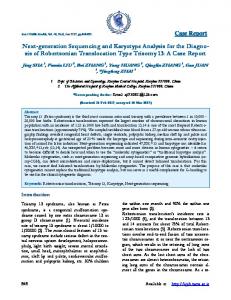
Figure 2. Time course (fungal) SIQ analyses compared with conventional clinical microbiology data of septic patients. The anti-infective treatment regime and (fungal) SIQ scores for species identified via NGS of the respective plasma samples are reported for a time course of 28 days (indicated by the x-axis) for patients S16 (A), S25 (B), and S35 (C).Only species identified by SIQ-score analyses are indicated at the left side. Red colored boxes reveal ranking of highest SIQ scores for the respective species in every patient. Pertinent (clinical microbiology) laboratory results are marked using arrows to indicate the day the clinical specimen was obtained. (A) A 73-year old male patient presented with a tumor of his bile duct with the need for a palliative resection. The surgical procedure included resections of the bile duct as well as the gallbladder and was followed by a double bypass procedure (biliodigestive anastomosis and gastrojejunal anastomosis). Four days after the initial operation the patient suffered from septic shock due to a duodenal ulcer perforation with the need for a total pancreatectomy. Shortly after, the patient suffered from another small bowel leakage, so that an additional small bowel resection had to be performed. Blood cultures at sepsis onset were shown to be negative, and meropenem (MEM) was administered in terms of an empiric antibiotic therapy. However, the patient suffered from a therapy-refractory course of the disease and C. albicans could be isolated from abdominal drainage fluids 6 days after sepsis onset. Accordingly, an additional antifungal treatment with fluconazole (FLC) was initiated. Due to the development of candidemia at 14 days after sepsis onset, this antifungal treatment regime was secondarily escalated towards caspofungin (CFG). These findings were in good agreement with next generation sequencing (NGS) diagnostics in plasma, since the SIQ-score was positive for C. albicans at the same timepoint. Abbreviations: NGS, next generation sequencing; SIQ, sepsis indicating quantifier; MEM, meropenem; IPM:CIL, imipenem/cilastatin; FLC, fluconazole; DOR, doripenem; CFG, caspofungin; BC, blood culture; CVC, central venous catheter; TS, tracheal secretion; (B) A 65-year old male patient suffered from a Klatskin tumor with the need for a right-sided hemihepatectomy. Due to an abscess at the resection site, the patient suffered from septic shock with the need for an interventional drainage 22 days after the initial operation. The further course was complicated by the development of a right-sided pleural empyema as well as recurrent intra-abdominal abscesses, which were both treated with repeated placements of interventional drainages. Empiric antibiotic therapy at sepsis onset included imipenem/cilastatin (IMP:CIL) in terms of a monotherapy. Culture-based microbiological diagnostics revealed no bacterial growth, whereas C. glabrata could be detected in both fluids of already positioned drainages as well as fresh puncture materials respectively. Based on these microbiological findings, the patient was classified as infected, so that an administration of caspofungin was started at 14 days after sepsis onset. Blood cultures remained negative for fungi at all time points. Contrariwise, a next generation sequencing (NGS)-based diagnostic approach in plasma samples of septic patients was able to support the presence of an invasive fungal infection already at 7 days after sepsis onset, since the SIQ-score was shown to be positive for C. glabrata at this time point. Unfortunately, a further evaluation of the patient’s course of the disease beyond 14 days after sepsis onset was not possible, since the patient denied further participation in the study.
Int. J. Mol. Sci. 2017, 18, 1796
11 of 32
Abbreviations: CFG, caspofungin; IMP:CIL, imipenem/cilastatin; na, not available; nd, not detectable; NGS, next generation sequencing; SIQ, sepsis indicating; (C) A 71-year-old female patient presented with a right pleural empyema caused by a liver abscess with the need for a video-assisted thoracoscopy (VATS). One day after VATS, the patient suffered from an acute abdomen with septic shock due to a perforation of the sigmoid colon, so that a removal of the sigmoid colon had to be performed. A second explorative laparotomy was necessary at 10 days after sepsis onset, due to a messy drainage fluid with a suspicion of another bowel leakage. However, during the revision surgery no clear focus could be found. Empiric anti-infective treatment consisted of imipenem/cilastatin (IMP:CIL) in combination with fluconazole (FLC), which was further supplemented by vancomycin (VAC) for 2 days in the early phase after sepsis onset. Anti-infective treatment was stepwise deescalated, so that the patient was free of any antibiotics or antimycotics at 12 days after sepsis onset. In the further course of the disease, the administration of caspofungin (CFG) was started at 20 days after sepsis onset, since the patient did not recover well and drainage fluids were shown to be positive for Candida spp. repeatedly starting from 3 days after sepsis onset. In parallel, next generation sequencing (NGS)-based diagnostics revealed a positive SIQ-score for C. glabrata also at 3 days after sepsis onset, whereas blood cultures were found to be negative for fungi throughout the whole observation period. The end of the 28 day-observation period was further characterized by an insufficiency of the stump by Hartmann as well as the development of severe pneumonia with the key bacteria Pseudomonas aeruginosa and Enterococcus faecalis, so that another antibiotic treatment phase with piperacillin/tazobactam as well as inhaled tobramycin was initiated. Abbreviations: BC, blood culture; BL, bronchoalveolar lavage; CFG, caspofungin; FLC, fluconazole; IMP:CIL, imipenem/cilastatin; n.a, not available; NGS, next generation sequencing; SIQ, sepsis indicating quantifier; TBC, inhaled tobramycine, TS, tracheal secretion; TZP, piperacilline/tazobactam; VAC, vancomycin.
Int. J. Mol. Sci. 2017, 18, 1796
12 of 32
Int. J. Mol. Sci. 2017, 18, 1796
12 of 31
2.4. Antifungal Therapy 2.4. Antifungal Therapy In total, 21 of 50 (42.0%) patients received an antifungal therapy during study participation. Of the In total, 21 of 50 (42.0%) patients received an antifungal therapy during study participation. Of 17 patients without any fungal isolates, 2 (11.8%) patients received an empiric antifungal therapy. the 17 patients without any fungal isolates, 2 (11.8%) patients received an empiric antifungal therapy. Of the remaining 33 patients with fungal isolates, 19 (57.6%) patients received an antifungal therapy, Of the remaining 33 patients with fungal isolates, 19 (57.6%) patients received an antifungal therapy, which was initiated in terms a specific therapy in 15 (78.9%) patients. Conversely, treatment was which was initiated in terms a specific therapy in 15 (78.9%) patients. Conversely, treatment was initiated in terms of an empiric therapy in the remaining 4 (21.1%) cases, which was stopped later on initiated in terms of an empiric therapy in the remaining 4 (21.1%) cases, which was stopped later on in all of these patients. In 7 (33.3%) patients, the initial antifungal therapy was changed during the in all of these patients. In 7 (33.3%) patients, the initial antifungal therapy was changed during the course of the disease. Antifungals used for the first and second line therapy are reported in Table 4. course of the disease. Antifungals used for the first and second line therapy are reported in Table 4. Table 4. First- and second-line antifungal therapy (n = 21). Table 4. First- and second-line antifungal therapy (n = 21). First-Line Antifungal First-Line Antifungal TherapyTherapy Fluconazole Fluconazole Caspofungin Caspofungin Liposomal amphotericin B Liposomal amphotericin B Used as empiric therapy Used as empiric therapy Fluconazole Fluconazole Caspofungin Caspofungin Change in antifungal therapy Change in antifungal therapy Second-Line Antifungal Second-Line Antifungal TherapyTherapy Caspofungin Caspofungin Fluconazole Fluconazole
n =n 21 (%) = 21 (%)
7 (33.3) 7 (33.3) 1313 (61.9) (61.9) 1 (4.8) 1 (4.8) 7 (33.3) 7 (33.3) 1 (14.3) 1 (14.3) 6 (85.7) 6 (85.7) 5 (23.9) 5 (23.9) n =n 5= (%) 5 (%) 4 (80.0) 4 (80.0) 1 (20.0) 1 (20.0)
given number(with (with the the corresponding percentage value). DataData are are given as as number corresponding percentage value).
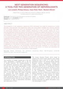
2.5. (1,3)-β-D-Glucan (BG) 2.5. (1,3)-β-D-Glucan (BG) Plasma concentrations of BG were comparable between the three subgroups throughout the Plasma concentrations of BG were comparable between the three subgroups throughout the entire study period (Figure 3) and therefore failed to be of diagnostic value for the prediction of a entire study period (Figure 3) and therefore failed to be of diagnostic value for the prediction of fungal infection. Even in patients suffering from candidemia, plasma concentrations of BG were not a fungal infection. Even in patients suffering from candidemia, plasma concentrations of BG were not increased reliably. increased reliably.
Figure 3.3.Plasma concentrations of β-of D-glucan (BG) in(BG) patients septicwith shock.septic Plasmashock. concentrations Figure Plasma concentrations β-D-glucan in with patients Plasma of BG were measured in patients suffering from septic shockfrom with septic a fungal infection squared box), concentrations of BG were measured in patients suffering shock with (grey a fungal infection a fungal colonization (grey plane box) or(grey without any fungal findings (white box). Plasma samples (grey squared box), a fungal colonization plane box) or without any fungal findings (white box). were collected the onset of septic shock and 1shock day (T1), days1(T2), days2 (T3), days (T4), Plasma samplesatwere collected at the onset(T0), of septic (T0),2 and day 7(T1), days 14 (T2), 7 days 21 days (T5), and 28 days (T6) afterwards. Data in box plots are given as median, 25th percentile, 75th (T3), 14 days (T4), 21 days (T5), and 28 days (T6) afterwards. Data in box plots are given as median, percentile with the 10th as well as 90th percentile at the end of the whiskers. 25th percentile, 75th percentile with the 10th as well as 90th percentile at the end of the whiskers.
Int. J. Mol. Sci. 2017, 18, 1796
13 of 32
2.6. Galactomannan (GM) Plasma concentrations of GM remained below the cut-off value of 1:320), whereas colonized patients (n = 22) were shown to have positive test results in 81.8% (n = 18) of cases. Patients suffering from a fungal infection (n = 11) also revealed positive test results in 81.8% (n = 9) of cases, but unfortunately two patients presenting with candidemia (at sepsis onset) failed to show a positive anti-Candida antibody titer. 2.8. Inflammation as Well as Infection Marker Levels Plasma levels of acute phase proteins (such as C-reactive protein or procalcitonin), leukocytes as well as general inflammation marker levels (such as IL-2, TNF-α) in patients without any fungal isolates, suffering from a fungal colonization or a fungal infection are presented in Table S1. With regard to fungal immunity, special attention should be given to the plasma levels of INF-γ, IL-4, -6, -10, -17 as well as MR-proADM. Plasma levels of the pro-inflammatory cytokine INF-γ were shown to be significantly elevated in patients suffering from a fungal infection in comparison to both control groups, starting from seven days after sepsis onset (T3). This increase in INF-γ was paralleled by a significant release of the immunosuppressive cytokines IL-10 and -4 in infected patients (Table S1). Plasma levels of IL-6 were shown to be significantly elevated in patients suffering from a fungal infection in comparison to septic patients with a fungal colonization or without any fungal findings at different time points especially in the early course of the disease (e.g., at T0, T1) (Table S1). In parallel, IL-17A was also shown to be significantly increased in septic patients suffering from a fungal infection in comparison to septic patients with a fungal colonization or without any fungal findings within the first 7 days after sepsis onset (Figure 4A). Therefore, IL-17A was found to be a suitable tool for early identification of patients with a fungal infection as assessed by receiver operating characteristic (ROC)-analyses (ROC-area under the curve (AUC) for patients with a fungal infection vs. non-infected patients (i.e., patients without any fungal isolates + colonized patients) e.g., at T0: 0.714; Cut-Off 14.165 pg/mL → Sens. 0.818; 1-Spec. 0.323; T1: 0.776; Cut-Off: 14.22 pg/mL → Sens. 0.818; 1-Spec. 0.29; T2: 0.865 Cut-Off 15.00pg/mL → Sens. 0.818; 1-Spec. 0.194, etc.) (Figure 4B). The same holds true for plasma levels of MR-proADM, which were shown to be significantly increased in infected patients in comparison to both colonized patients and those without any fungal findings (Figure 5A). Accordingly, MR-proADM was also shown to be a suitable tool for the identification of patients with a fungal infection as assessed by a receiver operating characteristic (ROC)-analyses (ROC-area under the curve (AUC) for patients with
Int. J. Mol. Sci. 2017, 18, 1796
14 of 32
a fungalInt.infection vs.18, non-infected patients (=see above) e.g., at T0: 0.738; Cut-Off: 6.9914nmol/L → J. Mol. Sci. 2017, 1796 of 31 Sens. 0.727; 1-Spec. 0.333; T1: 0.755; Cut-Off: 8.53 nmol/L → Sens. 0.727; 1-Spec. 0.212; T2: 0.774; 0.738; Cut-Off: 6,99 nmol/L Sens. 0.727; 1-Spec. 0.333; T1: 0.755; Cut-Off: 8.53 nmol/L Sens. 0.727; Int. J. Mol. Sci. 2017, 18, 1796 Cut-Off 14 of 31 0.212; T2:→ 0.774; nmol/L0.273, Sens. 0.818; 1-Spec. 0.273, etc.) (Figure 5B). Cut-Off1-Spec. 5.10 nmol/L Sens. 0.818;5.10 1-Spec. etc.) (Figure 5B). 0.738; Cut-Off: 6,99 nmol/L Sens. 0.727; 1-Spec. 0.333; T1: 0.755; Cut-Off: 8.53 nmol/L Sens. 0.727; 1-Spec. 0.212; T2: 0.774; Cut-Off 5.10 nmol/L Sens. 0.818; 1-Spec. 0.273, etc.) (Figure 5B).
(A)
(B)
Figure 4. Plasma concentrations of interleukin (IL)-17A in patients with septic shock. Legend: (A)
Figure 4. Plasma concentrations interleukin (IL)-17A in patients septic withshock septic shock. Legend: (A) of Plasma concentrations of IL-17A were measured in patients suffering from(B) with a fungal (A) Plasma concentrations of IL-17A were measured in patients suffering from septic shock with infection squared box), a fungal colonization (grey plane box) orwith without fungal findings Figure 4.(grey Plasma concentrations of interleukin (IL)-17A in patients septicany shock. Legend: (A) a fungal(white infection (grey box), a fungal colonization (grey plane box) without any fungal box). Plasmasquared samples were collected at the of septic shock (T0), and shock 1or day (T1), a2 fungal days Plasma concentrations of IL-17A were measured inonset patients suffering from septic with 7 days (T3),Plasma 14 days box), (T4), 21 days (T5), and 28 days (T6) afterwards. Data shock in box are given findings(T2), (white box). samples were collected at the onset of or septic (T0), and 1 day (T1), infection (grey squared a fungal colonization (grey plane box) without any plots fungal findings as(white median, 25th percentile, percentile theand 10th28as well as 90th percentile the (T1), end of the box). Plasma were collected at the onset ofdays septic shock (T0), and 1atData day 2 days 2 days (T2), 7 days (T3), 14samples days75th (T4), 21 dayswith (T5), (T6) afterwards. in box plots are whiskers. Concerning symbolism and higher orders of significance: * p