ANTICANCER RESEARCH 28: 295-304 (2008)
Immunohistochemical Expression Patterns of Neural and Neuroendocrine Markers, the Neural Growth Factor Receptors and the ‚-Tubulin II and IV Isotypes in Human Thymus MARIA BAI1, ALEXANDRA PAPOUDOU-BAI2, GEORGIOS KARATZIAS2, MICHAIL DOUKAS2, ANNA GOUSSIA1, KALLIOPI STEFANAKI3, DIMITRA RONTOGIANNI4, YOTANNA DALAVANGA2, NIKI JOHN AGNANTIS1 and PANAGIOTIS KANAVAROS2
Departments of 1Pathology and 2Anatomy, Histology and Embryology, Medical Faculty, University of Ioannina; 3Department of Pathology, Agia Sophia Childrens' Hospital, Athens; 4Department of Pathology, Evangelismos Hospital, Athens, Greece
Abstract. Increasing evidence suggests that neuroimmune networks play key roles in the thymic histophysiology and pathology. Prompted by this, we analyzed by immunohistochemistry the distribution of human thymic cells expressing major neural and neuroendocrine markers and neural growth factor (NGF) receptors in combination with the expression patterns of various cytokeratins. Additionally, since some ‚tubulin isotypes are preferentially expressed in neuronal cells, the immunotopographical distribution of thymic cells expressing ‚-tubulin II, III and IV was analyzed. Thymic epithelial cells (TECs) expressed protein gene product 9.5 (PGP 9.5), chromogranin A (CHRA), synaptophysin (SYN), neuronspecific enolase (NSE), tyrosine hydroxylase (TH), CD56, CD57, neurofilaments (NF) (140-160 kDa), NGF receptors (TrKA and p75), ‚-tubulin II and IV isotypes and cytokeratin 7, 8, 10, 13, 14, 18 and 19. PGP 9.5 was preferentially expressed in cortical TEC whereas SYN, CHRA, NSE, TH and NF 140160 kDa were preferentially expressed in medullary TECs and Hassal corpuscles. Variable levels of expression of ‚-tubulin II and IV were observed in all TEC subtypes whereas ‚-tubulin III was undetectable in TECs. Subcapsular and cortical TECs display higher expression of ‚-tubulin IV and lower expression of ‚-tubulin II in comparison to those observed in medullary TEC and Hassal corpuscles. The diversity of the immunotopographical distibution and the expression of neural and neuroendocrine markers, the NGF receptors TrKA and
Correspondence to: Dr. Maria Bai, Associate Professor, Department of Pathology, Medical Faculty, University of Ioannina, 45110, Ioannina, Greece. Tel: +302651099415, +302651097627, Fax: +302651097895, e-mail:
[email protected] and
[email protected] Key Words: Thymus, neuroendocrine differentiation, neural growth factor receptors, tubulin.
0250-7005/2008 $2.00+.40
p75, and the ‚-tubulin II and IV isotypes in the distinct subtypes of TEC may reflect the diversity of their biological functions and/or their different stages of differentiation. The present results provide further immunohistological evidence that numerous neural and neuroendocrine factors may be required for the development and function of the human thymic microenvironment. The thymus supports the production of self-tolerant T-cells with both competent and regulatory functions. In the process of T-cell differentiation an essential role is played by the thymic microenvironment which includes the socalled thymic stromal cells (epithelial cells, fibroblasts, macrophages and dendritic cells) (1). Among thymic stromal cells, the thymic epithelial cells (TECs) (2-5) have been regarded as the main drivers of thymocyte development and maturation through cell to cell contacts and the production of soluble factors (e.g. cytokines, hormones, growth factors, neurotransmitters) (1, 6-10). The thymus is a crucial site for cross-talk between the immune and neuroendocrine system (4, 7, 11, 12). Growing evidence suggests that TECs are involved in thymocyte education to neuroendocrine principles by expressing a vast repertoire of neuroendocrine-related proteins which are the source of self-antigens presented to pre T-cells by the major histocompatibility complex (13, 14). This presentation could be responsible for the establishment of central tolerance to neuroendocrine functions (13-15). This concept is supported by immunohistological studies which revealed that human TECs express a wide spectrum of neural and neuroendocrine-related proteins such as protein gene product 9.5, tyrosine hydroxylase, chromogranin A, synaptophysin, neurofilaments, A2B5 (CQ gaglioside), oxytocin, vasopressin, neuropeptide Y, somatostatin, substance P and bombesin (7, 16-24). Importantly, recent
295
ANTICANCER RESEARCH 28: 295-304 (2008) investigations revealed that mouse and human medullary TECs play a central role in the establishment of selftolerance by their ability to promiscuously express a remarkable spectrum of antigens characteristic of other tissues (25-28). Indeed, on the basis of RNA expression data, medullary TECs were found to promiscuously express a highly diverse set of genes, including many tissue-specific antigens, disease-associated autoantigens and cancergermline genes (25-28). It was suggested that the concept of promiscuous gene expression may be important for the understanding of human autoimmune diseases and immunotherapy of tumors (27). Neurotrophins (NTs) are a family of growth factors involved in the development and maintenance of the vertebrate nervous system and display different activities in non-neural cells, including cells of the immune system (29, 30). The NT family consists of nerve growth factor (NGF), brain derived neurotrophic factor (BDNF), neurotrophin-3 (NT3) and several other (29, 30). NTs interact with two different types of cell-surface receptors, the trk family of receptor tyrosine kinases and the low-affinity p75 receptor [30]. NT-receptors are involved in various cellular functions including development, apoptosis and the cell cycle (29-31). The immunohistological detection of the NT-receptors TrkA and p75 in subtypes of human TEC was suggested to be of potential importance because of the involvement of these cells in thymocyte development and selection (32-34). Microtubules are essential components of the cytoskeleton and play key roles in processes, such as spindle formation during mitosis, intracellular transport and cell motility (35). Microtubules are composed of polymers of alpha and beta-tubulin heterodimers; both alpha and beta-tubulins are encoded by several genes each and exist as multiple isotypes in cells (35, 36). At the tissue level, the ‚-I isotype is ubiquitous, the ‚-II is encountered predominantly in the brain and at lower levels in other somatic tissues, the ‚-III is expressed in neurons and Sertoli cells, and the ‚-IV in many cells and tissues (‚-IVa is found predominantly in the brain and the ‚-IVb in most tissues) (35, 36). Immunohistological studies described the distribution of ‚-tubulin isotypes in various normal human tissues (36-39) but, to the best of our knowledge, there is no detailed immunohistological information regarding the expression of ‚-tubulin isotypes II, III and IV in human thymus. Despite numerous studies, the antigenic characteristics of human TECs await further clarification. In particular, there is paucity of multiparametric immunohistological information regarding the distribution of human thymic cells expressing major neural and neuroendocrine markers and the NGF receptors in combination with the expression patterns of cytokeratins (CK). Therefore, we analyzed the immuno-topographical distribution of human thymic cells
296
expressing protein gene product 9.5 (PGP 9.5), neuronspecific enolase (NSE), chromogranin A (CHRA), synaptophysin (SYN), tyrosine hydroxylase (TH), neurofilaments (NF 68 kDa, 140-160 kDa and 200 kDa), CD56, CD57, glial fibrillary acid protein (GFAP), peripherin, Tau protein, and the neural growth factor (NGF) receptors TrKA and p75. In order to acheive a more elaborate identification of the cells expressing the above proteins, immunostaining was performed on serial sections with different cytokeratins, S100 protein, CD1a and CD68 and, in some cases, double immunostaining was also performed. Additionally, since some ‚-tubulin isotypes are preferentially expressed by neuronal cells (35, 36), the immunotopographical distribution of human thymic cells expressing ‚-tubulin II, III and IV was analyzed. Furthermore, since PGP 9.5 and the NGF-receptors TrKA and p75 have been related to cell cycle status in various cell types (31, 40-42), their expression was analyzed in comparison to the expression patterns of the proliferationassociated proteins Ki-67, cyclin A and cyclin B1 (43).
Materials and Methods Materials. Sixteen thymuses from 2 adolescents, 8 infants and 6 newborns removed during surgery for reasons other than thymic pathology were retrieved from the files of the Departments of Pathology of Agia Sophia Childrens' Hospital, Athens, Greece. Immunohistochemistry. Immunostainings were performed on formalin-fixed, paraffin-embedded tissue sections by the streptavidin-biotin peroxidase labeled (LSAB) procedure using the LSAB kit (DAKO SA, Glostrup, Denmark) (43). In some cases, double immunostaining using alkaline-phosphatase/anti-alkaline phosphatase (APAAP) and LSAB procedures was also performed (43). The antibodies and their dilutions and sources are presented in Table I. A step of microwave oven heating was performed prior to incubation with some antibodies (44, 45). Appropriate positive control slides from various tissues were included in all cases. Positive reactions noted occasionally in nerve fibres provided an internal positive control of the reaction specificity of some neural and neuroendocrine markers. Negative controls were included and were subjected to the same immunohistochemical method with omission of the primary antibody. For evaluation of immunostaining, a x40 objective lens was used and the groups of immunopositivity were determined using cut-off levels as follows: rare isolated cells (+/–); fewer than 10% positive cells: low expression (+); 10-25% positive cells: intermediate expression (++); and more than 25% positive cells: high expression (+++) (43). To acheive a better identification of cells expressing neural markers, neuroendocrine markers, the NGF receptors TrKA and p75 and the ‚-tubulin isotypes we used a) morphological criteria (cell size and shape, and nucleus size and shape), b) immunostaining on serial sections with different CK for epithelial cells, S100 protein and CD1a for dendritic cells and, CD68 for monocytes/macrophages (33, 46) and c) in some cases, double immunostaining using pan-CK or S100 protein with PGP 9.5, SYN, CHRA and TrkA.
Bai et al: Expression of Neural/¡euroendocrine Markers, NGF receptors and ‚-Tubulin Isotypes in Thymus
Table I. Antibodies used for immunohistochemical analysis. Antibody Pan-Cytokeratin Cytokeratin 7 Cytokeratin 8 Cytokeratin 10 Cytokeratin 13 Cytokeratin 14 Cytokeratin 18 Cytokeratin 19 Cytokeratin 20 Tyrosine hydroxylase CD56 S-100 PGP 9.5 TrkA NF68 NF140-160 NF200 P75 Pan-‚-tubulin ‚-Tubulin II ‚-Tubulin IV ‚-Tubulin III CD68 Peripherin CD57 Actin Vimentin Chromogranin A Synaptophysin Tau protein GFAP NSE CD1a
Clone
Dilution
MNF116 OV-TL 12/30 35‚H11 LHP1 KS-1A3 LL002 DC10 BA17 KS20.8 1B5 123C3.D5 Polyclonal (rabbit) 10A1 Polyclonal (rabbit) NF68.04 (8A1) NF160.05 (1B3) NF200.06 (2D2) NGFR5 DM-1B JDR3B8 ONS1A6 SDL3D10 KP1 PJM50 NK-1 1A4 V9 LK2H10 Snp88 Tau-2 Polyclonal (rabbit) MIG-N3 CD1a007
1:50 1:50 1:50 1:50 1:50 1:50 1:50 1:50 1:50 1:10 1:30 1:2000 1:50 1:200 1:200 1:100 1:200 1:200 1:150 1:200 1:200 1:200 1:50 1:50 1:50 Ready-to-Use Ready-to-Use Ready-to-Use Ready-to-Use Ready-to-Use Ready-to-Use Ready-to-Use Ready-to-Use
Results Cells expressing neural markers, neuroendocrine markers, the NGF receptors TrKA and p75 and the ‚-tubulin II and IV isotypes were identified within the stromal compartment of thymus tissue. A sizable fraction of these cells were TECs as was revealed by combining morphological evaluation (cell and nucleus, size and shape), and immunostaining with CK (on serial sections and, in some cases, also by double immunostaining). The immunotopographical distribution and the expression levels of individual proteins was variable in the subtypes of TEC (subcapsular, cortical, medullary and Hassal corpuscles) (Table II, Figure 1). On that basis the results were as follows. Expression of neural and neuroendocrine markers. In subcapsular TECs, rare isolated cells were positive for PGP 9.5 and CD57. In cortical TECs, rare isolated cells were positive
Source Dako SA, Glostrup, Denmark Dako SA, Glostrup, Denmark Dako SA, Glostrup, Denmark NovoCastra, Newcastle-upon-Tyne, UK NovoCastra, Newcastle-upon-Tyne, UK Bio-Genex, CA, USA Dako SA, Glostrup, Denmark Dako SA, Glostrup, Denmark Dako SA, Glostrup, Denmark NovoCastra, Newcastle-upon-Tyne, UK Cell Marque Corp, CA, USA Dako SA, Glostrup, Denmark NovoCastra, Newcastle-upon-Tyne, UK Santa Cruz, CA, USA NeoMarkers, CA, USA NeoMarkers, CA, USA NeoMarkers, CA, USA NeoMarkers, CA, USA Bio-Genex, CA, USA Bio-Genex, CA, USA Bio-Genex CA, USA Bio-Genex, CA, USA Dako SA, Glostrup, Denmark NovoCastra, Newcastle-upon-Tyne, UK NovoCastra, Newcastle-upon-Tyne, UK Bio-Genex, CA, USA Bio-Genex, CA, USA Bio-Genex, CA, USA Bio-Genex, CA, USA Bio-Genex, CA, USA Bio-Genex, CA, USA Bio-Genex, CA, USA Biocare Medical, CA, USA
for NSE and CD57 and intermediate expression of PGP 9.5 was found. In medullary TECs, rare isolated cells were positive for PGP 9.5, CD56, CD57 and NF 140-160 kDa and low expression of CHRA, SYN, TH and NSE was observed. In Hassal corpuscles, rare isolated cells were positive for CD57 (only at the outer layer of the corpuscles), low expression of CHRA, SYN and TH (only at the outer layer of the corpuscles) was found and intermediate expression of NSE and NF140-160 kDa (in the entirety of the corpuscles) was detected (Table II). Immunostaining for Tau protein was not interpretable because of intense background staining. Expression of the neural growth factor receptors. In subcapsular and cortical TECs, low expression of TrkA was observed. In medullary TECs, low expression of p75 and intermediate expression of TrkA was detected. In Hassal corpuscles, rare isolated cells were positive for TrkA and p75 (only at the periphery of the corpuscles) (Table II).
297
ANTICANCER RESEARCH 28: 295-304 (2008)
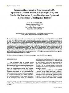
Figure 1.
298
Bai et al: Expression of Neural/neuroendocrine Markers, NGF receptors and ‚-Tubulin Isotypes in Thymus
Table II. Expression of neural, neuroendocrine and other markers in thymic epithelial cells. Subcapsular Cortical Medullary
PGP 9.5 NSE Chromogranin A Synaptophysin Tyrosine hydroxylase CD56 CD57 NF 68 kDa NF 140-160 kDa NF 200 kDa GFAP Peripherin TrkA P75 Pan-CK (MNF116) CK 7 CK 8 CK 10 CK 13 CK 14 CK 18 CK 19 CK 20 Pan-‚-tubulin ‚-Tubulin II ‚-Tubulin III ‚-Tubulin IV Actin Vimentin
+/– – – – – – +/– – – – – – + – +++ ++ – – – + – ++ – +++ – – +++ – +
++ +/– – – – – +/– – – – – – + – ++ – – – – +/– – + – +++ +/– – +++ – +
+/– + + + + +/– +/– – +/– – – – ++ + ++ + + – + + + ++ – +++ ++ – + – +++
Hassal corpuscles – ++ + + + – +/ – ++ – – – +/– +/– +++ ++ ++ + ++ ++ + ++ – +++ ++ – ++ – ++
Expression of the ‚-tubulin isotypes. In subcapsular TECs, rare isolated cells were positive for ‚-tubulin II and high expression of ‚-tubulin IV was found. In cortical TECs, rare isolated cells were positive for ‚-tubulin II and high expression of ‚-tubulin IV was observed. In medullary TEC, intermediate expression of ‚-tubulin II and low expression of ‚-tubulin IV was detected. In Hassal corpuscles, intermediate expression of ‚-tubulin II and ‚tubulin IV was found. In contrast, ‚-tubulin III was undetectable in TECs (Table II). Expression of cytokeratins. Pan-CK and CK19 were expressed in all TEC subtypes. Subtypes of TEC expressed CK7, 8, 13, 14 and 18, whereas CK10 was observed only in Hassal corpuscles and CK20 was undetectable in TECs (Table II). Comparison of the combined expression profiles of the distinct subtypes of TEC. Among the dinstict sybtypes of TEC, PGP 9.5 was preferentially expressed in cortical TECs whereas NSE, SYN, TH and NF 140-160 kDa were preferentially expressed in medullary TECs and Hassal corpuscles. Medullary TECs resembled Hassal corpuscles with respect to the immunoexpression patterns of NSE, CHRA, SYN, TH, NF 140-160 kDa and CK 8, 13 and 18, while these expressions were more restricted or undetectable in subcapsular and cortical TECs. Subcapsular and cortical TECs displayed higher expression of ‚-tubulin IV and lower expression of ‚tubulin II in comparison to those observed in medullary TECs and Hassal corpuscles (Table II). Expression profile of thymocytes Most cortical and medullary thymocytes expressed vimentin and pan-‚-tubulin. CD1a was expressed in most cortical thymocytes. Scattered thymocytes expressed CD57 and CD56 (CD57-positive cells outnumbered CD56-positive cells).
→ Figure 1. a) PGP 9.5 immunopositive cortical TEC (magnification x400). b) Synaptophysin immunopositive cells in Hassal corpuscles (magnification x400). c) Chromogranin immunopositivity in medullary TECs and Hassal corpuscles (magnification x400). d) NF 140-160 kDa immunopositivity in Hassal corpuscles (magnification x400). e) NSE immunopositivity in medullary TECs and Hassal corpuscles (magnification x400). f) Tubulin IV immunopositivity in cortical and medullary TECs and in Hassal corpuscles (magnification x400). g) Pancytokeratin immunopositivity in medullary TECs and Hassal corpuscles (magnification x400). h) Double S100 protein/synaptophysin immunostaining (magnification x400). S100 protein-immunopositive dendritic cells (brown). Synaptophysin-immunopositive cells in Hassal corpuscles (red). i) Double S100 protein/chromogranin immunostaining (magnification x400). S100 protein-immunopositive dendritic cells (brown). Chromogranin immunopositivity in medullary TECs and Hassal corpuscles (red). j) Double S100 protein/TrkA immunostaining (magnification x400). S100 protein-immunopositive dendritic cells (brown). TrkA immunopositivity in medullary TEC (red).
Associations with cell cycle proteins. Some studies suggest a relation between PGP 9.5 expression and cell proliferation in various cell types (40-42) and there is evidence TrkA mediates NGF-induced proliferation in epithelial cells (31, 47, 48). Therefore, the expression of PGP 9.5 and TrkA in TECs was analyzed in 7/16 cases in comparison to the expression patterns of the proliferation-associated proteins Ki-67, cyclin A and cyclin B1 in the same cells, using the respective antibodies as described elsewhere (43). We were not able to demonstrate parallel expression patterns between cell proliferation-associated proteins and PGP 9.5 or TrkA since low expression of Ki-67, cyclin A and cyclin B1 was detected in both cortical and medullary human TECs, while PGP 9.5 was preferentially expressed in cortical TECs, and TrkA exhibited intermediate expression in medullary TEC and low expression in cortical TEC (Table II).
299
ANTICANCER RESEARCH 28: 295-304 (2008) Discussion In the present study, we observed that medullary TECs and Hassal corpuscles preferentially express NSE, CHRA, SYN, TH, NF 140-160 kDa, CD56 and CD57 while these proteins were undetectable or rarely expressed in subcapsular and cortical TECs. The immunohistological detection of a wide spectrum of neural and neuroendocrine-related proteins in human medullary TECs and Hassal corpuscles (present study; 7, 16-19, 23, 24, 49) might be related to the ability of the medullary epithelium to promiscuously express a remarkable spectrum of antigens characteristic of other tissues (25-28, 50). Interestingly, the mutual comparison of RNA profiles of medullary TECs with cortical TECs, dendritic cells and thymocytes revealed in each case a much higher number of genes being promiscuously expressed in medullary TECs compared with the other populations (27, 50). Additionally, analysis at the protein level showed that strong promiscuous expression of particular tissue antigens was confined to a minor fraction of medullary TECs ranging from 1-5% (50). This might be related to our findings showing that most neural and neuroendocrine-related proteins are expressed in less than 10% of medullary TECs. Further immunohistological studies are required to determine the topographical distribution, the expression levels and the probable clinical significance of promiscuously expressed antigens in human TECs. The preferential location of various neural and neuroendocrine-related markers in distinct sybtypes of human TECs (for example: PGP 9.5 in cortical TEC; NSE, SYN, TH, NF 140-160 kDa and parathyroid hormone-related peptide in medullary TECs; substance P, bombesin and betaendorphin in subcapsular TECs; oxytocin and vasopressin in subcapsular and medullary TECs) (present study; 16-19, 21, 23), might provide a basis for a histogenetic classification of neuroendocrine thymic tumors (51). This suggestion is supported by the findings that differential expression of various cytokeratins can be used for the histogenetic classification of the thymic epithelial tumors (52). The expression of a wide spectrum of neural and neuroendocrine-related markers by human and rodent TECs led some authors to the assumption that these cells are derived from the neural crest (10, 53-55). However, recent evidence suggests that the mouse thymic epithelium is derived from the endoderm (56) and that the mouse cortical and medullary TECs have a common progenitor (57, 58). In view of these recent findings, it is tempting to hypothesize that TECs originate in endoderm and some of them acquire neural and neuroendocrine features during TEC differentiation. The present findings that PGP 9.5 is expressed throughout the cortex are in keeping with previous findings in human thymuses (17), whereas the PGP 9.5-
300
immunoreactive rat TECs were preferentially located at the inner cortex (59). Given the importance of TECthymocyte interaction, this difference might be of functional significance since in the inner cortex double negative thymocytes undergo proliferative clonal expansion, while in the outer cortex the double negative become double positive thymocytes (6). Interestingly, some studies suggest a relation between PGP 9.5 expression and cell proliferation (40-42). For example, Haley et al. (41) observed that PGP 9.5 immunoreactive cells in developing human lung were usually negative for proliferating cell nuclear antigen (PCNA) which is expressed by actively dividing cells. Since PGP 9.5 acts as ubiquitin carboxyl-terminal hydrolase which releases ubiquitin from their ligand proteins (60), it was suggested that PGP 9.5 deconjugates ubiquitin from the cyclindependent kinase (CDK) inhibitors thereby promoting cell cycle arrest (42). In contrast, Gianbianco et al. (40) observed the maximum PGP 9.5 expression during the proliferation phases of glioma cell lines. We could not demonstrate a relation between PGP 9.5 and cell proliferation since in the present and in our previous study low expression of the proliferation for the reasons stated earlier. The present findings that TrkA is expressed in subcapsular, cortical and medullary TECs are in line with a previous study in normal human thymus from fetuses and newborns (32). However, in another study, TrkA was described only in subcapsular and medullary TECs of human fetal thymuses (33). In this study, TrkA in cortical TECs was detected only in thymic hyperplasia associated with myasthenia in adolescents and young adults (33). Moreover, TrkA was not detected in rat or mouse cortical TECs under normal conditions (61-64); however, expression of TrkA in rat cortical TECs was observed during thymus regeneration following acute involution induced by cyclophoshamide (64) and after induction of thymocyte apoptosis by corticosteroids (63). Taken together, the above data from human, rat and mouse thymuses indicate variations in the TrkA expression in cortical TECs. These variations might be due to the cell-cycle dependent expression of TrkA (31) and/or to soluble factors released by the thymocytes since depletion of thymocytes in rat thymus results in down-regulation of TrkA in subcapsular and medullary TECs whereas absence of thymocytes induces up-regulation of TrkA in cortical TECs (63). Interestingly, accumulating data indicate that NGFreceptors are involved in cell cycle regulation and apoptosis of epithelial cells (31, 47, 48, 65). For example, TrkA mediates NGF-induced proliferation in breast carcinoma cells (48) and in epithelial cells of the hair follicle (47). We could not demonstrate a relation between TrkA and cell proliferation for the reasons stated earlier.
Bai et al: Expression of Neural/neuroendocrine Markers, NGF receptors and ‚-Tubulin Isotypes in Thymus
In the present study, variable levels of ‚-tubulin II and IV expression were detected in all human TEC subtypes. Subcapsular and cortical TECs displayed higher expression of ‚-tubulin IV and lower expression of ‚-tubulin II in comparison to those observed in medullary TECs and Hassal corpuscles. The widespread and heterogeneous distribution of ‚-tubulin II and IV in TEC is in keeping with immunohistochemical findings in other human epithelial cells. Roach et al. (39) observed ‚-tubulin II in the stratum granulosum of the skin and very low levels of ‚-tubulin IV in the skin and the pancreas, Dozier et al. (37) detected ‚-tubulin II and I+IV (with predominance of ‚-tubulin II) in normal and malignant breast epithelium and Ranganathan et al. (66) observed heterogeneous ‚-tubulin II and IV expression in benign prostate hyperplasia and carcinoma glands. Expression of ‚-tubulin III was undetectable in human TEC in the present study,. This is in keeping with immunohistochemical findings in other human epithelial cells. Indeed, Dozier et al. (37) did not detect ‚-tubulin III in normal or malignant breast epithelium and Draberova et al. (38) identified ‚-tubulin III in normal neuronal cells of the central and peripheral nervous systems but not in normal epithelial cells of various organs (lung, large intestine, skin, salivary glands and urinary bladder). However, Roach et al. (39) studied large intestine, skin, oviduct and pancreas by immunohistochemisty and detected low levels of ‚-tubulin III in columnar epithelial cells of the colon. Moreover, Ranganathan et al. (66) observed ‚tubulin III immunohistochemical expression in benign prostate hyperplasia and carcinoma glands. The above findings, taken together, suggest that expression of ‚-tubulin III is very rare in normal human epithelial cells, but it may be upregulated in some epithelial neoplasias. It could be assumed that the diversity of the immunotopographical distibution and the expression levels of ‚-tubulin II, III and IV isotypes in human TEC subtypes may denote different stages of differentiation and/or different functional significance. The latter can be supported by the finding that retinal rod and tracheal cells express ‚-tubulin II and IV isotypes, but appear to use only ‚-tubulin IV for their ciliary axonemal microtubules (67). Further studies are required to clarify the functional significance of the differential expression of beta-tubulin isotypes in human TEC subtypes. It would also be interesting to examine whether the ‚-tubulin isotype composition is related to the biological behavior of thymic epithelial tumors since Ranganathan et al. (66) reported a marked increase in ‚-tubulin II immunostaining from benign prostatic hyperplasia to carcinoma in 77% of the patients, suggesting that the expression of this isotype is related to the malignant status in prostate epithelial tumors. It is concluded that the diversity of the immunotopographical distibution and the expression levels of neural
markers, neuroendocrine markers, the NGF receptors TrKA and p75, ‚-tubulin II and IV isotypes and various cytokeratins in the distinct subtypes of TEC may reflect the diversity of their biological functions and/or their different stages of differentiation. The results presented herein provide further immunohistological evidence that numerous neural and neuroendocrine factors may be required for the development and the function of the human thymus microenvironment. Moreover, our findings might be helpful for the further understanding of the histogenesis and the classification of the neuroendocrine thymic tumors.
Acknowledgements The authors are grateful to Dr. Tania Markopoulou for helpful discussions.
References 1 Anderson G, Jenkinson WE, Jines T, Parnell SM, Kinsella FAM, White AJ, Pongracz JE, Rossi SW and Jenkinson EJ: Establishment and functioning of intrathymic microenvironments. Immunol Rev 209: 10-27, 2006. 2 Brelinska R: Thymic epithelial cells in age-dependent involution. Microsc Res Tech 62: 488-500, 2003. 3 Bodey B, Bodey B Jr, Siegel SE and Kaiser HE: Molecular biological ontogenesis of the thymic reticulo-epithelial cell network during the organization of the cellular microenviroment. In Vivo 13: 267-294, 1999. 4 Bodey B, Bodey BJr, Siegel SE and Kaiser HE: The role of the reticulo-epithelial (RE) cell network in the immunoneuroendocrine regulation of intrathymic lymphopoiesis. Anticancer Res 20: 1871-1888, 2000. 5 Von Gaudecker B, Kendall MD and Ritter MA: Immunoelectron microscopy of the thymic epithelial microenvironment. Microsc Res Tech 38: 237-249, 1997. 6 Blackburn CC and Manley NR: Developing a new paradigm for thymus organoneogenesis. Nat Rev Immunol 4: 278-289, 2004. 7 Bodey B: Neuroendocrine influence on thymic haematopoiesis via the reticulo-epithelial cellular network. Expert Opin Ther Targets 6: 57-72, 2002. 8 Garcia-Suarez O, Perez-Perez M, Germana A, Esteban I and Germana G: Involvement of growth factors in thymic involution. Microsc Res Tech 62: 514-523, 2003. 9 Hannestad J, Monjil DF, Diaz-Esnal B, Cobo J and Vega JA: Age-dependent changes in the nervous and endocrine control of the thymus. Microsc Res Tech 63: 94-101, 2004. 10 Mentlein R and Kendall MD: The brain and thymus have much in common: a functional analysis of their microenvironments. Immunol Today 21: 133-140, 2000. 11 Moll UT: Functional histology of the neuroendocrine thymus. Microsc Res Tech 38: 300-310, 1997. 12 Savino W and Dardenne M: Neuroendocrine control of thymus physiology. Endocrine Rev 21: 412-443, 2000. 13 Geenen V, Kecha O and Martens H: Thymic expression of neuroendocrine self-peptide precursors: role in T-cell survival and self-tolerance. J Neuroendocrinol 10: 811-822, 1998.
301
ANTICANCER RESEARCH 28: 295-304 (2008) 14 Hansenne I: Thymic transcription of neurohypophysial and insulin-related genes: impact upon T-cell differentiation and self-tolerance. J Neuroendocrinol 17: 321-327, 2005. 15 Geenen V: Thymus-dependent T-cell tolerance of neuroendocrine functions: principles, reflections, and implications for tolerogenic/negative self-vaccination. Ann NY Acad Sci 1088: 284296, 2006. 16 Batanero E, de Leeuw FE, Jansen GH, van Wichen DF, Huber J and Schuurman HJ: The neural and neuro-endocrine component of the human thymus. II. Hormone immunoreactivity. Brain Behav Immun 6: 249-264, 1992. 17 De Leeuw FE, Jansen GH, Batanero E, van Wichen DF, Huber J and Schuurman HJ: The neural and neuro-endocrine component of the human thymus. I. Nerve-like structures. Brain Behav Immun 6: 234-248, 1992. 18 Gessi M, Monego G, Lauriola L, Maggiano N and Ranelletti FO: Parathyroid hormone-related peptide and parathyroid hormone-related peptide receptor 1 expression in human thymus. J Histochem Cytochem 53: 955-962, 2005. 19 Kranz A, Kendall MD and von Gaudecker B: Studies on rat and human thymus to demonstrate immunoreactivity of calcitonin gene-related peptide, tyrosine hydroxylase and neuropeptide Y. J Anat 191: 441-450, 1997. 20 Marx A, Wilisch A, Schultz A, Greiner A, Magi B, Pallini V, Schalke B, Toyka K, Kirchner T and Muller-Hermelink HK: Expression of neurofilaments and of a titin epitope in thymic epithelial tumors. Implications for the pathogenesis of myasthenia gravis. Am J Pathol 148: 1839-1850, 1996. 21 Piantelli M, Maggiano N, Larocca LM, Ricci R, Ranelleti FO, Lauriola L and Capelli A: Neuropeptide-immunoreactive cells in human thymus. Brain Behav Immun 4: 189197, 1990. 22 Raica M, Encica S, Motoc A, Cimpean AM, Scridon T and Barsan M: Structural heterogeneity and immunohistochemical profile of Hassall corpuscles in normal human thymus. Ann Anat 188: 345-352, 2006. 23 Robert F, Geenen V, Schoenen J, Burgeon E, De Groote D, Defresne MP, Legros JJ and Franchimont P: Colocalization of immunoreactive oxytocin, vasopressin and interleukin-1 in human thymic epithelial neuroendocrine cells. Brain Behav Immun 5: 102-115, 1991. 24 Silva AB, Aw D and Palmer DB: Evolutionary conservation of neuropeptide expression in the thymus of different species. Immunology 118: 131-140, 2006. 25 Derbinski J, Gabler J, Brors B, Tierling S, Jonnakutty S, Hergenhahn M, Peltonen L, Worn J and Kyewski B: Promiscuous gene expression in thymic epithelial cells is regulated at multiple levels. J Exp Med 202: 33-45, 2005. 26 Gillard GO and Farr AG: Features of medullary thymic epithelium implicate postnatal development in maintaining epithelial heterogeneity and tissue-restricted antigen expression. J Immunol 176: 5815-5824, 2006. 27 Gotter J, Brors B, Hergenhahn M and Kyewski B: Medullary epithelial cells of the human thymus express a highly diverse selection of tissue-specific genes colocalized in chromosome clusters. J Exp Med 199: 155-166, 2004. 28 Taubert R, Schwendemann J and Kyewski B: Highly variable expression of tissue-restricted self-antigens in human thymus: implications for self-tolerance and autoimmunity. Eur J Immunol 37: 838-848, 2007.
302
29 Tessarolo L: Pleotropic functions of neurotrophins in development. Cytokine Growth Factor Rev 9: 125-137, 1998. 30 Vega JA, Garcia-Suarez O, Hannestad J, Perez-Perez M and Germana A: Neurotrophins and the immune system. J Anat 203: 1-19, 2003. 31 Lopez-Sanchez N and Frade JM: Control of the cell cycle by neurotrophins: lessons from the p75 neurotrophin receptor. Histol Histopathol 17: 1227-1237, 2002. 32 Hannestad J, Garcia-Suarez O, Huerta JJ, Esteban I, Naves FJ and Vega JA: TrkA neurotrophin receptor protein in the rat and human thymus. Microsc Res Tech 249: 373-379, 1997. 33 Parrens M, Labouyrie E, Groppi A, Dubus P, Carles D, Velly JF, de Mascarel A and Merlio JP: Expression of NGF receptors in normal and pathological human thymus. J Neuroimmunol 85: 11-21, 1998. 34 Pescarmona E, Pisacane A, Pignatelli E and Baroni CD: Expression of epidermal and nerve growth factor receptors in human thymus and thymomas. Histopathology 23: 39-44, 1993. 35 Luduena RF: Multiple forms of tubulin: different gene products and covalent modifications. Int Rev Cytol 178: 207275, 1998. 36 Katsetos CD, Herman MM and Mork SJ: Class III ‚-tubulin in human development and cancer. Cell Motil Cytoskeleton 55: 77-96, 2003. 37 Dozier JH, Hiser L, Davis JA, Thomas NS, Tucci MA, Benghuzzi HA, Frankfurter A, Correira JJ and Lombert S: Beta class II tubulin predominates in normal and tumor breast tissues. Breast Cancer Res 5: R157-R169, 2003. 38 Draberova E, Lukas Z, Ivanyi D, Viklicky V and Draber P: Expression of class III beta-tubulin in normal and neoplastic human tissues. Histochem Cell Biol 109: 231-239, 1998. 39 Roach MC, Boucher VL, Waiss C, Ravdin PM and Luduena RF: Preparation of a monoclonal antibody specific for the class I isotype of ‚-tubulin: The ‚ isotypes of tubulin differ in their cellular distributions within human tissues. Cell Motil Cytoskeleton 39: 273-275, 1998. 40 Gianbianco I, Bianchi R, Ceccarelli P, Pula G, Sorci G, Antonioli S, Bocchini V and Donato R: Neuron-specific protein gene product 9.5 (PGP 9.5) is also expressed in glioma cell lines and its expression depends on cellular growth state. FEBS 290: 131-134, 1991. 41 Haley KJ, Drazen JM, Osathanondh R and Sunday ME: Comparison of the ontogeny of protein gene product 9.5, chromogranin A and proliferating cell nuclear antigen in developing human lung. Microsc Res Tech 37: 62-68, 1997. 42 Tokunaga Y, Imai S, Torii R and Maeda T: Cytoplasmic liberation of protein gene product 9.5 during the seasonal regulation of spermatogenesis in the monkey (Macaca fuscata). Endocrinology 140: 1875-1883, 1999. 43 Kanavaros P, Stefanaki K, Rontogianni D, Papalazarou D, Arvanitis D, Vamvouka C, Gorgoulis V, Siatitsas I, Agnantis NJ and Bai M: Immunohistochemical expression of p53, p21/waf1, Rb, p16, cyclin D1, p27, Ki67, cyclin A, cyclin B1, bcl2, bax and bak proteins and apoptotic index in normal thymus. Histol Histopathol 16: 1005-1012, 2001. 44 Kanavaros P, Stefanaki S, Vlachonikolis J, Eliopoulos G, Kakolyris S, Rontogianni D, Gorgoulis V and Georgoulias V: Expression of p53, p21/waf-1, bcl-2, bax, Rb and Ki-67 proteins in Hodgkin's lymphomas. Histol Histopathol 15: 445-453, 2000.
Bai et al: Expression of Neural/neuroendocrine Markers, NGF receptors and ‚-Tubulin Isotypes in Thymus
45 Stefanaki S, Rontogianni D, Kouvidou C, Bolioti S, Delides G, Sotsiou F and Kanavaros P: Immunohistochemical expression of bcl2, p53, mdm2 and p21/waf-1 proteins in thymomas. Histopathology 30: 549-555, 1997. 46 Savchenko AS, Hasegawa G and Naito M: Development and maturation of thymic dendritic cells during human ontogeny. Cell Tissue Res 325: 455-460, 2006. 47 Botchkarev VA, Botchkareva NV, Peters EM and Paus R: Epithelial growth control by neurotrophins: leads and lessons from the hair follicle. Prog Brain Res 146: 493-513, 2004. 48 Chiarenza A, Lazarovici P, Lempereur L, Cantarella G, Bianchi A and Bernardini R: Tamoxifen inhibits nerve growth factorinduced proliferation of the human breast cancerous cell line MCF-7. Cancer Res 61: 3002-3008, 2001. 49 Bodey B, Bodey B Jr, Siegel SE and Kaiser HE: Novel insights into the function of the thymic Hassall’s bodies. In Vivo 14: 407-418, 2000. 50 Kyewski B, Derbinski J, Gotter J and Klein L: Promiscuous gene expression and central T-cell tolerance: more than meets the eye. Trends Immunol 23: 364-371, 2002. 51 Moran CA and Suster S: Neuroendocrine carcinoma (carcinoid tumor) of the thymus: a clinicopathologic analysis of 80 cases. Am J Clin Pathol 114: 100-110, 2000. 52 Kuo TT: Cytokeratin profiles of the thymus and thymomas: histogenetic correlations and proposal for a histological classification of thymomas. Histopathology 36: 403-414, 2000. 53 Bodey B, Bodey BJr, Kemshead JT, Siegel SE and Kaiser HE: Identification of neural crest-derived cells within the cellular microenvironment of the human thymus employing a library of monoclonal antibodies raised against neuronal tissues. In Vivo 10: 39-47, 1996. 54 Botham CA, Jones GV and Kendall MD: Immunocharacterisation of neuroendocrine cells of the rat thymus gland in vitro and in vivo. Cell Tissue Res 303: 381-389, 2001. 55 Jones GV, Botham CA, Clarke AG and Kendall MD: Immunoreactivity of neural crest-derived cells in thymic tissue developing under the rat kidney capsule. Brain Behav Immun 12: 163-180, 1998. 56 Gordon J, Wilson VA, Blair NF, Sheridan J, Farley A, Wilson L, Manley NR and Blackburn CC: Functional evidence for a single endodermal origin for the thymic epithelium. Nat Immunol 5: 546-553, 2004. 57 Bleul C, Corbeaux T, Reuter A, Fisch P, Monting JS and Boehm T: Formation of a functional thymus initiated by apostnatal epithelial progenitor cell. Nature 441: 992-996, 2006.
58 Rossi SW, Jenkinson WE, Anderson G and Jenkinson EJ: Clonal analysis reveals a common progenitor fot thymic cortical and medullary epithelium. Nature 441: 988-991, 2006. 59 Brelinska R, Ostalska D and Zabel M: Subtypes of thymic epithelial cells defined by neuroendocrine markers. Histochem Cell Biol 114: 239-244, 2000. 60 Wilkinson KD: Regulation of ubiquitin-dependent processes by deubiquitinating enzymes. FASEB J 11: 1245-1256, 1997. 61 Garcia-Suarez O, Germana A, Hannestad J, Perez-Perez M, Esteban I, Naves FJ and Vega J.A: Changes in the expression of the nerve growth factor receptor TrkA and p75LNGFR in the rat thymus with ageing and increased nerve growth factor plasma levels. Cell Tissue Res 301: 225-234, 2000. 62 Garcia-Suarez O, Germana A, Hannestad J, Ciriaco E, SilosSantiago I, Germana G and Vega JA: Involvement or the NGF receptors (Trka and p75lngfr) in the development and maintenance of the thymus. Ital J Anat Embryol 106: 279-285, 2001. 63 Perez-Pinera P, Garcia-Suarez O, Prieto J.G, Germana A, Hannestad J, Ciriaco E, del Valle ME and Vega JA: Thymocyte depletion affects neurotrophin receptor expression in thymic stromal cells. J Anat 208: 231-238, 2006. 64 Yoon S, Lee HW, Baek SY, Kim BS and Lee SA: Upregulation of TrkA neurotrophin receptor expression in the thymic subcapsular, paraseptal, perivascular, and cortical epithelial cells during thymus regeneration. Histochem Cell Biol 119: 5568, 2003. 65 Tabassum A, Khwaja F and Djakiev D: The p75 (NTR) tumor suppressor induces caspase-mediated apoptosis in bladder tumor cells. Int J Cancer 105: 47-52, 2003. 66 Ranganathan S, Salazar H, Benetatos CA and Hudes GR: Immunohistochemical analysis of ‚-tubulin isotypes in human prostate cercinoma and benign prostatic hypertrophy. The Prostate 30: 263-268, 1997. 67 Renthal R, Schneider BG, Miller NM and Luduena RF: ‚IV is the major ‚-tubulin isotype in bovine cilia. Cell Motil Cytoskeleton 25: 19-29, 1993.
Received October 2, 2007 Revised November 7, 2007 Accepted November 12, 2007
303