Nov 8, 1983 - Westbury,. NY) diluted. 1:10 in MEM. This ...... Baron and Proctor. [1], with the advantage of fewer monocyte-macrophages. (1 vs 7 %) in purified.
Journal
of Leukocyte
Biology
36:505-520
(1984)
Immunological Activation of Polymorphonuclear Neutrophils for Fungal Killing: Studies With Murine Cells and Blastomyces dermatitidis In Vitro Elmer
Brummer,
Alan
M. Sugar,
and David
A. Stevens
Division of Infectious Diseases, Department of Medicine, Santa Clara Valley Medical Center, and Institute for Medical Research, San Jose, and the Department of Medicine, Stanford University Medical School, Stanford, California The interaction of elicited murine polymorphonuclear neutrophils (PMN) and the thermally dimorphic fungal pathogen Blastomyces dermatitidis in vitro was studied.The PMN elicited intraperitoneally with thioglycollate, in normal mice or mice immune to B dermatitidis, failed to reduce colony forming units (CFU) of B dermatitidis in the inoculum in a 4-hr in vitro assay, even in the presence of 10% fresh immune serum. In contrast, PMN elicited intraperitoneally in immune mice by injection of nonviable B dermatitidis cells significantly reduced inoculum CFU (60 ± 5%) under the same conditions. Furthermore, nonviable B dermatitidis intraperitoneally (i.p.) in normal mice or nonviable Candida albicans p. in immune mice failed to elicit peritoneal exudate cells that reduced inoculum CFU in this system. These results support the concept that PMN, elicited in a site by means of an immunological reaction, acquired enhanced microbicidal activity. The fungicidal activity of immunologically elicited PMN was shown to be most effective at high effector to target cell ratios (1,000:1), maximal within 2 hr of coculture, and significantly enhanced in the presence of fresh immune serum compared to heat-inactivated immune serum, normal mouse serum, or fetal bovine serum. Such PMN also had significantly enhanced fungicidal activity against C albicans compared to normal PMN. Fungicidal activity was abrogated in the presence of catalase, implicating hydrogen peroxide generation as the killing mechanism in the activated cells.
Key words:
Received Reprint San
Jose,
polymorphonuclear of fungal infection
November requests: CA
© 1984 Alan
8,
1983;
E. Brummer, 95128.
R. Liss,
Inc.
accepted Dept.
neutrophils,
March of Medicine,
13,
activation,
fungal
killing,
immunology
1984. Sta.
Clara
Valley
Med.
Ctr.,
751
5. Bascom
Avenue,
506
Brummer,
Sugar,
and
Stevens
INTRODUCTION It has an important The
been well and vital
function
of F
documented role in the
and
C3
receptors
sequence after phagocytosis anism of PMN [16, 18] have activity is impressive and pathogens schenckii
(eg, Candida [8] Histoplasma
donovani
[24],
,
that polymorphonuclean body’s defense against on PMN
some
degree
different peripheral the
blood
of this
[26]
has
been
of heterogeneity
organism
and
the
[17];
Trypanosoma
most
[23]
with
dermatitidis. nonviable
B dermatitidis mice, did not
Since nonviable in immune
convenient
been
micnoor-
pneumophilia by PMN. source
of PMN
en route from the PMN [22], shows
postulated
that
they
may
be
and
of elicited dermatitidis.
in vitro.
murine The
It was
thioglycollate
killed
striking
PMN and yeast form that
Candida
PMN
albicans
forming units (CFU) of B dermatitidis in vitro. in immune mice with nonviable B dermatitidis high levels of fungicidal activity against B cells in nonimmune have this effect on
support the concept that microbicidal activity in immunological inflammatory reaction sites. AND
and
it has
in vivo mice
colony elicited exhibited
MATERIALS
secretory
a few
Legionella to killing
of mature PMN of the total body
the interaction Blastomyces
but failed to reduce inoculum However, when PMN were cells intaperitoneally, they cells
the
31]. This concept is supported by evidence that activity can be modulated by lymphokines [7,21].
extracellular
or immune
5%
However,
[2] and resistant
common
population 3 to
however,
30,
[15]).
endospores or relatively
we investigated fungal pathogen
is usually
in normal
19],
Cryptococcus neoformans [10, 29], Sporothrix [12], and pathogenic protozoa (eg, Leishmania
,
studies cited above. This to tissue sites, constituting
from tissue PMN [22, blood PMN microbicidal
elicited
,
,
albicans [18] capsulatum
In the present study thermally dimorphic
[13
,
Trichomonas
Peripheral in the marrow
in phagocytosis
play in 9].
[32] and the hydrogen peroxide-generating killing mechbeen thoroughly studied..The range of PMN microbicidal includes most pathogenic bacteria [9] several fungal
ganisms, such as Coccidioides immitis [14], have been reported to be resistant used bone
neutrophils (PMN) infection [reviewed
of PMN
can
mice PMN,
or C albicans these findings
be significantly
enhanced
METHODS
Animals Pathogen-free Jose,
CA)
BALB/cByJIMR
8 to 12 weeks
of age
male were
mice (Institute for Medical
used
in these
Research,
San
experiments.
B dermatitidis B dermatitidis
(ATCC
26199),
a strain
shown
to be virulent
in mice
[11],
was
used throughout these studies. Log-phase yeast-form B dermatitidis was transferred from 72-hr-defined liquid medium cultures [5,11] to blood agar plates. Inocula were prepared resuspended, pended number than
from
48-hr growth harvested from and counted with a hemacytometer.
inocula of CFU 2 years
Candida
were
were plated per milliliter. nonviable,
in quadruplicate B dermatitidis and
served
blood
agar, washed twice Appropriate dilutions
on blood (yeast-form) as killed
in saline, of resus-
agar plates to determine stored at 70#{176}Cfor -
B dermatitidis
cells.
nitrogen
(YNB)
the more
albicans C albicans
Detroit, MI)
(Sh 27 strain) at 32#{176}C from
was grown
in yeast
stock cultures maintained
base
on Sabouraud’s
broth
(Difco,
agar slants at
Immune
Activation
of PMN’s
4#{176}C.C albicans passaged twice in YNB washed twice in saline and counted with exclusion
of methylene
plating
blue
1 ml of appropriate
Media
and
culture essential
U/mi)
and
streptomycin
(FBS)
were
purchased
culture
Killing
broth, then cultured a hemacytometer.
(0. 1 %).
Colony
on blood
507
for 4 days at 32#{176}C,was Viability was assessed by
forming
units
were
determined
by
agar.
Reagents
The tissue minimal
(PBS),
stain dilutions
for Fungal
medium
penicillin-i00
media and reagents: medium (MEM), (10,000 from
(CTCM) meg
Dulbecco’s RPMI-1640
mcg/ml),
GIBCO
heat-inactivated
Laboratories,
consisted
streptomycin
and
of per
phosphate medium,
Grand
RPMI-1640,
milliliter.
fetal
Island, 10%
buffered penicillin
NY. FBS
Metnizamide
saline (10,000
bovine
serum
Complete (v/v)
tissue
100
and
U
(2-(3-acetamido-5-N-
methylacetamido-2,4,6-tniiodobenzamido)-2-deoxy-D-glucose) (Sigma Chemical Company, St. Louis, MO) was made up as a 35.3% solution in distilled water, which was isotonic. The required concentrations of metnizamide for gradients were made by diluting the 35.3% solution in PBS. Catalase (Sigma, St. Louis, MO) was dialyzed against 0.2 M phosphate buffer, pH 6.5 for 24 hr prior to use. Activity of catalase was assayed by the 0-dianisidine (Sigma, St. Louis, MO) method using standard techniques. Immunization
of Mice
Mice were As reported
sites. rendered
mice
correlated
resistant
with
proliferative [23]. These weeks
given 20,000 previoiusly to fatal
significant
responses mice are
CFU of B dermatitidis [23], resolution of this pulmonary
delayed
type
challenge
subcutaneously infection over with
at two a 4-week
B dermatitidis.
hypersensitivity
reactions,
Resistance and
to B dermatitidis antigens as well as specific referred to as immune mice and were used
dorsal period
lymphocyte
serum antibodies in experiments
4
I ml of thioglycolgiven to normal
or
postinfection.
PMN
late
To elicit peritoneal exudate cells (PEC) broth (Clinical Standards Laboratories,
immune harvested.
mice intraperitoneally (ip) When killed B dermatitidis
enriched Carson,
for PMN, CA) was
4 hr, and in some cases cells, or C albicans
24 hr, before PEC were cells, were used to elicit
PEC, 500 meg wet weight organisms (approximately 10 x 106 multicellular units, also referred to as MCU or yeast cell aggregates) were given ip in 0.5 ml saline 24 hr prior to harvest. The PEC were collected by lavage of the peritoneum with 10 ml of MEM Products,
containing MeGaw
10 U per ml of preservative-free Park, IL). The PEC were washed
and counted with a hemacytometer. The 40 x 106 cells) on the first metrizamide
15.5% gradient
metnizamide) one was
on the second centnifugation CTCM,
and
and washed
gradient as above, the
cells
centrifuge preparations stained with Wright’s
PEC were gradient
centrifuged (400 x g for once in MEM, resuspended (1.5 ml of 15.5% over 1.5 pelleted cells were washed
counted of cell stain or
with
hepanin once with
(American Scientific MEM, resuspended,
purified [6] by layering (1.5 ml of 14.5% over 20 mm). The pellet in 1 ml of MEM,
1 ml (301.5 ml of
of cells from and layered
ml of 17.5% metrizamide). once with MEM, resuspended
a hemacytometer.
layers and pellets from for nonspecific esterase
In selected metrizamide [5]. Two
experiments,
After in cyto-
gradients were hundred cells per
508
Brummer,
Sugar,
number
of PMN,
slide were counted phages recorded.
and
the
Cocultures
and
B dermatitidis
of PMN
In most flat-bottom
into
experiments microtest
and
Stevens
lymphocytes,
and
monocytes-macro-
or C albicans
5 x 10⁶ cells
0. 1 ml of plate wells
per milliliter CTCM were Flow Laboratories, Hamden,
(Linbro,
0. 1 ml of B dermatitidis or C albicans (5,000 to 10,000 CFU/ml added. In other experiments, the number of munine cells per culture ofB dermatitidis CFU per culture was varied. Cultures were incubated atmosphere of 5 % CO2 and air at 37#{176}Cfor 4 hr, unless otherwise were
harvested
described
using
previously
distilled
water,
and
[5]. The
percent
the
number
reduction
of CFU
of inoculum
per
of CTCM) was or the numbers in a humidified noted. Cultures
well
CFU
dispensed CT) and
was
determined
as
calculated
by
the formula (1 (coculture CFU/inoculum CFU)) x 100. Microscopic examination of harvested cultures indicated, as previously described [5], that reduction of CFU in cocultures was not due to clumping, eg, the mean number of cells per MCU was similar in control and experimental cultures. -
Serum Mice
were
anesthetized,
a pouch
was
formed
dissection, the brachial artery was severed, pipette. Blood was allowed to clot at room was collected. Complement incubation in a water bath 10% (v/v) complement that immune
contained
to have showed dermatitidis
a front leg and torso by
was collected for 2 hr, and
in fresh immune or normal serum at 56#{176}Cfor 30 mm. Unless otherwise
with a pasteur then the serum
was inactivated by indicated, cultures
fresh immune serum in CTCM. Fresh mouse serum was shown activity in a cytotoxicity assay, as previously described [4]. We mouse serum, but not normal mouse serum, reacted with B
antigens
dermatitidis
between
and blood temeprature
[23],
and
that
activity
was
removed
by
absorption
with
B
cells.
Sephadex
G-10
Column
Lymphocytes
Fractionation
were
separated
from
metnizamide
gradient
subpopulations
using
Sephadex G-10 (Pharmacia, Uppsala, Sweden) columns as previously described [3]. Briefly, 27 x 106 cells were incubated on a 2-ml Sephadex G-l0 column for 45 mm at 37#{176}C, then eluted with 3.5 ml of 37#{176}C medium. Fifteen percent of applied cells were eluted, and stained cytocentnifuge preparations showed that this population consisted of 80% lymphocytes, 14% PMN, and 6% monocytes-macrophages. Treatment
With
Antimouse
Monoclonal Coffman, DNAX, for granulocytic by
cytotoxicity
B dermatitidis
antibody specific Palo Alto, CA. series studies
1:10
cells here.
as described
at 5 x 10⁶ cells/ml The cells were then rabbit complement diluted
Granulocyte
was Pellet-i
above
Antibody
for munine Documentation determined cells
were
granulocytes of the
at DNAX elicited
incubated
was a kind gift from Bob specificity of this antibody
(unpublished),
from
immune
at 4#{176}C for
mice
and
confirmed
with
nonviable
1 hr with
the
antibody,
and 10 j.tl of a 1:30 dilution of antibody in MEM per 106 cells. pelleted by centnifugation andd resuspended in 4 ml of Low-Tox (Accurate Chemical and Scientific Corporation, Westbury, NY)
in MEM.
This
was
incubated
at 37#{176}Cfor
1 hr.
The
cells
were
then
Immune
Activation
centrifuged,
resuspended
as described. was omitted
Control cells from the MEM
of PMN’s
in CTCM,
and
for Fungal
assayed
were processed dilution in the
509
Killing
for their
ability
to kill B dermatitidis
in identical fashion, first incubation.
except
the
antibody
RESULTS Purification
of PMN by ip injection of immune mice with 500 meg of the mean yield of PEC at 24 hr was 19.8 ± 3.2 x l0 with six mice per experiment. This population of cells
When PEC were elicited nonviable B dermatitidis cells, per mouse in four experiments
for PMN (55.5%), gradient further PMN
was enriched metrizamide (63%)
(Table
1). When
pelleted
and when enrichment
cells
PEC were was found
(pellet-i)
were
fractionated on in the pelleted
fractionated
the first fraction
on a second
metri-
zamide gradient, the second pellet contained mainly PMN (84%). The percent recovery from metrizamide gradients of originally applied cells is given in Table 1. Thirty-four percent of applied PEC were recovered in pellet-i. Although enrichment for PMN was achieved in pellet-i, 50% of applied PMN were lost in layer-i and layer-lA (gradient between layer-i and pellet-i). When pellet-i cells were applied to the second gradient, 20% of the applied cells (6% of original PEC) were recovered in peliet-2. Even though purification of PMN was obtained with this method, there was
considerable
loss
of PMN.
The PEC elicited 4 hr after ip injection of normal or immune thioglycollate (12 x 106 cells per mouse) consisted of 84% PMN, composed 58%
of 88%
PMN
(66%
of applied
of originally applied PEC.
When
PEC).
PEC
after thioglycollate treatment, the yield was in pellet-i and pellet-2 was 63% and 34%, of PMN recovery
in pellet-2 from PEC from PEC elicited
significance
of these
in subpopulations Reduction When fractionated
of Inoculum PEC
92% immune
remains
PMN
and
mice
24 hr
13 x 106 PEC per mouse and the recovery respectively (data not shown). Recovery
to be determined,
but
may
efficient than cells. The
reflect
differences
[25].
CFU
elicited
contained
collected from
elicited with thioglycollate was thus more in immune mice with killed B dermatitidis
differences of PMN
Pellet-2
were
mice with 1 ml of and pellet-l was
by
PMN
in immune
mice
with
gradients
and
enriched
on metrizamide
also increased (Table 2). For example,
nonviable for
B dermatitidis
cells
were
fungicidal
activity
was
PMN,
percent reduction of inoculum
TABLE 1. Cellular Composition of Fractions Obtained Cells (PEC) 24 hr After ip Injection and Centrifugation
From
Immune
Mouse
on Metrizamide
CFU
paralleled
Peritoneal
Exudate
Gradients Recovery
Polymorphonuclear Cells PEC Layer-l
leukocytes
55.5 11.0
Percent
Monocytes-
lymphocytes
macrophages
(percent original
± 20.5
28.5
±
19.0
16.0 ±
1.4
±
1.4
21.0
±
5.6
68.0
±
7.0
±
7.7
of PEC)
-
30
Layer-lA
34.5
±
14.8
25.0
±
5.3
31.5
Pellet-I
63.0
±
4.2
28.0
±
7.0
9.0
± 2.8
34
16
Pellet-2
84.5
±
4.9
16.2
±
6.7
0.7
± 0.3
6
510
Brummer,
Sugar,
and
Stevens
TABLE 2. Reduction of Inoculum Colony Forming Units (CFU) by Populations PEC Obtained 24 hr After ip Injection and Metrizamide Gradient Separation Percent B dermatitidis
CFU
+
(Inoculum
CFU)”
Culture
medium
Layer-P’cells Layer-lA’
cells
per
well
of Im mune
Mouse
reduction
of inoculum
P
C
±
SD
dSeeTablc
ofquadruplicate
cultures
at 4 hrs.
0.05.
37%
killing),
highly
cells,
significantly
and
purified
than did pellet-i active than high iments are given
pellet-I
(63% PMN (81 % PMN),
fraction
(P < pellet-2,
However,
6%, pellet-I experiments
of cells being tested; PEC (55 % PMN and and 14% killing), layer 1-A (34.5% PMN
of PMN in the population Fig. I), layer-l (11 % PMN
most
assay.
at 0 hrs.
I.
the percentage 56% killing, and
cultures
0.001) despite
and 80% in relative
reduced inoculum a higher percent
killing)(Table absence
CFU PMN,
2). Pellet-2, the of other phagocytic
of B dermatitidis exhibited less
in the 4-hr killing (60%)
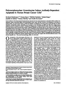
(Table 2). This suggests that intermediate density PMN may be more density PMN [25]. The combined results from three separate experin Figure 1. Unfractionated PEC reduced inoculum CFU by 56 ±
by 73 ± 4%, to be described,
and pellet-i pellet-2 cells
by 60 ± were more
5%. efficient
However, in some later in killing than in this
series.
In contrast to PMN elicited as described above, PMN elicited in normal or immune mice in 4 hr or 24 hr by thioglycollate, and purified on metrizamide gradients, failed to reduce inoculum CFU of B dermatitidis, but killed C albicans under
the same We found
experimental conditions. that a subpopulation from
pellet-i
(elicited
with
titidis cells from immune mice) enriched for lymphocytes column fractionation could not kill B dermatitidis in this
nonviable
B derma-
(80%) by Sephadex assay. Furthermore,
(1:1) the pellet-i cells with the G- 10 lymphocyte-enriched layer lA cells (which are enriched for monocytes), did
not
subpopulation, significantly
G-lO mixing or with enhance
killing in the mixture beyond that calculated arithmetically. Nor did mixing 1: 1 the pellet-i cells (which contain lymphocytes and monocytes) with the nonkilling pellet-i cells elicited by thioglycollate enhance killing in the mixture beyond that calculated arithmetically (data not shown). These experiments suggest that lymphocytes are ineffective, and lymphocytes and/or monocytes are nonenhancing to PMN killing during and Effect
the elicited
coculture
of Effector Having
nonviable
period,
by nonviable
even
to Target
established B dermatitidis
when
these
cells
are
obtained
from
immune
mice
B dermatitidis. Cell that
Ratio
the pellet-i
cells were
fraction
enriched
of PEC
for PMN
elicited
in immune
and had enhanced
mice
by
fungicidal
Immune
Activation
of PMN’s
for
Fungal
Killing
511
LI.
C)
-J
z LI.
0 I-.
0 w
Fig.
1.
(PEC)
Reduction elicited
experiments.
of
inoculum
by nonviable Pellet-I
colony
forming
B dermatitidis
cells
are
most
units
(CFU)
in immune
effective
(P