Behavioral Neuroscience 2007, Vol. 121, No. 6, 1258 –1271
Copyright 2007 by the American Psychological Association 0735-7044/07/$12.00 DOI: 10.1037/0735-7044.121.6.1258
Impaired Processing of Local Geometric Features During Navigation in a Water Maze Following Hippocampal Lesions in Rats Peter M. Jones, John M. Pearce, Vanessa J. Davies, Mark A. Good, and Anthony McGregor Cardiff University Hippocampal damage impairs navigation with respect to information provided by the shape of an arena. Recent evidence has suggested that normal rats use local geometric information, as opposed to a global geometric representation, to navigate to a correct corner. One implication of this pattern of results is that hippocampal lesions may impair processing of 1 or more of the local geometric features of an environment. The authors therefore investigated the effects of hippocampal cell loss in rats on navigation to a hidden goal with respect to a variety of local cues in an environment with a distinctive shape. Rats with lesions of the hippocampus were impaired in discriminating a right-angled corner from its mirror image. However, they were able to use cues provided by an acute-angled corner (Experiment 1) or a local polarizing cue (Experiment 2). In contrast, lesioned rats were impaired in discriminating long versus short walls (Experiment 3). Results indicate that the hippocampus plays a role in disambiguating locations by processing (metric) information related to the distance between corners. Keywords: spatial learning, water maze, geometry, hippocampus, navigation
2001), and humans (Garrad-Cole, Lew, Bremner, & Whitaker, 2001; Hermer & Spelke, 1994, 1996). These findings imply that animals can use geometric information provided by the shape of their environment to find a hidden goal, but they do not reveal the nature of this information. Cheng (1986) and Gallistel (1990; see also Cheng & Newcombe, 2005, for a review) proposed that animals rely on a global geometric representation of the overall shape of their environment to identify where a goal is located, but Pearce et al. (2004) have argued that animals rely on more local geometric information. In the study by Cheng (1986), rats may, for example, have ignored the shape of the arena and looked for a corner where a short wall was, say, to the left of a long wall. Alternatively, they may have searched for a long wall and then headed for the corner at a particular end. To determine whether animals rely on local or global geometric cues when navigating with reference to the shape of their environment, Pearce et al. (2004) first trained rats to find a submerged platform in one corner of a rectangular pool. Steps were taken to ensure that the only cues that could be used to find the platform were provided by the shape of the environment. The rats were then placed in a kite-shaped pool constructed from the same walls with two right-angled corners. On being placed into the new environment, rats showed a preference for searching in the right-angled corner that was congruent, rather than incongruent, with the corner that contained the platform during training in the rectangle. Pearce et al. argued that if rats identified the location of the platform in the rectangle by reference to its overall shape, then the difference between the shape of the rectangle and the kite should have prevented subjects from discriminating between the two rightangled corners in the latter environment. In contrast, if rats were to rely initially on a local geometric cue, such as a corner where a short wall is to the left of a long wall (i.e., sense information), then they should be able to identify this cue in the kite and head toward the correct right-angled corner. On the basis of these findings, they
Recent evidence has shown that the hippocampus is a vital brain structure for successful navigation to a goal hidden at a fixed location in an arena with a distinctive shape (Lever, Wills, Cacucci, Burgess, & O’Keefe, 2002; McGregor, Hayward, Pearce, & Good, 2004; O’Keefe & Burgess, 1996; Pearce, Good, Jones, & McGregor, 2004; Tommasi & Save, 2005). Interpretation of the role played by the hippocampus in this form of navigation clearly relies on the conceptual analysis of learning displayed by normal rats in this paradigm. Over the past 20 years, a considerable body of research has examined how animals orient with reference to the shape of their environment. One of the first of these investigations was by Cheng (1986), who trained rats in a reference memory task to find food hidden in one corner of a rectangular arena. Despite the presence of distinctive landmarks in each corner, the rats made rotational errors by searching in the diametrically opposite corner to the one in which the food was hidden. Similar results have been found not only with rats (Margules & Gallistel, 1988; Pearce, Ward-Robinson, Good, Fussel, & Aydin, 2001), but also with fish (Sovrano, Bisazza, & Vallortigara, 2003), pigeons (Kelly, Spetch, & Heth, 1998), chicks (Tommasi, Vallortigara, & Zanforlin, 1997), nonhuman primates (Gouteux, Thinus-Blanc, & Vauclair,
Peter M. Jones, John M. Pearce, Vanessa J. Davies, Mark A. Good, and Anthony McGregor, School of Psychology, Cardiff University, Cardiff, Wales. Peter M. Jones is now at the School of Psychology, University of Nottingham, Nottingham, England. This work was funded by a grant from the Biotechnology and Biological Sciences Research Council. We thank Jo Haddon, Sandy McGregor, and Katy Bowen for help in conducting the study. Correspondence concerning this article should be addressed to Anthony McGregor, who is now at the Department of Psychology, Durham University, South Road, Durham DH1 3LE, England, or to Mark A. Good, School of Psychology, Cardiff University, Cardiff CF10 4AT, Wales. E-mail:
[email protected] or
[email protected] 1258
SHAPE CUES AND THE HIPPOCAMPUS
concluded that rats navigate with reference to local rather than global geometric cues that derive from the shape of the environment (see also Esber, McGregor, Good, Hayward, & Pearce, 2005; McGregor, Jones, Good, & Pearce, 2006; Tommasi & Polli, 2004). The hippocampus has long been associated with processing spatial information (O’Keefe & Nadel, 1978, for review). Consistent with a role for this structure in navigation with respect to the shape of the environment, Pearce et al. (2004; see also McGregor et al., 2004) showed that rats with damage to the hippocampus were unable to identify the correct corner of a rectangle in which a goal was hidden. If it is accepted that navigation by normal rats in such an environment is controlled by local rather than global geometric cues, then it follows that the hippocampal lesions may impair navigation by disrupting the processing of local geometric features. The purpose of the present study was to evaluate this hypothesis by identifying the nature of the local geometric cues that require an intact hippocampus for use in navigation.
Experiment 1 Geometric information provided by an environment may encompass several dimensions, including information about the angles and the distances between features, that may provide cues for orientation and navigation (Collett, Cartwright, & Smith, 1986; Gallistel, 1990; Poucet, 1993; Scho¨ne, 1984). To establish the generality of our conclusion that normal rats use local geometric features to navigate to a hidden goal in a rectangular pool, we first determined whether normal rats use local geometric features in a kite-shaped pool. We then examined whether rats with hippocampal lesions were able to use these local features and also whether hippocampal cell loss affected rats’ ability to navigate with respect to all local geometric cues. There were four groups in Experiment 1: sham-R, hippocampal-R, sham-A, and hippocampal-A. The sham-R group received a sham lesion before it was trained to find a submerged platform in one right-angled corner of a kite-shaped pool. On the completion of this training, rats were given a test trial in a rectangular pool that was constructed from the same walls as the kite, so that two of the corners in the rectangle were equivalent to the correct right-angled corner in the kite and two were equivalent to the incorrect right-angled corner. In keeping with the local geometric cue hypothesis (Pearce et al., 2004), if rats showed a preference for the corners in the rectangle that were equivalent to the correct corner in the kite, then this would indicate they were navigating by means of local rather than global geometric cues. The main question of interest in the present study was to assess how a group with hippocampal lesions (hippocampal-R group) would perform given the same training as the sham-R group. If the hippocampal-R group failed to discriminate between the two rightangled corners in the kite, then it would demonstrate that the hippocampus is involved in navigation with respect to local geometric cues. By comparing the detailed performance of the two groups in the kite, we also hoped to gain some understanding of nature of the local cues that could be used by the sham, but not the hippocampal, group. The second aim of Experiment 1 was to examine whether rats with hippocampal lesions are impaired in their ability to use all local geometric cues provided by the shape of the arena. Thus, the two remaining groups were required to find a submerged platform
1259
in the apex of the kite (sham-A and hippocampal-A groups). This corner was chosen to maximize the sensitivity of the behavioral measure because the acute-angled corner was thought to provide a salient feature. The sham-A group received sham lesions before this training, whereas the hippocampal-A group received lesions of the hippocampus. If damage to the hippocampus impaired the rats’ ability to navigate with respect to local geometric cues provided by the angles between the walls, then both the hippocampal-R and hippocampal-A rats would be unable to navigate to the correct corner as well as the control groups. However, if the impairment produced by damage to the hippocampus is limited to a specific type of geometric cue, such as discriminating a right-angled corner from its mirror image, then we would expect only hippocampal-R rats to be impaired. This pattern of results would also indicate that the impairment in the hippocampal-R group has not occurred because the lesions have some general, theoretically uninteresting effect, such as disrupting navigation in any featureless environment. Pilot experiments in a rectangular pool have revealed that rats with hippocampal lesions tend to swim along the edge of the wall unless they receive special training. Accordingly, the present experiment included a pretraining phase to encourage the hippocampal groups to navigate in a similar fashion to the control groups (see also McGregor et al., 2004; Pearce et al., 2004). All four groups were first trained to find and then climb on a submerged platform with a beacon attached to it that was some distance from the edge of a circular pool. They were then trained to find the platform with the beacon attached to it in the corner of the kite that would be used for the main stage of the experiment. On completion of this pretraining, rats were then required to find the platform without the beacon attached to it for the principal phase of the experiment.
Method Subjects. The 44 subjects were experimentally naive male Hooded Lister rats supplied by Joint Services, Cardiff University (Cardiff, Wales). They weighed between 280 –330 g before surgery and were given unrestricted access to food and water in their home cages. All rats were housed in pairs in a room in which the lights were turned on at 0600 and turned off at 2030. The rats were tested for a minimum of 5 days a week during the period when the lights were on. At the start of the experiment, the rats were assigned at random to the four groups, with the constraint that there were 12 rats in each of the two groups with hippocampal lesions. Apparatus. The dimensions of the experimental room were 4.0 m ⫻ 3.0 m ⫻ 2.3 m high. A white circular pool made of fiberglass was mounted on a platform 0.6 m above the floor in the center of the room. It was 2 m in diameter and 0.6 m deep and was filled to a depth of 0.3 m with a mixture of water and 0.5 L of white food coloring (E308; Roehm and Haas, Ltd., Dewsbury, England). The water was maintained at a temperature of 25 °C (⫾ 2 °C). At the end of each day, the pool was drained and cleaned and was refilled the following morning. A white circular ceiling 2 m in diameter with a 30-cm diameter hole in the center was fixed 1 m above the rim of the pool. A wide-angled video camera was fixed 5 cm above the hole in the ceiling, 1.35 m above the water. The camera image was relayed to a PC equipped with tracking software
1260
JONES, PEARCE, DAVIES, GOOD, AND MCGREGOR
(Watermaze Software, Edinburgh, Scotland) and to recording equipment located in an anteroom. Eight 45-W, 22.5-cm diameter spotlights were recessed into the circular hung ceiling. The lights were arranged at equal distances in a 1.6-m diameter circle, with its center directly above the center of the pool. The spotlights were illuminated throughout the experiment. The pool was permanently enclosed within a light blue curtain hung from the ceiling on a runner at a distance of 25 cm beyond the edge of the pool. The curtain was 1.4 m deep so that it hung from the ceiling to 20 cm below the rim of the pool. In addition to the spotlights used to illuminate the pool, the room was also lit with four 1.5-m strip lights. The strip lights were attached end to end in pairs on opposite walls of the room 0.75 m above the floor and parallel to it. An arena in the shape of a kite, with two right-angled corners, was created using four white polyurethane boards, which were each 0.59 m high and 2 mm thick. Two of the boards were 1.8 m in length, and the other two were 0.9 m in length. The boards were suspended vertically in the pool from 2-cm square bars that were attached to the top edge and that extended beyond the ends of the boards by 6 cm and rested on the edge of the circular pool. As a result of this arrangement, the tops of opposite walls were either 33 or 35 cm above the surface of the water, and the positions of the boards that were lower in the water varied randomly from session to session. The four corners of the kite made contact with the edge of the pool. The circular escape platform was 10 cm in diameter, and its surface was 2 cm below the surface of the water. The center of the platform was always 25 cm from the corner in which it was located, on a line that bisected the corner. A beacon could be attached to the platform, 2.5 cm from its edge, at a point that was furthest from the corner in which the platform was located. The beacon was a plastic rod with a diameter of 1 cm, alternating black and white hoops, and with a width of 1 cm along its entire length. A white plastic disk, 3 cm in diameter and 0.5 cm thick, was attached to the top of the rod 23 cm above the surface of the water. Surgery. After a minimum of 7 days in the laboratory, the rats received surgery. Each rat was deeply anaesthetized with a mixture of oxygen and isoflurane. It was then placed into a stereotaxic frame (Kopf Instruments, Tujunga, CA) using atraumatic ear bars, and anaesthetic was reduced to a maintenance concentration. The scalp was incised along the midline, and a dental burr was used to remove the bone overlying the neocortex. A 2 l Hamilton syringe (Reno, NV), which was used to make injections of ibotenic acid, was attached to a moveable arm mounted on the stereotaxic frame. The plunger of the Hamilton syringe was attached to an electronic microdrive (Model KDS 310; KD Scientific, New Hope, PA), which regulated the volume and rate of infusion of the neurotoxin. Ibotenic acid (Biosearch Technologies, San Rafael, CA) was dissolved in sterile phosphate-buffered saline (pH 7.4) to produce a 63 mM solution and was infused at a rate of 0.03 l/min. Volumes of between 0.05 and 0.10 l were infused in 28 sites (for stereotaxic coordinates of the injection sites, see Coutureau, Galani, Gosselin, Majchrzak, & Di Scala, 1999). The neurotoxin was encouraged to diffuse away from the needle and into the tissue by leaving the Hamilton syringe in place for 2 min following each infusion. Rats in the sham-operated control groups underwent a similar surgical procedure, in which the skin was incised, the neocortex exposed, and the dura perforated with a 25 gauge Microlance 3 needle (Becton Dickinson, Drogheda, Ireland), but
no injection was given. At the end of both the lesion and sham procedures, the scalp incisions were sutured and the rats were placed in a recovery box maintained at 40 °C for 3– 4 hr. In addition, rats in the lesion groups received a subcutaneous 10-ml injection of saline and glucose solution. Once the rats had recovered sufficiently, they were transferred back to their home cages. A minimum of 14 days postoperative recovery was allowed before testing began. Behavioral training. Rats were transported 5 or 6 at a time in separate compartments of a light-tight carrying box that was placed on a work surface in the southwest corner of the anteroom. All rats were first trained for five sessions to swim to the platform with the beacon attached to it in the circular pool. There were four trials in each session. The platform was placed in the center of one of the four quadrants of the circular pool, and the rat was released from one of the four major compass points at the edge of the pool. The four possible quadrants, and four possible release points, were each selected once in every session in a randomly determined sequence. The trial began when the rat was placed gently into the pool facing the wall. Throughout the experiment, the experimenter watched the rat’s progress on the monitor in the anteroom and timed how long it took for it to climb onto the platform. If the rat did not find the platform after 60 s, then the experimenter placed a thumb about 5 cm in front of its snout and guided it to the platform. No prior training was needed for this treatment to be effective. The rat was allowed to remain on the platform for 20 s before being removed, dried gently in a towel, and returned to the carrying box. All rats in the carrying box were tested once before the next trial began. Following pretraining in the circular pool, all rats received four sessions, with four trials in each session, of pretraining in the kite-shaped arena with a beacon attached to the platform. The platform was located in the apex of the kite for the sham-A and the hippocampal-A groups and in a right-angled corner for the sham-R and hippocampal-R groups. For half the rats in each of the last two groups, the platform was in one right-angled corner of the kite, and for the remaining rats, it was in the other right-angled corner. After all the rats had received a single trial, the arena was rotated 90°, 180°, or 270° in a clockwise direction such that within a session, the acute-angled corner pointed once to each of the major compass points. Rats were released from the midpoints of each of the four walls in every session in a randomly determined sequence. The main phase of the experiment commenced immediately after the completion of pretraining. All four groups received 12 sessions of training in the kite that were identical to the sessions of the previous stage, except that the beacon was not attached to the platform. The sham-R and hippocampal-R groups then received a single test session with three training trials followed by a test trial for which they were placed in a rectangular arena for 60 s without the platform. The arena for this test trial was constructed from the same panels that were used to create the kite, and all rats were released from the center. During the training in the kite without the beacon, a record was taken on every trial of which corner the rat first swam to on being released into the pool. A corner was deemed to have been entered if the rat’s snout entered a search zone for that corner. Each zone was created by a circle with a radius of 15 cm, with its center 25 cm from the corner on a line that bisected the corner and two parallel tangents extending from the circle to the walls. During the
SHAPE CUES AND THE HIPPOCAMPUS
1261
test trial in the rectangle, a record was taken of the amount of time spent by the two groups in each of the four quadrants. Histology. After completion of behavioral testing, the rats were deeply anaesthetized using Euthatal (200 mg/kg sodium pentobarbitol) and perfused through the heart using physiological saline and 10% formol saline. The brains were then removed and stored in 10% formol saline solution for at least 4 hr. The formol saline solution was then replaced with 25% sucrose solution, and the brains were allowed to become saturated in the solution for 24 to 48 hr before being sectioned (40 m), mounted on slides, and stained with cresyl violet.
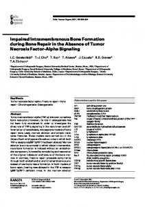
Results Histology. Of the 24 rats with hippocampal damage, 3 were found to have sustained less than 35% cell loss (range ⫽ 20 –35%) and were excluded from the analysis. All of the remaining lesioned rats (hippocampal-A, n ⫽ 10; hippocampal-R, n ⫽ 11) sustained bilateral damage to both the dentate gyrus and the CA subfields at all dorsal and ventral levels. The range of cell loss in the hippocampus was 65–100%. The majority of rats also sustained some cell loss in the subiculum (including the pre- and para-subiculum). This damage was most evident in ventral regions of the subiculum. There was no detectable cell loss in the post-subiculum or in the medial or lateral entorhinal cortex in any of the lesioned rats. The left-hand panel of Figure 1 shows reconstructions of the maximum (light shading) and minimum (dark shading) extent of the hippocampal lesions on a series of horizontal sections taken at distances in millimeters ventral to the surface of the brain (top to bottom: –3.1, –3.6, – 4.6, –5.6, – 6.6, –7.6, – 8.4 mm). Behavioral training. The mean escape latencies—the time taken to reach the platform after being released into the pool—are
Figure 1. Histology from Experiments 1 (left-hand panel), 2 (center panel), and 3 (right-hand panel). The maximum (gray) and minimum (black) extent of the hippocampal lesions at horizontal sections taken through the dorsoventral extent of the brain. The depicted sections are in millimeters from Bregma (clockwise from top left: 3.1, 3.6, 5.6, 7.6, 8.6, 6.6, and 4.6). From The Rat Brain in Stereotaxic Coordinates (Figures 91, 96, 99, 103, 107, 111, and 113), by G. Paxinos and C. Watson, 1986, San Diego, CA: Academic Press. Copyright 1986 by Elsevier. Adapted with permission.
Figure 2. The mean escape latencies (⫾ SEM) for the four groups in Experiment 1. The first five sessions show escape latencies during pretraining in a circular pool. The second four sessions show escape latencies during pretraining in the kite-shaped arena with a beacon attached to the platform. The final 12 sessions show escape latencies during training in the kite-shaped arena without a beacon attached to the platform. R ⫽ rightangle; A ⫽ apex.
shown for the four groups for the entire experiment in Figure 2. The results from the pretraining with the beacon attached to the platform are shown for the sessions in the circular pool in the left-hand side of the figure, and immediately to the right are the results from the pretraining sessions in the kite. Despite initial differences between the groups, by the end of the pretraining phase, the escape latencies for the four groups were similar and relatively short. A two-way analysis of variance (ANOVA) of individual mean escape latencies for the final pretraining session in the kite revealed that there was no effect of lesion, F(1, 37) ⫽ 1.54, p ⬎ .2, although rats trained to find the platform in the apex found the platform more quickly than did those trained to find the platform in one of the right-angled corners, F(1, 37) ⫽ 4.20, p ⬍ .05. There was no interaction between the main effects (F ⬍ 1). The right-hand portion of Figure 2 shows the mean escape latencies for the four groups during training in the kite with no beacon attached to the platform. By the end of this stage, the hippocampal-R group took considerably longer than any other group to find the platform, and there was rather little difference between the three remaining groups. To compare the final performance of the groups, we calculated individual mean escape latencies for the last six sessions of training. A two-way ANOVA revealed a significant effect of lesion, F(1, 37) ⫽ 28.15, p ⬍ .0001; training, F(1, 37) ⫽ 55.68, p ⬍ .0001; and their interaction, F(1, 37) ⫽ 10.88, p ⬍ .005. Subsequent tests of simple main effects revealed a significant difference between the hippocampal-R and the sham-R groups, F(1, 37) ⫽ 37.90, p ⬍ .001, but no difference between the hippocampal-A and the sham-A groups, F(1, 37) ⫽ 1.97, p ⬎ .17. The left-hand panel of Figure 3 shows, for every session of training in the kite without the beacon, the mean percentages of trials on which the hippocampal-R and sham-R groups swam directly to the correct corner. To analyze the terminal performance of the two groups, we calculated individual mean percentages of trials on which subjects swam to the correct corner for the final six
1262
JONES, PEARCE, DAVIES, GOOD, AND MCGREGOR
Figure 3. The mean percentages of trials on which rats swam directly to the correct corner for the right-angle (R) groups (left-hand panel) and the apex (A) groups (right-hand panel) during training in Experiment 1.
training sessions. A comparison of the results from both groups revealed that the sham-R group headed directly for the correct corner significantly more often than did the hippocampal-R group, U(10, 11) ⫽ 8.5, p ⬍ .005. During the 60-s test trial in the rectangular pool with no platform, the sham-R group spent 61.20% of the trial in the two correct quadrants, whereas the hippocampal-R group spent 54.46%. Chance performance was 50%. A comparison of individual percentages of time spent in the two correct quadrants revealed a significant difference between the groups, t(19) ⫽ 2.26, p ⬍ .05. One-sample tests confirmed that the sham-R group spent significantly more time in the correct quadrants than would be expected on the basis of chance, t(9) ⫽ 5.11, p ⬍ .01, but this was not the case for the hippocampal-R group, t(10) ⫽ 2.20. The results from the final six sessions of training and the test trial indicate that the sham-R group was more successful at finding the correct right-angled corner than the hippocampal-R group. To understand why the hippocampal lesions disrupted performance in
this way, we compared the performance of the two groups for each of the different points of release from the sides of the kite for the final six sessions of training. The top row of Figure 4 shows the mean percentages of trials on which the sham-R group headed directly for each corner of the kite from the four different release points (indicated by an arrow). The results have been normalized so that for all rats, the correct corner is on the left-hand side of the kite. The most striking feature of these results is that subjects swam directly to the correct corner more frequently when they were released from some locations rather than from others. For instance, the correct corner was chosen with much greater frequency when rats were released from the short wall adjacent to this corner than from the long wall opposite the corner. Pearce et al. (2004) observed a similar pattern of results. They concluded that one strategy adopted by rats in a kite was to swim in a particular direction when they were released from a short wall. In keeping with this suggestion, it is apparent that the sham-R group headed more often in the direction of the correct, as opposed
Figure 4. The mean percentages of trials on which group sham-R (top row) and group hippocampal-R (bottom row) swam to each corner of the kite during the final six sessions of training when released from the short walls (left-hand columns) and long walls (right-hand columns). Arrows indicate the release point. R ⫽ right angle; C ⫽ correct corner; A ⫽ apex; I ⫽ incorrect corner; O ⫽ other corner.
SHAPE CUES AND THE HIPPOCAMPUS
to the incorrect, corner when released from each of the two short walls. To analyze this outcome further, we compared the percentage of trials on which subjects headed in the direction of the correct corner when released from the two short walls with the percentage of trials on which it headed in the direction of the incorrect corner. Both percentages included the number of occasions on which subjects headed for the obtuse-angled corner if this corner lay between the point of release and the corner under consideration. There was a significant preference for heading in the direction of the correct corner, T(8) ⫽ 0. The bottom row of Figure 4 shows the equivalent results for the hippocampal-R group and indicates that the strategy just identified was used to a lesser extent in the hippocampal-R group than in the sham-R group. However, a similar analysis to the one above revealed no significant preference, when released from a short wall, for heading in the direction of the correct rather than the incorrect right-angled corner, T(8) ⫽ 10.5. In addition, when released from a short wall, the sham-R group headed in the direction of the correct corner significantly more often than did the hippocampal-R group, U(10, 11) ⫽ 26.0, p ⬍ .05. Therefore, perhaps, therefore, one effect of hippocampal lesions is to make it more difficult for rats to acquire strategies of heading in a particular direction when they are released from a wall of a certain length. Turning now to the trials on which when rats were released from the long walls, the sham-R group was able to discriminate between the two right-angled corners when released from the long wall adjacent to the correct corner, T(10) ⫽ 0, but not when it was released from the other long wall, T(9) ⫽ 22.0. One possible explanation for this pattern of results is that rats prefer to head for a corner that is near rather than distant from the point of release. If this is correct, then the hippocampal-R rats may have adopted a similar strategy because they, too, showed a discrimination between the two right-angled corners when they were released from the wall beside the correct corner, T(11) ⫽ 10.5, but not when they were released from the opposite wall T(8) ⫽ 12.5. In support of this conclusion, it is apparent from inspection of Figure 4 that when rats were released from one of the long walls, they had a tendency to swim first to one of the two adjacent corners. This may account for the high proportion of choices to the apex. It may also explain the reason why rats were able to discriminate correct from incorrect right-angle corners only when they were released from the long wall adjacent to the correct corner. The mean percentages of trials on which the sham-A and the hippocampal-A groups swam directly to each of the four corners of the kite can be seen in the right-hand panel of Figure 3. Both groups headed directly for the apex of the kite on the majority of trials. The preference for the apex at the outset of this stage presumably indicates that the presence of the beacon above the platform during the prior training in the kite did not prevent subjects from learning about the position of the goal relative to geometric cues. A comparison of the individual mean number of trials during the final six sessions on which subjects headed directly for the apex revealed no significant difference between the groups, U(10, 10) ⫽ 25.5, p ⬎ .064.
Discussion An important conclusion to be drawn from the experiment is that lesions to the hippocampus impaired the ability of the
1263
hippocampal-R group to identify the right-angled corner of the kite where the escape platform was located. Support for this conclusion can be found in the slower escape latencies by the hippocampal-R, relative to the sham-R, group during the final six sessions of training. Support can also be found in the greater frequency with which the sham-R group, versus the hippocampal-R group, headed directly for the correct corner after being released. Finally, the results from the test trial also confirmed that the hippocampal-R group was unable to discriminate between the correct and incorrect right-angled corners. Hippocampal lesions have been shown to impair the capacity to navigate with reference to cues provided by the shape of a rectangular arena (e.g., Pearce et al., 2004). The present results extend the generality of this effect by showing that hippocampal lesions can impair navigation in a kite-shaped environment. Another important conclusion to be drawn from the study derives from the finding that the sham-R group spent more time in the correct than incorrect quadrants during the test trial in the rectangle. Such an outcome can be most readily explained if it is assumed that during its training in the kite, the sham-R group relied on local cues to find the submerged platform. Presumably, at least some of these cues were also present in the rectangle and were responsible for the performance of the sham-R group during the test trial. The poor performance of the hippocampal-R group in the kite, and its inability to identify the correct corner in the rectangle, therefore implies that the hippocampal lesions were effective because they impaired rats’ capacity to use local geometric cues. Although some local geometric cues, such as the apex of the kite, were missing during the test trial, sham rats were still able to identify the correct local geometric properties of the rectangle during the test trial. Before discussing the nature of the local cues that may be used to navigate to a hidden goal, it is important to first establish that the deficits in learning to navigate with respect to cues provided by the shape of the arena in rats with hippocampal lesions are not symptomatic of a more general (nonspecific) disruption of learning. The results from the two groups trained with the platform in the apex of the kite in Experiment 1 indicate that an explanation of this type is unlikely to be correct. With the platform in this location, there was no evidence that the hippocampal-A group was impaired relative to the sham-A group. Thus, hippocampal lesions do not inevitably prevent subjects from learning to escape from a pool with a distinctive shape, nor do they encourage responses that interfere with heading directly for a particular location in such an environment. Instead, the results from the four groups together indicate that hippocampal lesions affect the ability of rats to navigate with reference to some local geometric cues but not others. On the basis of the results from the groups trained with the platform in the apex, it would seem that these lesions do not make it difficult to identify an acute-angled corner created by two long walls. However, learning about other geometric properties of an arena is presumably influenced by hippocampal cell loss. One possible local geometric cue that might have been used by the sham-R group to find the submerged platform is the relationship between the walls creating the correct corner (e.g., short wall to the left of the long wall). However, according to this explanation, rats would be expected to find the platform with equal ease, regardless of where they were released. In contrast to this prediction, the
JONES, PEARCE, DAVIES, GOOD, AND MCGREGOR
1264
sham-R group headed for the correct corner more frequently when the point of release was a short rather than long wall. On the basis of this finding and the results shown in Figure 4, it seems that one important strategy adopted by the sham-R group for finding the platform was to head in a particular direction with reference to a short wall. This interpretation raised the possibility that hippocampal lesions were effective either because they made it difficult for rats to learn to swim in a particular direction relative to some cue or because they made it hard to discriminate between a long and short wall.
Experiment 2 It has just been suggested that rats with hippocampal lesions find it difficult to head in a particular direction with reference to a local cue, such as a short wall. There are two reasons why the lesions may exert such an influence. They may make it difficult to tell the difference between short and long walls, or they may make it difficult to learn to head in a particular direction with reference to any local cue. The purpose of Experiment 2 was to evaluate the second of these alternatives. The final experiment examined the merits of the first alternative. Two groups were trained to find a submerged platform in a rectangular pool with one long black wall and three white walls (a hippocampal-BW group and a sham-BW group). The platform was located in a corner created by the long white wall and a short white wall. In keeping with the hippocampal-R group of Experiment 1, the hippocampal-BW group was required to discriminate between two right-angled corners constructed from white walls. On this occasion, however, the presence of the black wall might serve as a polarizing cue so that it would be possible to find the correct corner by heading in a certain direction relative to this wall. If damage to the hippocampus impaired the capacity to develop such a strategy, then the hippocampal-BW group would fail to discriminate between the correct and incorrect right-angled corners created by the three white walls. In contrast, if hippocampal lesions had relatively little impact on the acquisition of this type of strategy, then the hippocampal-BW group would demonstrate a clear discrimination between the two white right-angled corners. Two additional groups were included in the experiment to provide a control against which the results from the above groups could be assessed. A hippocampal-W and sham-W group were trained in the same way as the first two groups, except that the rectangle was constructed from four white walls. The results from Experiment 1, as well as those reported by Pearce et al. (2004) and McGregor et al. (2004), indicate that the hippocampal-W group would fail to discriminate between the geometrically correct and incorrect corners in the rectangle. There were two test sessions following the training just described. The first contained a trial in which all groups were placed in the pool and trained for 60 s in the absence of the platform. This test was intended to provide an additional indication of the degree to which the cues present during training were used to find the platform. The second test session was conducted with the sham-BW and hippocampal-BW groups and contained a trial in which subjects were placed in a rectangle with four white walls. This test was conducted to assess for these groups the degree to which geometric cues provided by the shape of the pool controlled searching for the platform. In keeping with the method adopted for
Experiment 1, all four groups received pretraining with a beacon attached to the platform in a circular pool and then in the rectangle.
Method Subjects, surgery, histology, and apparatus. The subjects were 32 experimentally naive rats from the same stock and housed in the same conditions as those in Experiment 1. Prior to training, they weighed between 310 and 345 g. Subjects were randomly assigned in equal numbers to the four groups at the start of the experiment. Two groups received lesions to the hippocampus, and two groups received sham lesions. Details concerning surgery and histology were the same as for Experiment 1. The experiment was conducted in the same room and with the same apparatus that was used for Experiment 1, with the exception that the rectangular arena was made up of either four white boards or three white boards and one long black board. The four corners of the arena made contact with the edge of the circular pool. Behavioral training. Behavioral training commenced 14 days after the completion of the surgery. The four groups received 4 days of pretraining in the circular pool and 4 days in the rectangular pool. The beacon was attached to the platform throughout this stage. The rectangular pool was composed of one long black wall and three white walls (two short and one long) for the sham-BW and hippocampal-BW groups, and from four white walls for the sham-W and hippocampal-W groups. For half the subjects in each group, the platform was always in a corner where a short white wall was to the left of a long white wall, and for the remaining rats, the platform was in the corner with the opposite properties. Within each session for the sham-W and hippocampal-W groups, the platform was located twice in one of the candidate corners and twice in the diagonally opposite corner. The order in which these corners were selected was random. The remaining procedural details were the same as for Experiment 1. The four groups then received 12 sessions of training in the manner just described but without the beacon. The next two sessions consisted of three training trials followed by a test trial in which subjects were placed in the rectangle without the platform for 60 s. The first test session involved the four groups that were placed for the test trial in a pool that was identical to the one used for the training trials. The second test session took place with just the sham-BW and hippocampal-BW groups for whom the pool during the test trials was constructed for the first time from four white walls. A record was taken for every training trial without the beacon of the corner that rats approached first after being released into the pool. A corner was said to have been chosen when the snout of the rat entered a search zone defined in Experiment 1. During the test trials, a record was taken of the time spent by rats in each of the four quadrants of the pool.
Results Histology. The center panel of Figure 1 shows the maximum and minimum extent of the hippocampal cell loss in the lesioned rats used in Experiment 2. Two rats were found to have sustained extra-hippocampal cell loss to the temporal cortex and were excluded from further analysis. All of the remaining lesioned rats (hippocampal-BW, n ⫽ 6; hippocampal-W, n ⫽ 8) sustained
SHAPE CUES AND THE HIPPOCAMPUS
1265
bilateral damage to both the dentate gyrus and the CA subfields at all dorsal and ventral levels (range ⫽ 70 –100%). As in Experiment 1, the majority of rats also sustained some cell loss in ventral subiculum but none in the post-subiculum or entorhinal cortex. Behavior training. The left-hand portion of Figure 5 shows the group mean escape latencies for the four sessions of pretraining in the circular pool, and immediately to the right are the equivalent results for the pretraining in the rectangle. By the end of each stage, there was little difference among the results of the four groups. A two-way ANOVA using individual mean escape latencies for the final session of pretraining in the rectangle revealed that the effects of lesion, F(1, 26) ⫽ 1.04, p ⬎ .32; training arena, F(1, 26) ⫽ 2.45, p ⬎ .13; and their interaction, F ⬍ 1, p ⬎ .5, were not significant. The mean escape latencies for each group for the 12 sessions of training without the beacon and the first test session can be seen in the right-hand side of Figure 5. By the end of training, rats swam more swiftly to the platform in the rectangle with black and white walls than in the rectangle with all white walls. In addition, in each type of rectangle, the mean escape latencies were longer for the hippocampal lesioned than the sham-operated group. A two-way ANOVA of individual mean escape latencies for the final six sessions combined revealed a significant effect of training arena, F(1, 26) ⫽ 39.65, p ⬍ .0001, and lesion, F(1, 26) ⫽ 67.31, p ⬍ .0001, but no significant interaction, F(1, 26) ⫽ 2.59, p ⬎ .1. The top panel of Figure 6 shows the mean percentage of trials in which the sham-BW group headed for the four corners of the rectangle for the 13 sessions of training that were given to this group without the beacon attached to the platform. The final two of these sessions contained three training trials and one test trial. The equivalent results for the hippocampal-BW group can be seen in the center panel of Figure 6. The corner containing the platform is referred to as the correct corner, the white corner without the platform as the incorrect corner, the corner diagonally opposite to the correct corner as the rotational corner, and the remaining
Figure 5. The mean escape latencies (⫾ SEM) for the four groups in Experiment 2. The first four sessions show escape latencies during pretraining in a circular pool. The second four sessions show escape latencies during pretraining in the rectangular arena with a beacon attached to the platform. The final 13 sessions show escape latencies during training in the rectangular arena without a beacon attached to the platform. BW ⫽ black and white; W ⫽ white.
Figure 6. The mean percentages of trials on which rats swam directly to each of the corners in the rectangular pool for the sham-BW (top panel) and hippocampal-BW (center panel) groups during the 13 sessions of training in Experiment 2. The bottom panel shows the mean percentages of trials on which groups sham-W and hippocampal-W swam directly to one of the correct corners during training in Experiment 2. BW ⫽ black and white; W ⫽ white.
1266
JONES, PEARCE, DAVIES, GOOD, AND MCGREGOR
corner as the other corner. Both groups acquired a strong tendency to head directly toward the correct corner as training progressed, although this effect was more marked in the sham-BW than in the hippocampal-BW group. Binomial tests showed, for the final six sessions of training, that both groups swam directly to the correct corner more often than would be expected by chance. Analyses of individual mean percentages of trials on which subjects headed directly for each of the four corners, for the final six sessions of training, revealed a significant difference for the sham-BW group, r2(3) ⫽ 19.6, N ⫽ 8, p ⬍ .001, and the hippocampal-BW group, r2(3) ⫽ 8.6, N ⫽ 6, p ⬍ .05. The correct corner was approached first significantly more often than any other corner by the sham-BW group, T(8) ⫽ 0, and the hippocampal-BW group, T(6) ⫽ 0. Evidence of a difference between the groups was revealed by the finding that the sham-W group approached the correct corner first significantly more often than did the hippocampal-BW group, U(8, 6) ⫽ 0.5. The remaining panel in Figure 6 shows the mean percentages of trials on which the sham-W and the hippocampal-W groups headed directly for a correct corner in the white rectangle for each of the 13 sessions of training that these groups received. A correct corner was regarded as the one containing the platform and the one that was diagonally opposite to it. An analysis of individual mean percentages of trials on which a correct corner was approached first, during the final six sessions, confirmed the impression in the figure that the sham-W group headed toward these corners more frequently than did the hippocampal-W group, U(8, 8) ⫽ 6.5, p ⬍ .001. In addition, binomial tests on the same data showed that the sham-W group swam to a correct corner more often than expected by chance, but this was not the case for the hippocampal-W group. The results from the first test trial are shown in the top panel of Figure 7. The left-hand pair of histograms shows the results for the sham-BW and hippocampal-BW groups. These groups were tested in an arena with one black and three white walls, and the results depict the mean percentage of time spent in the correct quadrant relative to the time spent in this quadrant plus the time spent in the adjacent quadrant that contained a corner with two white walls. The remaining histograms show for the sham-W and the hippocampal-W groups, who were tested in a white pool, the percentage of time that was spent in the two quadrants containing a correct corner. A two-way ANOVA of individual percentages of time spent in the correct quadrants revealed a significant effect of lesion, F(1, 26) ⫽ 11.80, p ⬍ .005, and a significant effect of training arena, F(1, 26) ⫽ 13.94, p ⬍ .001, but no significant interaction, F ⬍ 1, p ⬎ .7. Additional analyses using one-sample tests revealed that the sham-BW, t(7) ⫽ 8.14, p ⬍ .0001; hippocampal-BW, t(5) ⫽ 4.43, p ⬍ .01; sham-W, t(7) ⫽ 4.30, p ⬍ .01; but not the hippocampal-W, t(7) ⫽ 1.50, p ⬎ .18, groups spent more than half of the test trial in the correct region of the pool. The results from the second test trial are shown in the bottom panel of Figure 7. This test involved the sham-BW and the hippocampal-BW group and a rectangle with four white walls. The sham-BW group spent significantly more time in the two correct quadrants of the pool than did the hippocampal-BW group, t(12) ⫽ 4.01, p ⬍ .01. In addition, the sham-BW group spent significantly more than half of the test trial in the correct quadrant, t(7) ⫽ 6.75, p ⬍ .001, but this was not the case for the hippocampal-BW group, t(5) ⬍ 1.
Figure 7. The mean percentages of time (⫾ SEM) spent in the correct quadrants of the arena during Test Trial 1 (top panel) and Test Trial 2 (bottom panel) in Experiment 2. In Test Trial 1, rats were tested in the arena in which they were trained. In Test Trial 2, rats trained in a black and white (BW) arena were tested in a white (W) arena.
Discussion Experiment 2 evaluated the possibility that rats with hippocampal lesions were unable to learn to navigate in a specific direction from a distinctive wall. Rats with hippocampal lesions were able to discriminate between two right-angled corners that were both constructed from white walls in a rectangle with a single black wall. In contrast, lesioned rats failed to acquire a similar discrimination when all four walls of the rectangle were white. One interpretation of this finding is that the former group, on being placed in the pool, oriented with respect to the black wall and then headed in a particular direction to reach the goal. An implication of this analysis is that the failure of rats with hippocampal lesions to navigate successfully in an arena with four white walls is not a consequence of them being unable to learn to swim in a certain direction with respect to at least some features provided by the environment. Some comment is needed concerning the difference between the results from the hippocampal-BW and sham-BW groups during the training trials and the first test session. If the hippocampal-BW group was able to find the platform by swimming in a particular direction with reference to the black wall, then the sham-BW group would be expected to adopt the same strategy, and the performance of the two groups should have been similar in the black and white rectangle. It is possible, however, that the sham-BW group made use of the black wall as well as cues provided by the shape of the rectangle to find the platform. If it is assumed that damage to the hippocampus disrupts the ability to use this additional set of cues, then the difference between the groups
SHAPE CUES AND THE HIPPOCAMPUS
can be readily explained. In the black and white rectangle, the sham-BW group could refer to two types of cue to find the platform, whereas the hippocampal-BW group could refer to only one. An implication of this analysis is that if the black wall in the rectangle were replaced by a white wall, then the sham-BW group would continue to discriminate between the correct and incorrect corners— but to a lesser extent than in the original arena—and this discrimination would not be possible for the hippocampal-BW group. The results from the second test session confirmed these predictions.
Experiment 3 To navigate successfully in an arena with a distinctive shape, it would seem to be essential to be able to tell the difference between walls of different lengths. There are at least two strategies for finding a goal in one corner of a rectangle that would depend on this ability. Suppose the goal is in a corner where a short wall is to the left of a long wall, then it could be found either by searching for a corner with these properties or by heading to the left-hand end of a long wall. It hardly needs to be said that neither of these strategies would be successful if animals were unable to identify which wall is short and which is long. Experiment 3 was designed to assess whether lesions to the hippocampus affect this aspect of processing information provided by the shape of the environment. Four groups of rats, two with hippocampal lesions and two sham-operated controls, were trained to find a submerged platform that was located at the midpoint of one of the long walls of a rectangular pool. For one group with hippocampal lesions (hippocampal-W group) and one sham-operated control group (sham-W group), the four walls of the arena were white, so that the only cue that could be used to find the platform was the relative length of the walls. If hippocampal lesions impair the ability to appreciate the difference between walls of different lengths, then the hippocampal-W group would find it more difficult to locate the platform than the sham-W group. The remaining two groups, the hippocampal-BW and the sham-BW groups, were trained in a similar way, except that the long walls of the arena were white and the short walls were black. These groups could therefore discriminate between the short and long walls on the basis of their color, and the performance of these two groups was not expected to differ. Acquisition of the task was assessed primarily by recording where the rats swam to on being released into the pool, using circular search zones centered on the location of the platform and on equivalent locations at the midpoints of the other three walls. A series of test trials, during which rats were allowed to swim freely in the absence of a platform, was then used to determine the information used by the rats during training. The first test trial took place in the arena that was used for training. The second test trial took place in a rectangle with four white walls and was intended to reveal the degree to which the two groups trained in the black and white rectangle were able to use information about the lengths of the walls to find the platform. The remaining test was conducted with just the hippocampal-BW and sham-BW groups by placing them into a black and white square to assess the control acquired by the color of the walls when length information was removed. In keeping with the previous experiments, all groups initially received pretraining with a beacon attached to the platform.
1267
Method Subjects, surgery, histology, and apparatus. The subjects were 45 experimentally naive rats from the same stock and housed in the same conditions as for Experiment 1. Prior to surgery, they weighed between 300 and 350 g. Subjects were randomly assigned in equal numbers to the four groups at the start of the experiment. The hippocampal-W (n ⫽ 13) and hippocampal-BW (n ⫽ 12) received lesions to the hippocampus, and the sham-W (n ⫽ 10) and sham-BW (n ⫽ 10) group received a control operation. Details concerning surgery and histology were the same as for Experiment 1. The experiment was conducted in the same circular pool with the same platform and beacon as Experiment 1. A rectangular arena of the same dimensions as the one used in Experiment 1 was constructed from either four white walls or two long white walls and two short black walls. In addition, a square arena was made from alternating black and white walls that were each 1.41 m long and the same height as for the rectangular pool. Behavioral training. All rats received five sessions of pretraining in the circular pool in the same manner as for Experiment 1. They then received five sessions of pretraining in a rectangular pool with the platform located half way along one of the long walls, its center 25 cm from the wall. A beacon was attached to the platform for this stage. The platform was located beside one of the long walls for two randomly selected trials in each session and beside the other long wall for the remaining trials. Rats were released from each corner of the rectangle once within a session. The rats were released facing the corner, and the order of release point was randomized. The sham-W and hippocampal-W groups were trained in the white rectangle, and the sham-BW and hippocampal-BW groups were trained in the rectangle with short black walls and long white walls. Procedural details that have been omitted were the same as for Experiment 1. Training continued in the manner just described but without the beacon attached to the platform for the four groups for 15 sessions. Session 16 of the training phase consisted of three training trials followed by a 60-s test trial in which rats were placed in the pool that was used for their training but without the platform. Session 17 contained four training trials. Session 18 was conducted in the same manner as Session 16, except that all rats were tested in a rectangle constructed from four white walls. Sessions 19 –22 involved only the sham-BW and the hippocampal-BW groups. They received four training trials in Session 19 and three training trials and a 60-s test trial in the black and white square arena in Session 20. This test trial was repeated without any additional training trials in Sessions 21 and 22. Throughout the main phase of the experiment, which was conducted in the absence of the beacon, four circular search zones with a radius of 15 cm were identified for both the rectangular and the square arena using the tracking software. The centers of these zones were 25 cm from the wall at the midpoints of each of the four walls. The two search zones beside the long walls in the rectangle or the white walls in the square are referred to as the correct search zones, whereas the zones beside the short walls in the rectangle and the black walls in the square are referred to as the incorrect zones.
1268
JONES, PEARCE, DAVIES, GOOD, AND MCGREGOR
Results Histology. The reconstructions of the maximum and minimum extent of hippocampal lesions in the lesioned rats used in Experiment 3 are shown in the right-hand panel of Figure 1. Six of the 25 lesioned rats were found to have sustained less than 30% cell loss (range ⫽ 10 –30%) and were subsequently removed from further analysis. All of the remaining rats (hippocampal-BW, n ⫽ 10; hippocampal-W, n ⫽ 9) sustained bilateral cell loss to both dentate gyrus and CA subfields both dorsally and ventrally, with a range of 60 –100%. The majority of rats also sustained some cell loss to ventral subiculum, but no rat sustained damage to postsubiculum or entorhinal cortex. Behavior training. The left-hand portion of Figure 8 shows the mean escape latencies for the five sessions of pretraining in the circular pool, with the results for the pretraining in the rectangular pool shown immediately to the right. By the end of training in the rectangular pool, there were no differences between the groups. A two-way ANOVA of individual mean escape latencies for the final session of pretraining in the rectangle revealed that the effects of lesion, F(1, 35) ⬍ 1, p ⬎ .38; training arena, F(1, 35) ⫽ 3.27, p ⬎ .079; and their interaction, F ⬍ 1, p ⬎ .88, were not significant. The right-hand portion of Figure 8 shows the mean escape latencies for each group for each of the first 18 sessions of the training phase. All groups received three training trials followed by a single test trial in Sessions 16 and 17. Toward the end of training, the mean escape latencies were longer for the hippocampal-W group than for the other three groups, whose results did not differ substantially. A two-way ANOVA of individual mean escape latencies for the final six sessions shown in the figure revealed a significant effect of lesion, F(1, 35) ⫽ 9.38, p ⬍ .01, and a significant effect of training arena, F(1, 35) ⫽ 4.33, p ⬍ .05, but no interaction (F ⬍ 1). Figure 9 shows the mean percentages of trials on which the groups swam directly to a correct search zone for each of the 18
Figure 8. The mean escape latencies (⫾ SEM) for the four groups in Experiment 3. The first five sessions show escape latencies during pretraining in a circular pool. The second five sessions show escape latencies during pretraining in the rectangular arena with a beacon attached to the platform. The final 18 sessions show escape latencies during training in the rectangular arena without a beacon attached to the platform. W ⫽ white; BW ⫽ black and white.
Figure 9. The mean percentages of trials on which rats swam directly to a correct zone during training in Experiment 3. W ⫽ white; BW ⫽ black and white.
sessions of training without the beacon. It is evident that the pretraining with the beacon was effective because the performance of all the groups was better than would be expected on the basis of chance from the outset of this stage. Indeed, with the exception of the hippocampal-W group, the performance of the groups was close to ceiling throughout this stage. To compare the performance of the groups, we calculated individual mean percentages of trials on which subjects entered a correct search zone without first entering an incorrect zone for the final six sessions of training. Analysis of these results revealed a significant difference among the groups, H(3) ⫽ 15.16, p ⬍ .005. Fewer correct choices were made by the hippocampal-W group than by any other group, Us(9, 10) ⬍ 21, ps ⬍ .05, but the differences between the other three groups were not significant, Us(10, 10) ⬎ 31. In addition, binomial tests indicated that each group approached a correct search zone significantly more frequently than they did an incorrect zone. The mean percentages of time spent in the two correct and two incorrect search zones during the first test trial, which was conducted in the rectangle that was used for training, are shown for the four groups in the left-hand panel of Figure 10. The results from the test trial were analyzed with a three-way ANOVA with the effects of search zone, color of arena, and lesion. One important feature of the figure is that the discrimination between the correct and incorrect search zones was more marked in the sham than hippocampal groups, which was supported by a significant Lesion ⫻ Search Zone interaction, F(1, 35) ⫽ 22.81, p ⬍ .0005. Tests of simple main effects revealed that the sham groups combined spent significantly more time in the correct quadrants than did the hippocampal groups, F(1, 67) ⫽ 41.99, p ⬍ .005. Another important feature of the figure is that the discrimination between the two types of zone was more marked in the rectangle with black and white walls than in the rectangle with white walls, which was supported by a significant Color of Arena ⫻ Search Zone interaction, F(1, 35) ⫽ 17.71. Subsequent tests of simple main effects revealed that significantly more time was spent in the correct quadrants by the groups trained in the black and white rectangle than in the white rectangle, F(1, 67) ⫽ 32.90, p ⬍ .005. The three-way ANOVA also revealed that the main effects of lesion, training arena, and search zone, Fs(1, 35) ⬎ 15.34, ps ⬍ .0005,
SHAPE CUES AND THE HIPPOCAMPUS
1269
Figure 10. The mean percentages of time (⫾ SEM) spent in the correct and incorrect zones by four groups in Test Trial 1, which was conducted in the same arena as that used during training (left-hand panel), and Test Trial 2, which was conducted in a white rectangle (center panel). The right-hand panel shows the mean percentages of time (⫾ SEM) spent by sham-BW and hippocampal-BW rats in the correct and incorrect zones of the black and white square used in the three trials of Test 3. W ⫽ white; BW ⫽ black and white.
were significant, but the two remaining interactions were not ( ps ⬎ .09). The results from the second test trial, which was conducted in a white rectangle, are presented in the center panel of Figure 10. A similar ANOVA to the one just described confirmed that the discrimination between the correct and incorrect search zones was more marked in the sham than hippocampal groups by revealing a significant Lesion ⫻ Search Zone interaction, F(1, 35) ⫽ 16.92, p ⬍ .0005. Tests of simple main effects indicated that significantly more time was spent in the correct search zone by the two sham groups than the two hippocampal groups, F(1, 59) ⫽ 26.92. The ANOVA also revealed a significant effect of lesion and search zone, Fs(1, 35) ⬎ 10.30, but the effect of training arena and the remaining interactions were not significant ( ps ⬎ .08). The mean percentages of time spent in the correct and incorrect search zones of the black and white square by the sham-BW and hippocampal-BW groups are shown for each of the final three test trials in the right-hand panel of Figure 10. Both groups showed a similar, and pronounced, preference for the correct rather than the incorrect search zones. A three-way ANOVA revealed a significant effect of zone, F(1, 18) ⫽ 54.24, p ⬍ .0001, but the effect of lesion and trial number and all of the interactions were not significant ( ps ⬎ .08).
Discussion The results from the two groups trained in a white rectangle show that hippocampal lesions impair the ability of rats to locate a platform that was situated beside the midpoint of one of the long walls. This effect was observed during training and the first two test trials. One explanation for this finding is that the lesions made it difficult for rats to tell the difference between the long and short walls. An alternative explanation for this finding is that rats were able to discriminate between the long and short walls but had difficulty in navigating to the middle of a wall. The results from the first test trial with the groups trained in the black and white rectangle might appear to support this explanation. These groups did not need to discriminate between the walls on the basis of their lengths because they could be distinguished on the basis of their color, yet the test trial revealed a superior discrimination between the two types of wall in the sham-BW group as opposed to the hippocampal-BW group. However, it is possible that the superior performance of the sham-BW group was a consequence of it identifying the correct wall by its color and by its length, whereas
the hippocampal-BW group might have had to rely solely on the color of the walls for this discrimination. Support for this explanation can be found in the results from the three trials of the final test. In this test, the sham-BW and hippocampal-BW groups were placed in a square pool with alternating black and white walls. Both groups spent more time searching in the middle of the white walls than in the black walls, and there was no indication that the extent of this discrimination was different for the groups. On the basis of these findings, therefore, it would seem that hippocampal lesions do not make it difficult to identify the midpoint of a wall and that the results from the previous tests were a consequence of the lesions interfering with the ability to discriminate between long and short walls. Of course, this conclusion rests on the failure to detect a difference between the two groups in the final test. It was for this reason that subjects received three trials in this test to provide ample opportunity of any difference between their performance to be revealed.
General Discussion The aim of the experiments was to investigate further the nature of the deficit observed in rats with hippocampal lesions when they are required to navigate to one corner of a rectangular environment (McGregor et al., 2004; Pearce et al., 2004). The first experiment was successful in demonstrating the generality of the deficit when the rats were trained to find a platform in one right-angled corner of a kite-shaped pool. Further analysis showed that for shamoperated rats, successful performance depended on their point of release: They headed in the direction of the correct corner more often when they were released from a short wall. When released from a long wall, they tended to head for one of the two nearest corners. Rats with hippocampal lesions tended to head for one of the near corners from all release points. In a control condition, lesioned rats were unaffected in their ability to navigate to a platform hidden in the apex of the kite, demonstrating that lesions to the hippocampus did not affect the rats’ ability to navigate with respect to unique features generated by the intersection between the walls of an arena. There are two possible sources of information that the rats may have used to acquire the latter discrimination. The first is that the rats were able to discriminate the angles produced by the intersecting walls. The second possibility is that luminance differences produced by light reflection at this corner may have supported the discrimination. The important point derived from this experiment, however, is that rats with hippocampal
1270
JONES, PEARCE, DAVIES, GOOD, AND MCGREGOR
lesions were able to use local environmental cues to navigate to a hidden platform, and this is inconsistent with the lesion causing a nonspecific deficit in learning. Two possible explanations for the deficit in navigating with respect to the shape of the arena following hippocampal cell loss were tested in Experiments 2 and 3. First, we examined whether hippocampal cell loss affected rats’ ability to head in a particular direction with respect to some salient cue, such as a black wall. Second, we tested whether lesioned rats were impaired in their ability to discriminate long from short walls. The results of these experiments are most consistent with the second of these hypotheses. There are, of course, numerous theories of hippocampal function. Two theories that seem particularly relevant to the current study are those that postulate a role for the hippocampus in the formation of a cognitive map (e.g., O’Keefe & Nadel, 1978) and those that implicate the same structure in spatial pattern separation (e.g., Shapiro & Olton, 1994). It is difficult to explain the results of the experiments described here in terms of disruption of the formation of a cognitive map. Even though Gallistel (1990, p. 220) has stated that a cognitive map encodes “only the (Euclidian) shape of the environment,” there is little in the results of Experiment 1 to support the hypothesis that normal rats form a representation of the overall shape of the training environment (see also Esber et al., 2005; Pearce et al., 2004). First, if the rats had formed a global representation of the kite-shaped pool during training, then they would be expected to head directly to the correct corner from any release point. Our results suggested that the sham-R group learned to head in a particular direction when released from a short wall. Second, having been trained to discriminate between right-angled corners in a kite-shaped pool, the rats discriminated between corners with the same local spatial properties in a rectangle. It might be claimed that rats had both local and global representations of the spatial cues provided by the shape of the kite and that in the test in the rectangle they were only able to rely on the local cues. Some support for this suggestion may be found in the seemingly low amount of time spent in the correct quadrants of the rectangular pool (although sham rats in Experiment 1 performed similarly to rats both trained and tested in a rectangular arena in a study by Hayward, McGregor, Good, & Pearce, 2003, p. 117). Nevertheless, our account of navigation with respect to local cues does not preclude the use of local cues other than those near the correct corner. During training, for example, when released from a short wall, the rats may have learned to avoid the acuteangled corner as well as to turn in a particular direction from the short wall. In the test trial in the rectangle, the absence of the apex could have been a source of generalization decrement between the training and test arenas. However, poor generalization between arenas would not be facilitated by a change in the global representation but rather by a change in one of the numerous local cues that might have been used for navigation during training. Although some electrophysiological studies of hippocampal place cells and grid cells in the entorhinal cortex seem to demonstrate the importance of cues provided by environmental shape (e.g., Barry, Hayman, Burgess, & Jeffery, 2007; Fyhn, Hafting, Treves, Moser, & Moser, 2007) and others have demonstrated different populations of place cells are correlated with different proximal and distal cues (Cressant, Muller, & Poucet, 1997, 1999; Lenck-Santini, Rivard, Muller, & Poucet, 2005; Rivard, Li, Lenck-Santini, Poucet, &
Muller, 2004), none show how these spatial cues are represented psychologically. Therefore, evidence that animals are capable of representing their environments in terms of global cognitive maps must be provided by behavioral studies. So far, the evidence for such representations remains controversial. The results of Experiment 3 suggest that rats with hippocampal lesions are impaired in learning to discriminate between short and long walls. The mild impairment observed may have been the consequence of some sparing of the rats’ ability to discriminate wall length or may have been due to the degree of sensitivity of the test. Nonetheless, the ability to discriminate between wall lengths is an important component to navigating with respect to the shape of the environment (Gallistel, 1990). Consistent with the involvement of the hippocampus in processing information provided by landmarks and walls, previous research has shown that rats with hippocampal lesions show impaired memory for distance information between landmarks (although the ability to discriminate distances is not impaired by hippocampal lesions; Long & Kesner, 1996). Furthermore, unit recording studies have shown that hippocampal place cells draw on sensory information provided by the walls of an arena, independent of their visual identity (O’Keefe & Burgess, 1996). Our results suggest that hippocampal lesions may disrupt navigation with respect to the shape of the arena by impairing processing of, or memory for, the length of the walls. In conclusion, our results confirm previous research showing that rats with hippocampal lesions are impaired in navigating to a hidden goal using cues provided by the shape of the arena. In addition, our results suggest that normal rats navigate to a goal location with reference to local, as opposed to global, representations of the geometrical properties of an arena. We hypothesized therefore that hippocampal cell loss impairs processing of certain local geometric properties of the environment. We observed that rats with hippocampal lesions were able to use cues provided by the intersection of the walls and distinctive cues placed on the walls of the environment to navigate to a location. In contrast, rats with hippocampal lesions were impaired in learning to navigate to a hidden goal with reference to the length of the walls. This suggests that rather than impairing memory for the global geometric properties of an arena, hippocampal cell loss disrupts processing of local geometric feature(s) of the environment.
References Barry, C., Hayman, R., Burgess, N., & Jeffery, K. J. (2007). Experiencedependent rescaling of entorhinal grids. Nature Neuroscience, 10, 682– 684. Cheng, K. (1986). A purely geometric module in the rat’s spatial representation. Cognition, 23, 149 –178. Cheng, K., & Newcombe, N. S. (2005). Is there a geometric module for spatial orientation? Squaring theory and evidence. Psychonomic Bulletin & Review, 12, 1–23. Collett, T. S., Cartwright, B. A., & Smith, B. A. (1986). Landmark learning and visuospatial memories in gerbils. Journal of Comparative Physiology A: Sensory Neural and Behavioral Physiology, 158, 835– 851. Coutureau, E., Galani, R., Gosselin, O., Majchrzak, M., & Di Scala, G. (1999). Entorhinal but not hippocampal or subicular lesions disrupt latent inhibition in rats. Neurobiology of Learning and Memory, 72, 143–157. Cressant, A., Muller, R. U., & Poucet, B. (1997). Failure of centrally placed objects to control the firing fields of hippocampal place cells. Journal of Neuroscience, 17, 2531–2542.
SHAPE CUES AND THE HIPPOCAMPUS Cressant, A., Muller, R. U., & Poucet, B. (1999). Further study of the control of place cell firing by intra-apparatus objects. Hippocampus, 9, 423– 431. Esber, G. R., McGregor, A., Good, M. A., Hayward, A., & Pearce, J. M. (2005). Transfer of spatial behaviour controlled by a landmark array with a distinctive shape. The Quarterly Journal of Experimental Psychology: Comparative and Physiological Psychology, 58(B), 69 –91. Fyhn, M., Hafting, T., Treves, A., Moser, M. B., & Moser, E. I. (2007, March 8). Hippocampal remapping and grid realignment in entorhinal cortex. Nature, 446(7132), 190 –194. Gallistel, C. R. (1990). The organization of learning. Cambridge, MA: MIT Press. Garrad-Cole, F., Lew, A. R., Bremner, J. G., & Whitaker, C. J. (2001). Use of cue configuration geometry for spatial orientation in human infants (Homo sapiens). Journal of Comparative Psychology, 115, 317–320. Gouteux, S., Thinus-Blanc, C., & Vauclair, J. (2001). Rhesus monkeys use geometric and nongeometric information during a reorientation task. Journal of Experimental Psychology: General, 130, 505–519. Hayward, A., McGregor, A., Good, M. A., & Pearce, J. M. (2003). Absence of overshadowing and blocking between landmarks and the geometric cues provided by the shape of a test arena. Quarterly Journal of Experimental Psychology: Comparative and Physiological Psychology, 56(B), 114 –126. Hermer, L., & Spelke, E. S. (1994, July 7). A geometric process for spatial reorientation in young children. Nature, 370(6484), 57–59. Hermer, L., & Spelke, E. (1996). Modularity and development: The case of spatial reorientation. Cognition, 61, 195–232. Kelly, D. M., Spetch, M. L., & Heth, C. D. (1998). Pigeons’ (Columba livia) encoding of geometric and featural properties of a spatial environment. Journal of Comparative Psychology, 112, 259 –269. Lenck-Santini, P. P., Rivard, B., Muller, R. U., & Poucet, B. (2005). Study of CA1 place cell activity and exploratory behavior following spatial and nonspatial changes in the environment. Hippocampus, 15, 356 –369. Lever, C., Wills, T., Cacucci, F., Burgess, N., & O’Keefe, J. (2002, March 27). Long-term plasticity in hippocampal place-cell representation of environmental geometry. Nature, 416(6876), 90 –94. Long, J. M., & Kesner, R. P. (1996). The effects of dorsal versus ventral hippocampal, total hippocampal, and parietal cortex lesions on memory for allocentric distance in rats. Behavioral Neuroscience, 110, 922–932. Margules, J., & Gallistel, C. R. (1988). Heading in the rat: Determination by environmental shape. Animal Learning & Behavior, 16, 404 – 410. McGregor, A., Hayward, A. J., Pearce, J. M., & Good, M. A. (2004). Hippocampal lesions disrupt navigation based on the shape of the environment. Behavioral Neuroscience, 118, 1011–1021. McGregor, A., Jones, P. M., Good, M. A., & Pearce, J. M. (2006). Further evidence that rats rely on local rather than global spatial information to
1271
locate a hidden goal: Reply to Cheng and Gallistel (2005). Journal of Experimental Psychology: Animal Behavior Processes, 32, 314 –321. O’Keefe, J., & Burgess, N. (1996, May 30). Geometric determinants of the place fields of hippocampal neurons. Nature, 381(6581), 425– 428. O’Keefe, J., & Nadel, L. (1978). The hippocampus as a cognitive map. Oxford, England: Oxford University Press. Paxinos, G., & Watson, C. (1986). The rat brain in stereotaxic coordinates (2nd ed.). San Diego, CA: Academic Press. Pearce, J. M., Good, M. A., Jones, P. M., & McGregor, A. (2004). Transfer of spatial behavior between different environments: Implications for theories of spatial learning and for the role of the hippocampus in spatial learning. Journal of Experimental Psychology: Animal Behavior Processes, 30, 135–147. Pearce, J. M., Ward-Robinson, J., Good, M., Fussell, C., & Aydin, A. (2001). Influence of a beacon on spatial learning based on the shape of the test environment. Journal of Experimental Psychology: Animal Behavior Processes, 27, 329 –344. Poucet, B. (1993). Spatial cognitive maps in animals: New hypotheses on their structure and neural mechanisms. Psychological Review, 100, 163– 182. Rivard, B., Li, Y., Lenck-Santini, P. P., Poucet, B., & Muller, R. U. (2004). Representation of objects in space by two classes of hippocampal pyramidal cells. Journal of General Physiology, 124, 9 –25. Scho¨ne, H. (1984). Spatial orientation. Princeton, NJ: Princeton University Press. Shapiro, M. L., & Olton, D. S. (1994). Hippocampal function and interference. In D. L. Schacter & E. Tulving (Eds.), Memory systems (pp. 141–146). London: MIT Press. Sovrano, V. A., Bisazza, A., & Vallortigara, G. (2003). Modularity as a fish (Xenotoca eiseni) views it: Conjoining geometric and nongeometric information for spatial reorientation. Journal of Experimental Psychology: Animal Behavior Processes, 29, 199 –210. Tommasi, L., & Polli, C. (2004). Representation of two geometric features of the environment in the domestic chick (Gallus gallus). Animal Cognition, 7, 53–59. Tommasi, L., & Save, E. (2005). Generalization in place learning across geometrically different environments is impaired by hippocampal lesions in rats. Neurobiology of Learning and Memory, 84, 1– 8. Tommasi, L., Vallortigara, G., & Zanforlin, M. (1997). Young chickens learn to localize the centre of a spatial environment. Journal of Comparative Physiology A: Sensory Neural and Behavioral Physiology, 180, 567–572.
Received June 6, 2007 Revision received July 25, 2007 Accepted July 27, 2007 䡲