Send Orders for Reprints to
[email protected] Current Drug Targets, 2015, 16, 000-000
1
Impairment of Liver Glycogen Storage in the db/db Animal Model of Type 2 Diabetes: A Potential Target for Future Therapeutics? Mitchell A. Sullivan1,5, Brooke E. Harcourt2,3, Ping Xu4, Josephine M. Forbes2 and Robert G. Gilbert1,5,* 1
Tongji School of Pharmacy, Huazhong University of Science and Technology, Wuhan, Hubei 430030, China; 2Glycation and Diabetes Complications, Mater Research-UQ, Translational Research Institute, Woolloongabba, QLD 4102, Australia; 3Centre for Hormone Research, Murdoch Childrens Research Institute, Royal Children’s Hospital, Melbourne, VIC 3052, Australia; 4State Key Laboratory of Proteomics, Beijing Proteome Research Center, National Engineering Research Center for Protein Drugs, National Center for Protein Sciences, Beijing Institute of Radiation Medicine, Beijing, 102206, P. R. China; 5The University of Queensland, Centre for Nutrition and Food Sciences, Queensland Alliance for Agriculture and Food Innovation, Brisbane, QLD 4072, Australia
Please provide corresponding author(s) photograph size should be 4" x 4" inches
Abstract: After the discovery of the db gene in 1966, it was determined that a blood-borne satiety factor was produced excessively, but was not responded to, in db/db mice. This model for type 2 diabetes is widely used since it phenocopies human disease and its co-morbidities including obesity, progressive deterioration in glucose tolerance, hypertension and hyperlipidaemia. Db/db mice, unlike their non-diabetic controls, have consistently elevated levels of liver glycogen, most likely due to hyperphagia. In transmission electron micrographs, liver glycogen usually shows a composite cauliflower-like morphology of large “α particles” (with a wide range of sizes) made up of smaller “β particles” bound together. New studies have explored the size distribution of liver glycogen molecules and found that α particles in db/db mice are more chemically fragile than those in healthy mice, and can readily break apart to smaller β particles. There is evidence that smaller glycogen particles have a higher association with glycogen phosphorylase, a key enzyme involved in glycogen degradation, as well as being degraded more rapidly in vitro; therefore the inability to form stable large glycogen α particles is predicted to result in a faster, less controlled degradation into glucose. The implications of this for glycaemic control remain to be fully elucidated. However, “rescuing” the more fragile diabetic glycogen to decrease hepatic glucose output in type 2 diabetes, may provide a potential therapeutic target which is the subject of this review.
Keywords: Db/db, diabetes, drug targets, glycogen, glycogenolysis, liver, molecular structure. 1. INTRODUCTION 1.1. Glucose Homeostasis Glucose is an important cellular energy source that must be tightly regulated to maintain blood glucose homeostasis. Conditions that result in insufficient maintenance of blood glucose concentrations, such as diabetes, place great pressure on health systems and result in significant mortality and morbidity worldwide. This is not just a problem in developed countries with high levels of obesity, but also in developing ones: a recent survey in China showed that the level of diabetes in that country is not only higher than in the US but growing rapidly [1]. An equilibrium between endogenous glucose production (EGP) and glucose utilization maintains homeostasis. Insulin, epinephrine and glucagon are the predominant hormones regulating this process with metabolites (such as glucose) also playing a crucial role [2]. *Address correspondence to this author at The University of Queensland, Centre for Nutrition and Food Sciences, Queensland Alliance for Agriculture and Food Innovation, Brisbane, QLD 4072, Australia; Tel: +61 7 3365 4809; Fax: ??????????; E-mail:
[email protected] 1389-4501/15 $58.00+.00
Insulin-producing pancreatic β-cells sense glucose concentrations in the blood and adjust the amount of insulin released [3]. Insulin is secreted following food digestion, thereby promoting the uptake and utilization of glucose in insulin sensitive cells and tissues. In the liver, increased glucose concentrations result in the synthesis of glycogen and the inhibition of glycogenolysis and gluconeogenesis [4]. By contrast, fasting, and the resultant decline in blood glucose initiate the production of glucagon from pancreatic α-cells. The glucagon peptide promotes a rapid increase in gluconeogenesis and glycogenolysis, resulting in the restoration of blood-glucose levels [4]. To ensure that the net flux is in the appropriate direction, key enzymes in opposing metabolic pathways, glycolysis and glycogenesis, respectively, must be regulated [4]. Insulin-independent uptake of glucose in the hepatocytes is achieved via the GLUT-2 glucose transporter. This transporter allows the entry of glucose even when there are high concentrations of glucose in the hepatic sinusoids. This glucose is then phosphorylated to glucose-6-phosphate, where it can either undergo glycolysis, to be used for ATP prodcu© 2015 Bentham Science Publishers
2 Current Drug Targets, 2015, Vol. 16, No. 8
tion, or undergo further reactions to become UDP-glucose, a precursor for glycogen synthesis. Storage of intracellular glucose occurs in the form of glycogen. This is a large highly branched polymer of glucose (or more precisely of anhydroglucose) containing a small but significant amount of protein [5, 6], which are enabled when efficient energy sources are needed. Glycogen storage occurs primarily in the liver and in skeletal muscles and to lesser extent in organs such as the heart and kidney [7-9]. Mobilization of glycogen stores within skeletal muscle enables their utilization solely by muscle while conversely hepatic glycogen stores which are mobilized can be utilized throughout the body, providing an energy source more efficient than lipids. 1.2. The Discovery of the db/db Mouse and Leptin The spontaneous diabetes (db) mutation in mice, occurring in an inbred mouse strain (C57BL/Ks) from the Jackson Laboratory, was first reported in 1966. This mutation is inherited as an autosomal recessive unit with complete penetrance, with homozygote (db/db) mice being infertile, having an increased fat accumulation, hyperglycemia and a shortened life span. Heterozygotes (+/db) female mice however cannot be distinguished physiologically from the wild-type (+/+) mice [10]. This mutation exhibits a similar phenotype to the autosomal recessive obese gene (ob), reported approximately 17 years earlier (from the inbred C57BL/6 strain at the Jackson Laboratory) [11], which also results in infertility, increased fat accumulation and hyperglycemia; however the lifespan of ob/ob mice is longer than that of db/db mice. While causing similar phenotypes, the ob mutation is on chromosome 6 and results in the inability to produce the satiety factor, now called leptin [12, 13]; the db gene however, is located on chromosome 4 and results in a dysfunctional leptin receptor [14, 15]. The elegant experiments that led to the elucidation of the mechanism behind these phenotypes has been outlined in a review by Coleman [16]. In summary, the determination that ob/ob mice lacked a certain blood-borne satiety factor and that db/db mice, while producing an excessive amount of this factor were unable to effectively respond to it, was determined using a number of parabiosis experiments. This involved the surgical combining of pairs of mice which, upon healing established cross circulation, allowing any potential blood-borne factors to be exchanged. One of the key insights, that db/db mice contain an excessive amount of a satiety factor in the blood, but are resistant to it, was deduced from the parabosis of db/db mice with wild type (+/+) mice [17]. When joined with db/db mice, wild type mice drastically reduced their food intake to such an extent that they died of starvation. After it was subsequently shown that ob/ob mice, when parabiotically combined with db/db mice also died of starvation [18], it was clear that ob/ob mice were able to respond to this satiety factor. When surgically joined with wild-type (+/+) mice, ob/ob mice began to consume approximately the same amount of food as wild type mice, leading to a reduction in obesity, insulinemia and blood glucose levels. This indicated that ob/ob mice did not produce the satiety factor observed both in wild type, and in elevated levels in db/db mice [18].
Sullivan et al.
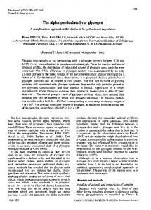
2. HEPATIC GLYCOGEN AND THE DB/DB MOUSE 2.1. Liver-Glycogen Structure Liver glycogen release acts as a blood glucose buffer. Shortly after eating there is a peak in blood glucose levels, causing the synthesis of glycogen in liver cells; these molecules can be degraded in times of fasting in order to restore blood glucose levels [19]. Glycogen is comprised of chains of glucose units that are connected to form large, highly branched molecules, with molecular weights ranging from small β particles of 106 up to large α particles that can reach molecular weights greater than 108 [20]. A sketch representing the three main levels of glycogen structure is given in Fig. (1); a transmission electron microscope (TEM) image of mouse-liver glycogen is given in Fig. (2). The events leading to α particle formation are still unclear; however it has been postulated recently that there is probably a protein “glue” that joins the smaller β particles together to form these large α particles [21, 22]. Subsequent studies [23, 24] have been consistent with this hypothesis, supporting (but not proving) the supposition that the link is a non-covalent interaction with a protein that is resistant to degradation. More proteomics studies (in addition to an earlier study [25] which, although pioneering, did not completely remove exogenous proteins) however are crucial to determine if indeed there is a protein glue responsible for the joining together of β particles, and, if so, what is the nature of that protein. The first indication that db/db (on the C57BL/KsJ background) mice may have a different glycogen structure to that of control mice was the observation that the activity of glycogen synthase, the enzyme responsible for the addition of glucose molecules onto a growing glycogen chain, was higher in the supernatants after sucrose density centrifugation (used to fractionate glycogen into different sizes) of db/db mouse glycogen compared to the controls. As the majority of glycogen synthase is bound to glycogen (diabetic and healthy glycogen having the same affinity for glycogen synthase [26]), this indicated that there were a higher proportion of glycogen molecules that did not sediment; as these particles are probably smaller molecules. This suggested db/db samples had a higher proportion of small molecules compared to non-diabetic (db/+) glycogen [26, 27]. This was then supported by further sucrose density centrifugation experiments, where it was observed that the glycogen from db/db mice was missing the “heavier” fraction that was present in the control mice [26]. One limitation with this observation however was that only a small number of mice were compared (3 separate experiments of just 1 non-diabetic and 1 diabetic mouse each time). Also while qualitatively useful, as sucrose density centrifugation separates based on the size, density and the shape of particles, it is unable to produce quantitative size distributions. The situation changed when unambiguous size distributions of liver glycogen were obtained for the first time [20, 28]. This used size exclusion chromatography (SEC; sometimes also termed gel-permeation chromatography, GPC, and also sometimes termed HPLC-SEC). This technique separates based on size (hydrodynamic volume) and thus, with careful calibration, is able to produce quantitative
Glycogen Molecular Structure and New Potential Drug Targets for Diabetes
Current Drug Targets, 2015, Vol. 16, No. 8
3
Fig. (1). The three main levels of glycogen structure.
distributions of glycogen. The result is the number or weight (depending on which type of detector is used) of molecularly separated glycogen particles as a function of size. The SEC weight distributions of glycogen from db/db mice (females on the C57BL/6J background) were compared to that of controls (db/+, +/+). Putting together data obtained using techniques which were improved over the last few years [21, 24, 28, 29], it was found that while db/db mice were able to synthesize α particles, these particles were significantly more fragile in dimethyl sulfoxide
(DMSO) than those in the healthy controls, and readily disintegrated to β particles. DMSO is a solvent which disrupts hydrogen bonds. (It is noted that the first study of this size distribution [28] used only DMSO as SEC eluent, which as stated showed only β particles in db/db mice; this led to the conclusion that α particles were absent in these mice, whereas in fact α particles are present in both db/db and controls when the SEC elution is performed in water, which does not cause degradation). While the reason for the fragile nature of the db/db α particles is unknown, a few possibilities were discussed. Firstly, if there is a protein “glue” holding α particles together, it is possible that in db/db mice the confirmation or quantity of this protein is different, resulting in the observed fragility. However it is also possible that there is a difference in the structure of the db/db glycogen, making this hypothesized protein “glue” less effective [23] at holding the particles together. Further experiments are needed to elucidate this. Also the question still remains as to whether an increased fragility of α particles would play a role in the pathology of type 2 diabetes. It may be that conditions inside the diabetic liver are such that, for example, the interiors of diabetic α particles are more easily penetrated by degradation enymes during glycogenolysis, such that they are akin to separate β particles. It has been suggested that a population of small β particles would be more susceptible to enzymatic degradation, due to the increased surface area to volume ratio and thus exposed chain-ends available to be hydrolyzed (see Fig. 3 for a schematic depicting this hypothesis) [28].
Fig. (2). TEM image of mouse-liver glycogen, which consists of a distribution of sizes ranging from small β particles up to large α particles. From the authors’ laboratory.
This hypothesis is supported by a study that showed that glycocgen phosphorylase did indeed have a higher activity for smaller glycogen particles; while this was in the direction of synthesis, it still supports the idea of surface area to volume ratio being important for glycogen phosphorylase action, with
4 Current Drug Targets, 2015, Vol. 16, No. 8
Sullivan et al.
Fig. (3). Suggested physiological effect of having more fragile α particles. Alternatively, fragile α particles could impact hepatic glycogen release times, so that the liver not as capable of lowering blood glucose by extracting it and forming glycogen.
a similar trend expected to be seen in the direction of degradation [30]. Glycogen phosphorylase has also been shown to be more associated with [31], and have a higher in vitro activity for [32, 33], smaller glycogen particles. It has been reported that hepatic glycogen from db/db mice has a shorter exterior, interior and average chain-length to non-diabetic glycogen [26]. This finding was supported by early chain-length distributions obtained using fluorophore assisted carbohydrate electrophoresis (FACE) of diabetic and non-diabetic glycogen which suggested that db/db mouse glycogen had a slightly lower proportion of longer chains [28]. The physiological effects of having slightly shorter chains is not obvious, but it is expected [34] to play a role in the overall size of the glycogen and may also be important for the formation of α particles. 2.2. Liver-Glycogen Content Comparisons of the glycogen content of non-diabetic and db/db mice have been inconsistent in the past, with some studies reporting similar levels [35], while others reporting that diabetic mice having a 2-3 fold increase in hepatic glycogen levels [36, 37]. It was suggested that perhaps the discrepancies arose due to an inconsistency in the time of which tissue samples were collected [38]. Given the diurnal nature of glycogen metabolism in many animals such as mice [3841], it is possible that at some stages of this cycle the glycogen content is similar between diabetic and non-diabetic mice, whereas at other stages the diabetic mice have much more liver glycogen. This was shown to be the case, as can be seen in Fig. (4), with the glycogen content between db/db and control mice being similar sometimes during the diurnal cycle, while being much higher in the db/db mice at other time points [38]. While control mice have a steady decrease in liver-glycogen levels during the light period of a light/dark (day/night) cycle, db/db mice consistently have high levels. One suggested reason for this maintained level of hepatic glycogen is that db/db mice continue to eat during the light hours [38].
Fig. (4). A sketch, based on published data [38], of the hepatic glycogen content of control (blue) and db/db (red) mice over the course of one diurnal light/dark cycle.
2.3. Potential Effects of Brain-Derived Neurotrophic Factor on Hepatic Glycogen Metabolism in db/db Mice It has been shown that db/db mice (on a C57BL/KsJ related background) that have been administered brain-derived neurotrophic factor (BDNF), have a decrease in food intake, body weight and non-fasted blood glucose levels [42]. It was then shown that this treatment, when administered 5 times a week, also led to a decrease in the liver size of the db/db mice, from ~848 mg (compared to that of the control db/+, ~443 mg), down to ~410 mg [43]. This decrease in liver size led to a decrease in the total glycogen content, but not glycogen concentration, which remained the same as untreated db/db mice at ~3.4% of the wet liver weight. To properly understand how BDNF affects glycogen concentration, the diurnal nature of the glycogen metabolism would have to be taken into account. As far as we know the effects of BDNF on the glycogen structure of db/db mice have not been analyzed. It would be of interest to see if the treated mice de-
Glycogen Molecular Structure and New Potential Drug Targets for Diabetes
velop α particles that are not as fragile as those from the untreated db/db mice. 2.4. Human Glycogen Structure and Type 2 Diabetes While the fasting levels of muscle glycogen have been shown to be similar in healthy and type 2 diabetic humans [44-46], after food intake the rate of glycogen synthesis in skeletal muscle is impaired in type 2 diabetic patients [44]. There have been conflicting results on whether or not liverglycogen synthesis is impaired in type 2 diabetes, with one study [47] showing a considerable decrease in liver-glycogen production of ~54% and another more recent study showing no significant difference [44]. It was noted in the latter study that this discrepancy may be due to differences in the diabetic patients used for both studies, with the patients selected being “at an earlier stage of the disease” (based on differing HbA1c levels) in the more recent study. A considerable amount of research has focused on increasing muscle- and liver-glycogen uptake in individuals with type 2 diabetes, with attempts to increase glycogen synthesis via activation of glycogen synthase [48] or via inhibition of glycogen synthase kinase-3 [49, 50], an enzyme responsible for phosphorylating and inactivating glycogen synthase. Research has also been conducted to create treatments that decrease the degradation of glycogen, generally with the approach to inhibit the degradative enzyme glycogen phosphorylase [51].
Current Drug Targets, 2015, Vol. 16, No. 8
treatment, but (b) it is now possible [52] to obtain semiquantitative size distribution data from human liver samples which have been preserved in formalin, which may overcome this problem. While there are many questions to answer, the potential for increasing our understanding of the pathology of type 2 diabetes, and any associated new drug targets that may be developed as a result, make this a promising new angle of diabetic research. CONFLICT OF INTEREST The authors confirm that this article content has no conflict of interest. ACKNOWLEDGEMENTS This study was supported by the Chinese National Basic Research Programs (Grant No. 2011CB910600), the National Natural Science Foundation of China (Grant No. 31470809), the International Collaboration Program (Grant No. 2014DFB30020), an NHMRC CJ Martin fellowship (GNT1092451), and NHMRC Peter Doherty fellowship (GNT1072086), an Australian Research Council grant (DP130102461) and the 1000-Talents Program of the Chinese State Administration of Foreign Expert Affairs. REFERENCES
The only size-distribution data in the literature on human liver glycogen seems to be a very recent study [23] giving the chain-length distributions in samples from human volunteers, none of whom were diabetic, who were having liver surgery for a range of reasons (the study complied with requisite ethical requirements). There seems to be no current data on the molecular structure of human diabetic glycogen. Given the outlined differences observed between the structure of db/db and non-diabetic mouse-liver glycogen, and the possible effects this has on glycogen degradation and glucose homeostasis (see Fig. 3), there appears to be a large gap in our current understanding of glycogen’s role in Type 2 diabetes in humans.
[1]
CONCLUSION
[7]
An important goal for public-health world-wide is the development of improved drugs for diabetes management. The observed differences in the molecular structures of healthy and diabetic liver glycogen offer the potential of new types of drug targets. With the analysis of the structure of hepatic glycogen from diabetic mice still in its infancy, there are a range of questions still needed to be addressed. Here are some of the more pertinent of these: Is the difference in glycogen structure seen in db/db mice present across the whole diurnal cycle? Why are α particles more fragile in db/db mice? Do these fragile α particles break down faster in vivo, as hypothesized? Can a “healthy” glycogen structure be rescued in db/db mice with the administration of suitable type 2 diabetic drugs? Are the more fragile α particles seen in other models for type 2 diabetes and most importantly, in humans? It is noted in this last context that (a) ethical considerations make it extremely difficult to obtain fresh liver glycogen from a diabetic human subject who has not had any
5
[2] [3]
[4] [5]
[6]
[8] [9] [10] [11] [12] [13] [14] [15]
Xu Y, Wang L, He J, et al. Prevalence and control of diabetes in Chinese adults. JAMA 2013; 310(9): 948-59. Aronoff SL, Berkowitz K, Shreiner B, Want L. Glucose metabolism and regulation: beyond insulin and glucagon. Diabetes Spectrum 2004; 17(3): 183-90. Hazard B, Zhang XQ, Colasuonno P, Uauy C, Beckles DM, Dubcovsky J. Induced mutations in the starch branching enzyme ii (sbeii) genes increase amylose and resistant starch content in durum wheat. Crop Science 2012; 52(4): 1754-66. Nordlie RC, Foster JD, Lange AJ. Regulation of glucose production by the liver. Annu Rev Nutr 1999; 19: 379-406. Meyer F, Heilmeyer L, Haschke RH, Fischer EH. Control of phosphorylase activity in a muscle glycogen particle .1. Isolation and characterization of protein-glycogen complex. J Biol Chem 1970; 245(24): 6642-8. Rybicka KK. Glycosomes - The organelles of glycogen metabolism. Tissue Cell 1996; 28(3): 253-65. Reikeras O, Nordstrand K, Henden T. Effects of fasting and glucose insulin potassium on glycogen contents in heart, skeletalmuscle and liver. Scand J Clin Lab Invest 1988; 48(3): 285-8. Besford QA, Sullivan MA, Zheng L, Gilbert RG, Stapleton D, Gray-Weale A. The structure of cardiac glycogen in healthy mice. Int J Biol Macromolecules 2012; 51(5): 887-91. Darnton SJ. Glycogen metabolism in rabbit kidney under differing physiological states. Q J Exp Physiol Cogn Med Sci 1967; 52(4): 392-400. Hummel KP, Dickie MM, Coleman DL. Diabetes a new mutation mouse. Science 1966; 153(3740): 1127-8. Ingalls AM, Dickie MM, Snell GD. Obese, a new mutation in the house mouse. J Hered 1950; 41(12): 317-8. Dickie MM, Lane PW. Plus letter to Roy Robinson 7/7/70. Mouse News Lett 1957; 17: 52. Zhang YY, Proenca R, Maffei M, Barone M, Leopold L, Friedman JM. Positional cloning of the mouse obese gene and its human homolog. Nature 1994; 372(6505): 425-32. Tartaglia LA, Dembski M, Weng X, et al. Identification and expression cloning of a leptin receptor, OB-R. Cell 1995; 83(7): 1263-71. Chen H, Charlat O, Tartaglia LA, et al. Evidence that the diabetes gene encodes the leptin receptor: identification of a mutation in the leptin receptor gene in db/db mice. Cell 1996; 84(3): 491-5.
6 Current Drug Targets, 2015, Vol. 16, No. 8 [16] [17] [18] [19]
[20]
[21] [22]
[23] [24] [25] [26] [27] [28] [29] [30] [31] [32] [33] [34]
Sullivan et al.
Coleman DL. A historical perspective on leptin. Nat Med 2010; 16(10): 1097-9. Coleman DL, Hummel KP. Effects of parabiosis of normal with genetically diabetic mice. Am J Physiol 1969; 217(5): 1298-304. Coleman DL. Effects of parabiosis of obese with diabetes and normal mice. Diabetologia 1973; 9(4): 294-8. Alftren J, Penarrieta JM, Bergenstahl B, Nilsson L. Comparison of molecular and emulsifying properties of gum arabic and mesquite gum using asymmetrical flow field-flow fractionation. Food Hydrocolloids. 2012; 26(1): 54-62. Sullivan MA, Vilaplana F, Cave RA, Stapleton DI, Gray-Weale AA, Gilbert RG. Nature of alpha and beta Particles in Glycogen Using Molecular Size Distributions. Biomacromolecules 2010; 11(4): 1094-100. Sullivan MA, O’Connor MJ, Umana F, et al. Molecular Insights into Glycogen Alpha-Particle Formation. Biomacromolecules 2012; 13(11): 3805-13. Powell PO, Sullivan MA, Sheehy JJ, Schultz BL, Warren FJ, Gilbert RG. Acid hydrolysis and molecular density of phytoglycogen and liver glycogen helps with understanding the bonding in alpha (composite) glycogen molecules. PLoS One 2015; 10(3): e0121337. Deng B, Sullivan MA, Li J, Tan X, Zhu C, Schulz BL, et al. Molecular structure of glycogen in diabetic liver. Glycoconj J 2015; 32(3-4): 113-8. Sullivan MA, Powell PO, Witt T, Vilaplana F, Roura E, Gilbert RG. Improving size-exclusion chromatography for glycogen. J Chromatography A. 2014; 1332(1): 21-9. Stapleton D, Nelson C, Parsawar K, McClain D, Gilbert-Wilson R, Barker E, et al. Analysis of hepatic glycogen-associated proteins. Proteomics 2010; 10(12): 2320-9. Roesler WJ, Khandelwal RL. Kinetic properties of glycogen synthase and phosphorylase and structural aspects of glycogen in the db/db mouse liver. Diabetes 1986; 35(2): 210-6. Roesler WJ, Pugazhenthi S, Khandelwal RL. Hepatic glycogen metabolism in the db/db mouse. Mol Cell Biochem 1990; 92: 99106. Sullivan MA, Li J, Li C, Vilaplana F, Zheng L, Stapleton D, et al. Molecular structural differences between type-2-diabetic and healthy glycogen. Biomacromolecules 2011; 12(6): 1983-6. Gilbert RG, Sullivan MA. The molecular size distribution of glycogen and its relevance to diabetes (review). Aust J Chem 2014; 67(4): 538-43. Stetten MR, Stetten Jr. D. Influence of molecular size of glycogen on the phosphorylase reaction. J Biol Chem 1958; 232(1): 489-504. Barber AA, A. OS, Bueding E. Association of Enzymes with Rat Liver Glycogen Isolated by Rate-zonal Centrifugation. J Biol Chem 1967; 242(18): 4040-44. Orrell SA, Bueding E. A comparison of products obtained by various procedures used for the extraction of glycogen. J Biol Chem 1964; 239(12): 4021-6. Sullivan MA, Aroney STN, Li S, Warren FJ, Joo L, Mak KS, et al. Changes in glycogen structure over feeding cycle sheds new light on blood-glucose control. Biomacromolecules 2014; 15(2): 660-5. Melendez R, Melendez-Hevia E, Mas F, Mach J, Cascante M. Physical Constraints in the Synthesis of Glycogen That Influence Its Structural Homogeneity: A Two-Dimensional Approach. Biophys J 1998; 75(1): 106-14.
Received: June 13, 2015
[35]
[36] [37] [38]
[39] [40] [41] [42] [43]
[44]
[45]
[46] [47] [48] [49]
[50]
[51] [52]
Revised: July 23, 2015
Stearns SB, Benzo CA. Structural and chemical alterations associated with hepatic glycogen-metabolism in genetically diabetic (db) and in streptozotocin-induced diabetic mice. Lab Invest 1977; 37(2): 180-7. Coleman DL, Hummel KP. Studies with the mutation, diabetes, in the mouse. Diabetologia 1967; 3(2): 238-48. Chan TM, Young KM, Hutson NJ, Brumley FT, Exton JH. Hepatic-metabolism of genetically diabetic (db-db) mice .1. Carbohydrate-metabolism. Am J Physiol 1975; 229(6): 1702-12. Roesler WJ, Helgason C, Gulka M, Knandelwal RL. Aberrations in the diurnal rhythms of plasma-glucose, plasma-insulin, liverglycogen, and hepatic glycogen-synthase and phosphorylase activities in genetically diabetic (db/db) mice. Horm Metab Res 1985; 17(11): 572-5. Higgins GM, Berkson J, Flock E. The diurnal cycle in the liver I. Periodicity of the cycle, with analysis of chemical constituents involved. Am J Physiol 1932; 102(3): 673-82. Soilberger A. The control of circadian glycogen rhythms. Ann N Y Acad Sci 1964; 117: 519-54. Cohn C, Joseph D. Feeding habits and daily rhythms in tissue glycogens in rat. Proc Soc Exp Biol Med 1971; 137(4): 1303-6. Ono M, Ichihara J, Nonomura T, et al. Brain-derived neurotrophic factor reduces blood glucose level in obese diabetic mice but not in normal mice. Biochem Biophys Res Commun 1997; 238(2): 633-7. Tonra JR, Ono M, Liu X, et al. Brain-derived neurotrophic factor improves blood glucose control and alleviates fasting hyperglycemia in (C57BLKS-Lepr db/lepr db) mice. Diabetes 1999; 48(3): 588-94. Macauley M, Smith FE, Thelwall PE, Hollingsworth KG, Taylor R. Diurnal variation in skeletal muscle and liver glycogen in humans with normal health and Type 2 diabetes. Clin Sci (Lond) 2015; 128(10): 707-13. Stephenson MC, Leverton E, Khoo EYH, et al. Variability in fasting lipid and glycogen contents in hepatic and skeletal muscle tissue in subjects with and without type 2 diabetes: a H-1 and C-13 MRS study. NMR Biomed 2013; 26(11): 1518-26. He J, Kelley DE. Muscle glycogen content in type 2 diabetes mellitus. Am J Physiol Endocrinol Metab 2004; 287(5): E1002-E7. Krssak M, Brehm A, Bernroider E, et al. Alterations in postprandial hepatic glycogen metabolism in type 2 diabetes. Diabetes 2004; 53(12): 3048-56. Qian YM, Bolin DR, Conde-Knape K, et al. N-substituted sultam carboxylic acids as novel glycogen synthase activators. Medchemcomm 2013; 4(5): 833-8. Motawi TMK, Bustanji Y, El-Maraghy SA, Taha MO, Al Ghussein MAS. Naproxen and Cromolyn as New Glycogen Synthase Kinase 3 Inhibitors for Amelioration of Diabetes and Obesity: An Investigation by Docking Simulation and Subsequent In vitro/In vivo Biochemical Evaluation. J Biochem Mol Toxicol 2013; 27(9): 425-36. Eldar-Finkelman H, Ilouz R. Challenges and opportunities with glycogen synthase kinase-3 inhibitors for insulin resistance and Type 2 diabetes treatment. Expert Opin Investig Drugs 2003; 12(9): 1511-9. Loughlin WA. Recent Advances in the Allosteric Inhibition of Glycogen Phosphorylase. Mini Rev Med Chem 2010; 10(12): 1139-55. Sullivan MA, Li S, Aroney ST, et al. A rapid extraction method for glycogen from formalin-fixed liver. Carbohydr Polym 2015; 118(1): 9-15.
Accepted: July 24, 2015