THE JOURNAL
OF
BIOLOGICAL CHEMISTRY
Vol. 279, No. 1, Issue of January 2, pp. 805–811, 2004 Printed in U.S.A.
Implication of DNA Polymerase in Alignment-based Gap Filling for Nonhomologous DNA End Joining in Human Nuclear Extracts* Received for publication, July 21, 2003, and in revised form, October 8, 2003 Published, JBC Papers in Press, October 15, 2003, DOI 10.1074/jbc.M307913200
Jae Wan Lee, Luis Blanco‡, Tong Zhou, Miguel Garcia-Diaz§, Katarzyna Bebenek§, Thomas A. Kunkel§, Zhigang Wang¶, and Lawrence F. Povirk‡储 From the Department of Pharmacology and Toxicology, Virginia Commonwealth University, Richmond, Virginia 23298, ‡Centro de Biologı´a Molecular Severo Ochoa (CSIC-UAM), Universidad Auto´noma de Madrid, 28049 Madrid, Spain, the §Laboratory of Molecular Genetics and Laboratory of Structural Biology, NIEHS, Department of Health and Human Services, Research Triangle Park, North Carolina 27709, and the ¶Graduate Center for Toxicology, University of Kentucky, Lexington, Kentucky 40536
Accurate repair of free radical-mediated DNA doublestrand breaks by the nonhomologous end joining pathway requires replacement of fragmented nucleotides in the aligned ends by a gap-filling DNA polymerase. Nuclear extracts of human HeLa cells, supplemented with recombinant XRCC4-DNA ligase IV complex (XRCC4/ligase IV), were capable of accurately rejoining model double-strand break substrates with a 1- or 2-base gap, and the gap-filling step was dependent on XRCC4/ligase IV. To determine what polymerase was responsible for gap filling, end joining was examined in the presence of polyclonal antibodies against each of two prime candidate enzymes, DNA polymerases and , both of which were present in the extracts. For a DNA substrate with partially complementary 3ⴕ overhangs and a 2-base gap, antibodies to polymerase completely eliminated both gap filling and accurate end joining, whereas antibodies to polymerase had little effect. Immunodepletion of polymerase , but not polymerase , likewise blocked both gap filling and end joining, and both functions could be restored by addition of recombinant polymerase . Recombinant polymerase , and a truncated polymerase lacking the Brca1 C-terminal domain, were at least 10-fold less active in restoring gap filling to the immunodepleted extracts, and polymerase  was completely inactive. The results suggest that polymerase is the primary gap-filling polymerase for accurate nonhomologous end joining, and that the Brca1 C-terminal domain is required for this activity.
Although the nonhomologous end joining pathway of DNA double-strand break (DSB)1 repair is error-prone (1–3), it can nevertheless often restore the original sequence at DSB sites, even for DSBs with damaged ends and fragmented nucleotides (4). In the case of staggered, free radical-mediated DSBs, this accurate repair is accomplished by a mechanism involving * This work was supported by Grant CA40615 from the NCI, Department of Health and Human Services, National Institutes of Health. The costs of publication of this article were defrayed in part by the payment of page charges. This article must therefore be hereby marked “advertisement” in accordance with 18 U.S.C. Section 1734 solely to indicate this fact. 储 To whom correspondence should be addressed: Virginia Commonwealth University, P. O. Box 980230, Richmond, VA 23298-0230. Tel.: 804-828-9640; Fax: 804-828-8079; E-mail:
[email protected]. 1 The abbreviations used are: DSB, double-strand break; BRCT, Brca1 C-terminal; DNA-PK, DNA-dependent protein kinase; DNAPKcs, DNA-PK catalytic subunit; pol, DNA polymerase; PCV, packed cell volume; XRCC4/LigIV, XRCC4-DNA ligase IV complex; PMSF, phenylmethylsulfonyl fluoride; BSA, bovine serum albumin. This paper is available on line at http://www.jbc.org
alignment of residual complementarities in 3⬘ or 5⬘ overhangs, fill in of any remaining gaps, and ligation (5, 6). This process can be mimicked in vitro using whole-cell extracts of human or hamster cells, and it requires Ku, XRCC4, and DNA ligase IV but not DNA-dependent protein kinase catalytic subunit (DNAPKcs) (4, 7). The XRCC4-DNA ligase IV complex (XRCC4/ LigIV) is required not only for the final ligation, but for the gap-filling step as well (8). The gap-filling polymerase has not been definitively identified, but there is circumstantial evidence that it may be either DNA polymerase (pol ) (9, 10) or polymerase (pol ) (11). Immunodepletion studies reported herein suggest that, in an in vitro system based on human nuclear extracts, pol is primarily responsible for gap filling, and the BRCT domain of pol is required for this activity. EXPERIMENTAL PROCEDURES
DNA Substrates—The 1- or 2-base-gapped substrates (Fig. 1) were constructed by ligation of the 3⬘-hydroxyl oligomers pGCCGGACGCGACG or pGCCGGACGCGATCG, respectively, into a 5⬘ overhang generated in plasmid pSV56 by controlled exonucleolytic degradation with T4 DNA polymerase, as described previously (4, 12, 13). In each case the 5⬘-end-labeled (*) oligomer *pCGAGGAACGCGACG was ligated into the opposite end of the plasmid to produce internally labeled substrates with partially cohesive 3⬘ overhangs. Preparation of HeLa Nuclear Extracts—Confluent cultures of HeLa S3 cells grown on 20 plates of 150-mm were harvested by scraping the cells off the plates into 10 ml/plate phosphate-buffered saline. Cells were washed twice with ice-cold phosphate-buffered saline and resuspended in 4 packed cell volumes (PCV) of hypotonic buffer (10 mM KCl, 10 mM Tris-HCl, pH 7.9, 1 mM dithiothreitol, 1 g/ml pepstatin, 1 g/ml leupeptin, 1 g/ml soybean trypsin inhibitor, and 10 g/ml PMSF) on ice for 10 min. Cells were lysed by Dounce homogenizer (20 strokes), and the resulting nuclei were collected by centrifugation at 2,000 rpm for 10 min in Sorvall SS-34 rotor. The pelleted nuclei were washed in hypotonic buffer and centrifuged again at 2,000 rpm for 10 min and resuspended in 4 PCV of nuclear extraction buffer (50 mM Tris-HCl, pH 7.9, 0.42 M KCl, 5 mM MgCl2, 0.1 mM EDTA, 20% glycerol, 10% sucrose, 2 mM dithiothreitol, 1 g/ml pepstatin, 1 g/ml leupeptin, 1 g/ml soybean trypsin inhibitor, and 10 g/ml PMSF). The mixture was stirred for 30 min at 4 °C and centrifuged for 30 min at 15,000 rpm. The collected supernatant was mixed with 0.33 g of ammonium sulfate per ml of supernatant, and 10 l of 1 M NaOH per g of ammonium sulfate was added. This mixture was stirred for 20 min at 4 °C and centrifuged for 10 min at 13,300 rpm. The protein pellet was resuspended in 0.25 PCV of storage buffer (20 mM Tris-HCl, pH 7.9, 0.1 M KOAc, 0.5 mM EDTA, 20% glycerol, 1 mM dithiothreitol, 1 g/ml pepstatin, 1 g/ml leupeptin, 1 g/ml soybean trypsin inhibitor, and 10 g/ml PMSF) and was dialyzed against the same buffer overnight at 4 °C. The nuclear extracts were clarified by centrifugation at 85,000 ⫻ g and frozen in liquid nitrogen to be stored at ⫺80 °C (14). Whole-cell extracts of CHO-K1 and XR-1 cells were prepared as described previously (4, 15). Antibodies—Recombinant murine pol and human pol were expressed in Escherichia coli as described (16, 17), and ⬃300 g of each were purified from the insoluble fraction by PAGE, electroeluted, and
805
806
DNA Polymerase in Alignment-based Gap Filling
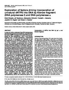
FIG. 1. End joining substrates and repair products. The 1-base-gapped substrate has -ACG 3⬘ overhangs on each end, such that annealing of the self-complementary CG results in a 1-base gap. Fill in of the gap followed by ligation results in an accurate head-to-tail end joining product that yields a labeled 43-base XhoI-BstXI fragment. The 2-base-gapped substrate has an -ATCG overhang on the unlabeled end and an -ACG overhang on the labeled end; fill in and accurate head-to-tail end joining yields a 44-base fragment. Accurate head-to-head end joining of either substrate yields a palindromic 24-base fragment. injected into rabbits. Blood was collected immediately before and then again 2 weeks after immunization, and IgG antibodies were purified by protein A affinity chromatography as prescribed by the vendor (Bio-Rad). Proteins—Full-length hexahistidine-tagged human pol was expressed in E. coli and purified as described previously (18). Similar procedures were used for expression and purification of a 39-kDa Cterminal fragment of pol (amino acids 242–575; details to be presented elsewhere). Hexahistidine-tagged human pol and pol were expressed in Saccharomyces cerevisiae and purified as described previously (10). Except where specifically noted, the bacterially expressed pol was used for all experiments. Human pol  was from Trevigen. End Joining and Gap Filling in Nuclear Extracts—Reaction mixtures contained HeLa nuclear extract (27 g of protein), 0.1 g of XRCC4/LigIV (Trevigen), 50 mM triethanolamine, 10 mM Tris-HCl, pH 7.9, 1 mM Mg(OAc)2, 40 mM KOAc, 0.5 mM dithiothreitol, 1 mM ATP, 50 M each of dNTP or dideoxynucleoside 5⬘-triphosphate (ddNTP), 50 g/ml BSA, and 20 ng of DNA substrate in a total volume of 20 l. For antibody inhibition studies, samples were preincubated in the presence of various concentrations of antibodies without DNA substrate for 30 min at 25 °C to allow antibody binding. In some cases, samples were similarly preincubated in the presence of wortmannin (10 M) in dimethyl sulfoxide (final concentration 2%). Following substrate addition, samples were incubated for 6 h at 37 °C and then deproteinized by treatment with proteinase K followed by phenol/chloroform extraction. DNA was digested with BstXI and XhoI and analyzed on 20% denaturing polyacrylamide gels (13). Intensities of bands corresponding to repair products and intermediates were quantitated by PhosphorImaging and are expressed as a fraction of the total PhosphorImager intensity in the lane, excluding any material that remained in the well. End joining assays with CHO-K1 and XR-1 whole-cell extracts were performed as described previously (4, 8), with incubation for 6 h at 25 °C. Assays for the activity of pol and pol were performed in 20 l of the same buffer containing 30 fmol of the 5⬘-end-labeled 15-mer *pGATCACAGTGAGTAC annealed to a 1.5-fold excess of the 33-mer ACTGGCCGTCGTTCTAATGTACTCACTGTGATC. Immunodepletion—Antibodies (1.35 or 2.7 g) were bound to 5 l (bead volume) of protein A-agarose beads (Sigma) by mixing at 900 rpm in antibody binding buffer (0.1 M KOAc, 20% glycerol, 0.2 mM EDTA, 0.5 mM dithiothreitol, 0.5 mM PMSF, 50 g/ml BSA) with a total volume of 250 l at 4 °C for 16 h in an Eppendorf Thermomixer. Antibody-bound beads were washed 3 times with 400 l of antibody washing buffer (10 mM Tris-HCl, pH 7.9, 65 mM KOAc, 0.25 mM EDTA, 0.5 mM dithiothreitol, 1 mM Mg(OAc)2) in order to remove unbound antibodies. HeLa nuclear extract (15 l) was directly added to the beads and mixed at 900 rpm for 1.5 h at 4 °C. Beads were briefly pelleted in a microcentrifuge; 10 l of supernatant was removed, and 5 l was used for each reaction.
Samples were incubated and processed as described above. For reconstitution, 70 ng of recombinant pol , truncated pol , pol , or pol  was added to each reaction mixture. Immunodepletion of purified polymerases was performed similarly except that instead of extract, 75 ng of pol  or 120 ng of pol or pol , in 15 l of extract storage buffer containing 0.1 mg/ml BSA, was added to protein A beads to which 2.7 g of antibodies had been adsorbed. Following immunodepletion and washing, 5 l of supernatant was added to a reaction containing 30 fmol of the labeled 33-mer/15-mer template/primer in 15 l of 50 mM TrisHCl, pH 8.5, 2.5 mM MgCl2, 1 mM dithiothreitol, 0.1 mg/ml BSA, 4% glycerol, 10 M dNTPs. Samples were incubated at 37 °C for 6 h, denatured, and analyzed on a denaturing 20% polyacrylamide gel. Western Blotting—The samples, including MagicMark size standards (Invitrogen), were heat-denatured (100 °C, 5 min), electrophoresed on 12% SDS-polyacrylamide gels, and transferred to nitrocellulose membranes (Bio-Rad). The membranes were blocked in 5% nonfat milk in Tris-buffered saline (50 mM Tris, pH 7.4, 200 mM NaCl, 0.1% Triton X-100) containing 0.2% Tween 20. The dilution for the primary rabbit antibody against human pol or mouse pol (initial concentration 1.3 mg/ml) was 1:8,000. For chemiluminescence detection, horseradish peroxidase-conjugated secondary goat anti-rabbit antibody was from Pierce and was used at a dilution of 1:20,000. Bound peroxidase signal was detected using a Supersignal West Pico Chemiluminescence kit (Pierce). For direct staining, alkaline phosphatase-conjugated goat antirabbit antibody was from Promega and was used at 1:2,000 dilution. Detected proteins were then stained with Western Blue substrate (Promega), according to the vendor’s instructions, and photographed. RESULTS
Gap Filling in Human Nuclear Extracts Is Dependent on XRCC4/LigIV—We showed previously that a model free radical-mediated DSB substrate, with partially complementary phosphoglycolate-terminated 3⬘ overhangs and a 1-base gap in each strand, could be accurately rejoined by CHO-K1 whole-cell extracts (4), and that XRCC4 and DNA ligase IV were required for the gap-filling step as well as for the final ligation (8). In order to bypass the phosphoglycolate removal step and thus improve the efficiency of end joining, an analogous 3⬘-hydroxyl terminated vector, with the same partially complementary -ACG 3⬘ overhangs and 1-base gap in each strand was constructed (Fig. 1). Incubation of this substrate in CHO-K1 whole-cell extracts yielded the expected 43-base XhoI/BstXI fragment (Fig. 2, lane A2), consistent with accurate end joining, and the repair was
DNA Polymerase in Alignment-based Gap Filling
FIG. 2. XRCC4/LigIV-dependent gap filling and accurate end joining of the 1-base (A) or 2-base (B)-gapped substrates in whole-cell extracts of normal CHO-K1 or XRCC4-deficient XR-1 cells. Substrates were incubated in the indicated extracts for 6 h, cut with XhoI and BstXI, and analyzed on DNA sequencing gels. Samples contained either all four dNTPs, or three dNTPs plus either ddTTP or ddATP, as indicated. Recombinant XRCC4/LigIV (0.2 g) was also added to some samples. The 1- and 2-base-gapped substrates yielded primarily the accurate end joining products of 43 and 44 bases, respectively. M, size markers; the 43-base marker has the same sequence as the expected accurate repair product.
significantly more efficient than for the comparable 3⬘-phosphoglycolate substrate (4, 8). As expected, ddTTP (lane A4), but not ddATP (lane A3), blocked end joining by trapping the filled in but unligated intermediate. In extracts of XRCC4-deficient XR-1 cells, neither gap-filling (lane A6) nor end joining (lane A5) could be detected, but both could be restored by addition of recombinant XRCC4-LigIV complex (lanes A7–A9). Addition of XRCC4/LigIV appeared to reduce the extent of exonucleolytic degradation (as indicated by formation of 11- to 14-mer fragments; compare lanes A7–A9 with lanes A5 and A6), suggesting that XRCC4/LigIV may help to sequester and protect DNA ends prior to end joining. In order to verify that gap filling was template-dependent, a similar substrate with a 2-base gap in one strand and a 1-base gap in the opposite strand was also constructed. As predicted (see Fig. 1), the predominant in vitro repair product was now 44 bases long (Fig. 2, lanes B2 and B7), and ddTTP trapped a 17-mer intermediate 2 bases longer than the starting substrate (lanes B4 and B9), whereas ddATP trapped an intermediate only 1 base longer (lanes B3 and B8), in each case consistent with the templated fill in of the gap. There was some 16-mer product detected with ddTTP, probably reflecting head-to-head annealing of -ACG overhangs of two different molecules of the labeled substrate (see Fig. 1). Consistent with this proposal, several much shorter (⬃20 –24-base) end joining products were also detected, the longest of which comigrated with a marker having the sequence of an accurate 24-base head-to-head end joining product (*pTCGAGGAACGCGACGTCGCGTTCC, not shown). As with the 1-base-gap substrate, both gap filling and accurate end joining in XR-1 extracts were completely dependent on addition of XRCC4/LigIV (lanes B5–B9). All these results strongly suggest that 1- or 2-base extension of these
807
FIG. 3. XRCC4/LigIV-dependent gap filling and accurate end joining in HeLa nuclear extracts. The 1-base-gapped substrate (see Fig. 1) was incubated for 6 h in nuclear extracts harvested from confluent monolayers of HeLa cells (A) or obtained from Promega (B). Reactions also contained recombinant XRCC4/LigIV (0.1 g), wortmannin, and/or dimethyl sulfoxide (DMSO, solvent for wortmannin), as indicated. M, size markers.
substrates in whole-cell extracts represents templated synthesis on DNA ends aligned and annealed by a complex that includes XRCC4/LigIV. Most of the available recombinant end joining proteins that have been cloned are of human origin. Although the nonhomologous DNA end joining pathway appears to be highly conserved among all vertebrates (4, 15), interaction between proteins of different species may be less than optimal. To avoid these complications, we sought a human system for further complementation experiments. Previous studies showed that whole-cell extracts of human lymphoblastoid cells contain all components necessary for accurate end joining of DSBs with complementary as well as partially complementary 3⬘ overhangs (4, 19, 20). However, in nuclear extracts of HeLa cells, end joining is almost completely dependent on addition of exogenous recombinant XRCC4/LigIV (14), presumably because it is present only at very low concentrations in these extracts. Thus, HeLa nuclear extracts can be used to assess the requirement for XRCC4/LigIV in gap filling in a human system. As shown in Fig. 3A, the results obtained with nuclear extracts harvested from confluent HeLa cells were essentially identical to those obtained in XRCC4/LigIV-deficient XR-1 extracts, with both gap-filling and accurate end joining being dependent on exogenous XRCC4/LigIV (compare lanes 7 and 8 with lanes 1 and 2). An attempt to assess the sensitivity of end joining to the DNA-dependent protein kinase (DNA-PK) inhibitor wortmannin was complicated by the inhibitory effect of the solvent (dimethyl sulfoxide) alone. In the presence of dimethyl sulfoxide no end joining was detected (lane 11), but there was some residual gap filling (lane 12), which was blocked completely by wortmannin (lane 10). Thus, as in human whole-cell extracts (4, 20), a phosphorylation event, probably catalyzed by DNAPK, appears to be required prior to gap filling. Similar XRCC4/ LigIV-dependent gap filling and end joining was seen in commercially available HeLa nuclear extracts (Promega, in vitro transcription grade). With these extracts (Fig. 3B) there was a trace of both end joining and gap filling with unsupplemented extracts (lanes 1 and 2), but addition of exogenous XRCC4/ LigIV increased the yield of both end points by more than 10-fold (lanes 3 and 4). Although the extent of accurate end
808
DNA Polymerase in Alignment-based Gap Filling
FIG. 4. Immunodetection of pol (A) and pol (B) in HeLa nuclear extracts. The indicated amounts of recombinant polymerases or nuclear extracts were heat-denatured, separated on a 12% polyacrylamide gel, and electroblotted. Blots were probed with polyclonal antibodies to pol or pol , and proteins were detected by chemiluminescence (A and upper panel in B) or Western Blue staining (lower panel in B). The 2nd lane in B contained 25 ng of pol .
joining was the same with extracts from either source (⬃3– 4%), the commercial extracts gave somewhat greater gap filling (⬃16% versus ⬃9%) and were therefore used for subsequent experiments. Nuclear Extracts Contain Both pol and pol —To determine whether pol and pol were present in HeLa nuclear extract, proteins in the extract were separated on polyacrylamide gels, and blots were probed with antibodies to each polymerase (Fig. 4). Although both antibodies detected multiple species in the extract, in each case there was a prominent band that comigrated with the corresponding histidine-tagged recombinant at approximately the expected molecular mass, 55 kDa for pol and 68 kDa for pol. Densitometric comparison of chemiluminescence signals produced by various quantities of extract and of each recombinant polymerase suggested that 5 l of nuclear extract (the amount used in each end joining assay) contained ⬃20 ng of pol and ⬃12 ng of pol . Because chemiluminescence-based detection with the pol antibodies resulted in relatively high background, a duplicate blot was subjected to direct alkaline phosphatase-based staining, which yielded less background but also resulted in detection of more nonspecific bands on the blot; nevertheless, 5 l of extract again appeared to be roughly equivalent to 12 ng of pol . There was no apparent cross-reactivity of the pol antibodies with recombinant pol . Gap Filling Is Blocked by Antibodies against pol but Not against pol —The polymerase that catalyzes gap filling for nonhomologous end joining is not known for certain, but the prime candidates are pol and pol . In order to assess the participation of these polymerases in end joining in nuclear extracts, polyclonal rabbit antibodies to each polymerase were generated and purified on a protein A affinity column. End joining reactions were then carried out with both the 1- and 2-base-gapped substrates in XRCC4/LigIV-supplemented human nuclear extracts, in the presence of various dilutions of antibodies to each polymerase (Fig. 5). Fill in of the 1-basegapped substrate was dramatically reduced by antibodies to pol , whereas preimmune antibodies, or antibodies to pol , had little effect. pol antibodies also completely blocked fill in of a 2-base gap, and again, preimmune and pol antibodies did not. These results suggest that, at least in this in vitro system, pol
FIG. 5. Inhibition of gap filling by antibodies to DNA polymerases. The 1- (A) or 2-base (B)-gapped substrates were incubated in XRCC4/LigIV-supplemented HeLa nuclear extracts in the presence of dATP, dCTP, dGTP, and ddTTP plus 0.9, 2.7, 8, or 24 g/ml of antibodies to pol or pol , or preimmune antibodies, as indicated. No Abs, no antibodies.
rather than pol is primarily responsible for alignmentbased gap filling. pol and pol Are Specifically Immunodepleted by Polyclonal Antibodies—To determine whether the respective antibodies could be used to specifically immunodeplete pol and pol , immunobeads bearing preimmune, pol or pol antibodies were prepared. Dilute solutions of pol (8 g/ml), pol (8 g/ml), and pol  (5 g/ml) were immunodepleted with each antibody, and then polymerase activity in the supernatant was assessed on a simple 33-mer/15-mer template/primer. As shown in Fig. 6A, immunodepletion with pol antibodies reduced pol activity dramatically, although not quantitatively, whereas preimmune antibodies or antibodies to pol had little effect. pol , despite its close homology to pol , was not immunodepleted by any of the antibodies. In several experiments, immunodepletion with pol antibodies consistently eliminated pol activity completely, but this enzyme appeared to be extremely sensitive to the immunodepletion procedure and lost most of its activity even in the absence of antibodies. However, a direct comparison of pol and pol activity on the same template/primer, without immunodepletion, showed that although pol was more processive, pol was only about 2-fold less efficient than pol in adding the 1st base (Fig. 6B). Gap Filling Is Restored by Recombinant pol —To determine whether the effect of pol antibodies on gap filling was due to inactivation of pol , the same antibodies were preadsorbed to protein A-agarose beads, and extracts were immunodepleted with the beads. End joining was then examined in immunodepleted extracts complemented with recombinant pol (Fig. 7). Like simple antibody addition, immunodepletion with pol antibodies, but not with preimmune antibodies, completely blocked accurate end joining (Fig. 7A, lane 4). In the presence of ddTTP, immunodepletion with pol antibodies blocked fill in of a 2-base gap (Fig. 7, B, lanes 5 and 9, and D, lane 4), whereas depletion with preimmune antibodies (Fig. 7, B–D) or antibodies to pol (Fig. 7D, lane 3) had relatively little effect. Addition of recombinant pol to extracts immunodepleted with pol antibodies restored both end joining (Fig. 7A, lane 5) and gap filling (Fig. 7, B, lanes 6 and 10, and D, lanes 6 and 7). pol expressed in bacteria or in yeast (Fig. 7D, lane 7, Y) were equally effective in restoring gap filling. In fact, gap filling and end joining in the pol -complemented extract were greater
DNA Polymerase in Alignment-based Gap Filling
809
at most only a trace of gap filling (⬃0.8 versus 16% for fulllength pol , as estimated by PhosphorImager intensity; Fig. 7C, lane 6). Thus, as expected, the BRCT domain of pol appears to be essential for efficient alignment-based gap filling. DISCUSSION
FIG. 6. A, specificity of polymerase immunodepletion by polyclonal antibodies. Solutions containing recombinant pol , pol , or pol  were depleted with immunobeads bearing antibodies to pol , antibodies to pol , or preimmune (P) antibodies. The no antibody control samples were either held at 4 °C in buffer (⫺) or exposed to protein A (A) beads only. Polymerase activity in the supernatant was then assessed using a 33-mer/15-mer template/primer. B, polymerization by pol and pol on the same template/primer in end joining buffer without immunodepletion. Samples (20 l) contained 2.5, 5, and 10 ng (left panel) or 25, 50, and 100 ng (right panel) of each polymerase and were incubated at 37 °C for 6 h.
than in the extracts depleted with preimmune antibodies or exposed to protein A beads alone, suggesting that pol was a limiting factor in gap filling. When pol -depleted extracts were supplemented with various amounts of recombinant pol , ⬃6 ng were required for half-maximal gap filling (Fig. 7E). Because undepleted, unsupplemented extracts contained ⬃12 ng of pol and augmentation with recombinant pol enhanced gap filling slightly, this result suggests that the efficiency of recombinant pol in gap filling was comparable with that of the endogenous enzyme in the extracts. All these results confirm the implication of pol as the primary polymerase for alignment-based gap filling in these extracts. Intriguingly, although pol antibodies did not block gap filling, addition of recombinant pol to the pol -depleted extracts resulted in a detectable level single-base gap filling (Fig. 7F, lane 4) and apparent inaccurate head-to-head end joining (lane 8); in some experiments (not shown) a trace of the accurate 44-base head-to-tail product was also detected. However, the extent of gap filling and accurate end joining was only about 5–10% of the level seen with pol . These results suggest that pol may be capable of substituting for pol in gap filling but with markedly lower efficiency. This difference in efficiency is much greater than was seen for polymerization on a simple template/primer (Fig. 6B). No gap filling was detected when pol  was added to pol -depleted extracts (Fig. 7D, lane 5), suggesting that pol  cannot substitute for pol in this capacity. Interactions among DNA repair proteins are often mediated by Brca1 C-terminal (BRCT) domains (21). To determine whether the BRCT domain of pol was required for alignmentbased gap filling, gap filling was examined in pol -immunodepleted extracts supplemented with a truncated form of pol which contained the polymerase domain but lacked the BRCT domain (Fig. 7C). Like pol , the truncated pol (T) produced
Accurate repair of DSBs with partially complementary 3⬘ overhangs, such as DSBs induced by radiation and other free radical-based genotoxins, requires alignment of residual complementarities in the overhangs, and fill in of gaps left by free radical-mediated nucleotide fragmentation (4, 5). The insensitivity of this gap filling to aphidicolin (8), and the efficient use of dideoxynucleotides (4, 6, 22), suggests that it is carried out by some member of the pol X family, which includes pol , pol , and pol (23, 24). Of these polymerases, pol and pol are the more likely candidates because they can catalyze polymerization on misaligned templates (10, 11) and because they contain BRCT motifs, whereas pol  does not. The mutational specificity of pol suggests that it can catalyze synthesis on a template/primer aligned by only a single terminal base pair (11), and a single base pair appears to be sufficient to promote alignment-based gap filling during end joining (6). BRCT motifs mediate interactions of many DNA repair proteins (21) and have specifically been implicated in alignment-based gap filling by the yeast proteins Pol4 and Dnl4䡠Lif1, which are homologous to mammalian pol /pol and to the XRCC4-LigIV complex, respectively (25). In recent experiments, we found that inactivating antibodies to pol  (gift of Dr. Sam Wilson, NIEHS) did not block gap filling and accurate end joining in human lymphoblastoid whole-cell extracts.2 pol has been regarded as a particularly likely participant in nonhomologous end joining because its inferred domain structure is so similar to that of terminal transferase, which is involved in the closely related pathway of V(D)J recombination (23). Thus, these two enzymes could interact with the end joining complex using very similar molecular contacts but in different situations, i.e. pol in DSB repair and terminal transferase in V(D)J recombination. pol interacts with Ku and appears to stabilize complexes of Ku, XRCC4/LigIV, and DNA (9). The extremely error-prone nature of pol -mediated DNA synthesis, particularly its propensity for template slippage, has also been taken as an argument for its participation in end joining (10), as this feature might account for the microhomology-based deletions and insertions often detected at repair joints. On the other hand, the more accurate (compared with pol ) template-directed synthesis catalyzed by pol (18) would appear to be more compatible with the largely accurate gap filling detected in in vitro end joining assays (4, 6, 7). Moreover, pol seems a more likely candidate for a gap-filling polymerase because of its preference for substrates with short gaps, and because like pol  and unlike pol it possesses 5⬘-deoxyribose-5-phosphate lyase activity (18). A significant fraction of free radical-mediated DSBs apparently arise by cleavage of abasic sites resulting from glycosylase-catalyzed release of oxidatively modified bases (26 –28). Some of these DSBs could bear 5⬘-terminal abasic sugars on 5⬘ ends of DSBs, and these sugars would have to be removed by a 5⬘ lyase or similar activity prior to religation of the break. In this report, we present evidence from an in vitro system that for a substrate with a 2-base complementarity, pol rather than pol or pol  is the primary effector of alignmentbased gap filling in human cells and that the BRCT domain of pol is required for this activity. Even though pol was clearly present in HeLa nuclear extracts, antibodies to pol did not significantly reduce gap filling or end joining in the extracts. In 2
K. V. Inamdar and L. F. Povirk, unpublished data.
810
DNA Polymerase in Alignment-based Gap Filling
FIG. 7. Inhibition of gap filling and end joining by immunodepletion of pol and reconstitution with recombinant pol . Extracts were depleted with immunobeads bearing the indicated antibodies (P, preimmune antibodies). The 2-base-gapped substrate was then incubated in the XRCC4/LigIV-supplemented extracts, with or without recombinant polymerases. A, reconstitution of end joining by recombinant pol in the presence of all four dNTPs. B, reconstitution of gap filling by pol. dTTP was replaced with ddTTP to trap the filled in, unligated 17-mer intermediate. C, lack of reconstitution of gap filling by a truncated pol (T) lacking the BRCT domain. ddTTP was present in all samples. D, inhibition of gap filling by immunodepletion with antibodies to pol but not pol , and reconstitution of gap filling by pol expressed in either bacteria () or yeast (Y) but not by pol . ddTTP was present in all reactions. E, extent of 2-base gap filling by various amounts of recombinant pol in the presence of ddTTP in pol -immunodepleted extracts. F, lack of reconstitution of gap filling and end joining by pol . In B, 1.35 g (low concentration) or 2.7 g (high concentration) of antibodies was adsorbed to the beads; in all other experiments 2.7 g were adsorbed.
contrast, at least for the 2-base-gapped substrate, antibodies to pol blocked gap filling and end joining completely. Western blotting and immunodepletion data strongly suggest that there is no cross-reactivity between pol and pol antibodies, but because purified recombinant pol was extremely sensitive to the immunodepletion procedure, it is possible that the extracts immunodepleted with pol antibodies may also have lacked active pol . Nevertheless, if pol and pol were equally competent in alignment-based gap filling, then supplementation of the depleted extracts with recombinant pol , which catalyzed synthesis on a simple template/primer almost as efficiently as pol, should have efficiently restored alignmentbased gap filling to the depleted extract. Although recombinant pol catalyzed a low level of gap filling and accurate end joining in pol -depleted extracts, it was at least 10-fold less efficient in this capacity than pol , even when both enzymes were expressed in yeast and purified by very similar procedures. Availability of mouse knockouts for the various members of the pol X family (29) may soon allow a test of which or if either of these polymerases is required for alignment-based gap filling of transfected substrates in vivo. Compared with deficiencies in essential end joining factors such as XRCC4 and ligase IV, the phenotypes of the pol and pol knockouts are relatively mild (29). This result could be explained by pol and pol having partially redundant functions, but even if one of these polymerases were essential for gap filling, a gap-filling defect might lead to less accurate repair joints yet have little effect on the overall extent of rejoining or on the cytotoxicity of DSBs. Recently, it was reported that a combination of Ku, XRCC4/ LigIV, and pol could carry out gap filling and end joining of a substrate with a cohesive 4-base 3⬘ overhang and a 1-base gap in one strand (9). In similar assays with purified proteins (data not shown), we were able to detect very weak gap-filling on the
1-base-gapped substrate by pol , but this activity was not XRCC4/LigIV-dependent and was not seen with the 2-basegapped substrate. Thus, in our hands the requirements and specificity of this activity of pol do not accurately reflect the requirements for alignment-based gap filling as seen in extracts. In the same assays, we did not detect any gap filling by pol with either substrate even in the presence of purified Ku and XRCC4/LigIV. The failure of these purified proteins to carry out alignment-based gap filling, despite the fact that each of them was active in complementing deficiency in the respective endogenous protein in cell extracts (4, 8), suggests that additional factor(s) must be required for this process. With most of the known participants in nonhomologous end joining having been cloned, purified, and reasonably well characterized, a working model of the end joining process (30) is beginning to emerge, as follows. The broken DNA ends initially bind Ku, which recruits XRCC4/LigIV, DNA-PKcs, and possibly additional factors including Artemis (31). DNA-PKcs, most likely in concert with other proteins, sequesters the DNA ends until synapsis of the two end-bound DNA-PK complexes activates DNA-PK (32), which phosphorylates Artemis (31), DNAPKcs (33, 34), and perhaps other components of the complex. By some as yet poorly defined process, 3⬘-terminal blocking groups are removed (perhaps by tyrosyl-DNA phosphodiesterase (35)), and overhangs are either annealed or trimmed by Artemis and/or other nucleases. Once a suitably aligned substrate is formed, gaps are filled by a repair complex that includes Ku, XRCC4/LigIV and, we believe, pol. Finally, both stands are ligated by XRCC4/LigIV. Questions that remain to be resolved include the following: (i) whether additional factors such as the Mre11-Rad50-Nbs1 complex are also essential to the process (14); (ii) which DNA-PK-catalyzed phosphorylations are essential for end joining and for its regulation; (iii) what factors determine whether ends are joined accurately or
DNA Polymerase in Alignment-based Gap Filling trimmed to produce terminal deletions; (iv) whether the end joining machinery is capable of choosing correct ends based on the complementarity of the overhangs; and (v) how the structure of the end joining complex allows pol access to the gaps while still maintaining juxtapositioning of the ends. The known structural features of Ku, XRCC4/LigIV, and pol do not readily suggest a conformation that would promote alignment-based gap filling. Ku forms a basket-like structure that has been proposed to encircle juxtaposed DNA ends and mold them into a continuous helix for gap filling and ligation (36). One side of the Ku-bound helix is largely exposed, in theory allowing access by a polymerase and then by ligase IV. However, if pol binding produces a 90° bend in gapped DNA, as does the closely related pol  (37), it seems unlikely that the helix-molding properties of Ku would be involved, at least at the gap itself. XRCC4 consists of a globular head containing a helix-turn-helix DNA-binding motif and a long ␣-helical tail, in the middle of which is the ligase IV-binding site (38). X-ray crystallography suggests that four XRCC4 tails can interact to form a dumbbell-like tetramer with two XRCC4 “heads” at each end. It has thus been proposed that the putative DNA binding domains of such a tetramer could interact with two DNA molecules and potentially position them for end joining (39). Our own modeling studies,3 however, suggest that it is not sterically possible to produce a structure in which the helix-turnhelix motifs of an XRCC4 dimer or tetramer are bound in the usual manner in the major grooves of two juxtaposed DNA ends. Thus, additional structural information, particularly on the conformation of gapped DNA with pol and on the putative interactions between the BRCT motifs of pol and XRCC4, will probably be required before a coherent structural model of alignment-based gap filling can be devised. Acknowledgments—We thank David A. Gewirtz and Sehmi Paik for assistance with Western blots. Note Added in Proof—Bertocci et al. (Bertocci, B., De Smet, A., Berek, C., Weill, J. C., and Reynaud, C. A. (2003) Immunity 19, 203–211) have shown that pol -deficient mice show abnormal B-cell differentiation, with impaired formation of V-J junctions in immunoglobulin light chains. While these results do not exclude a role for pol in DSB repair, they do provide a possible alternative function for this polymerase. REFERENCES 1. Phillips, J. W., and Morgan, W. F. (1994) Mol. Cell. Biol. 14, 5794 –5803 2. Lukacsovich, T., Yang, D., and Waldman, A. S. (1994) Nucleic Acids Res. 22, 5649 –5657 3. Rouet, P., Smih, F., and Jasin, M. (1994) Mol. Cell. Biol. 14, 8096 – 8106 4. Chen, S., Inamdar, K. V., Pfeiffer, P., Feldmann, E., Hannah, M. F., Yu, Y., Lee, J. W., Zhou, T., Lees-Miller, S. P., and Povirk, L. F. (2001) J. Biol. 3
J. W. Lee and G. E. Kellogg, unpublished observations.
811
Chem. 276, 24323–24330 5. Roth, D. B., and Wilson, J. H. (1986) Mol. Cell. Biol. 6, 4295– 4304 6. Pfeiffer, P., Thode, S., Hancke, J., and Vielmetter, W. (1994) Mol. Cell. Biol. 14, 888 – 895 7. Feldmann, E., Schmiemann, V., Goedecke, W., Reichenberger, S., and Pfeiffer, P. (2000) Nucleic Acids Res. 28, 2585–2596 8. Lee, J. W., Yannone, S. M., Chen, D. J., and Povirk, L. F. (2003) Cancer Res. 63, 22–24 9. Mahajan, K. N., S. A.Nick McElhinny, S. A., Mitchell, B. S., and Ramsden, D. A. (2002) Mol. Cell. Biol. 22, 5194 –5202 10. Zhang, Y., Wu, X., Yuan, F., Xie, Z., and Wang, Z. (2001) Mol. Cell. Biol. 21, 7995– 8006 11. Bebenek, K., Garcia-Diaz, M., Blanco, L., and Kunkel, T. A. (2003) J. Biol. Chem. 278, 34685–34690 12. Bennett, R. A. O., Gu, X.-Y., and Povirk, L. F. (1996) Int. J. Radiat. Biol. 70, 623– 636 13. Gu, X. Y., Weinfeld, M., and Povirk, L. F. (1998) Biochemistry 37, 9827–9835 14. Huang, J., and Dynan, W. S. (2002) Nucleic Acids Res. 30, 667– 674 15. Daza, P., Reichenberger, S., Go¨ ttlich, B., Hagmann, M., Feldmann, E., and Pfeiffer, P. (1996) Biol. Chem. Hoppe-Seyler 377, 775–786 16. Dominguez, O., Ruiz, J. F., Lain de Lera, T., Garcia-Diaz, M., Gonzalez, M. A., Kirchhoff, T., Martinez-A, C., Bernad, A., and Blanco, L. (2000) EMBO J. 19, 1731–1742 17. Garcia-Diaz, M., Dominguez, O., Lopez-Fernandez, L. A., de Lera, L. T., Saniger, M. L., Ruiz, J. F., Parraga, M., Garcia-Ortiz, M. J., Kirchhoff, T., del Mazo, J., Bernad, A., and Blanco, L. (2000) J. Mol. Biol. 301, 851– 867 18. Garcia-Diaz, M., Bebenek, K., Sabariegos, R., Dominguez, O., Rodriguez, J., Kirchhoff, T., Garcia-Palomero, E., Picher, A. J., Juarez, R., Ruiz, J. F., Kunkel, T. A., and Blanco, L. (2002) J. Biol. Chem. 277, 13184 –13191 19. Pastwa, E., Neumann, R. D., and Winters, T. A. (2001) Nucleic Acids Res. 29, E78 20. Baumann, P., and West, S. C. (1998) Proc. Natl. Acad. Sci. U. S. A. 95, 14066 –14070 21. Huyton, T., Bates, P. A., Zhang, X., Sternberg, M. J., and Freemont, P. S. (2000) Mutat. Res. 460, 319 –332 22. Thode, S., Schafer, A., Pfeiffer, P., and Vielmetter, W. (1990) Cell 60, 921–928 23. Aoufouchi, S., Flatter, E., Dahan, A., Faili, A., Bertocci, B., Storck, S., Delbos, F., Cocea, L., Gupta, N., Weill, J. C., and Reynaud, C. A. (2000) Nucleic Acids Res. 28, 3684 –3693 24. Ruiz, J. F., Dominguez, O., Lain de Lera, T., Garcia-Diaz, M., Bernad, A., and Blanco, L. (2001) Philos. Trans. R. Soc. Lond-Biol. Sci. 356, 99 –109 25. Tseng, H. M., and Tomkinson, A. E. (2002) J. Biol. Chem. 277, 45630 – 45637 26. Sutherland, B. M., Bennett, P. V., Sidorkina, O., and Laval, J. (2000) Proc. Natl. Acad. Sci. U. S. A. 97, 103–108 27. Blaisdell, J. O., and Wallace, S. S. (2001) Proc. Natl. Acad. Sci. U. S. A. 98, 7426 –7430 28. Tian, K., McTigue, M., and de los Santos, C. (2002) DNA Repair 1, 1039 –1049 29. Bertocci, B., De Smet, A., Flatter, E., Dahan, A., Bories, J. C., Landreau, C., Weill, J. C., and Reynaud, C. A. (2002) J. Immunol. 168, 3702–3706 30. Valerie, K., and Povirk, L. F. (2003) Oncogene 22, 5792–5812 31. Ma, Y., Pannicke, U., Schwarz, K., and Lieber, M. R. (2002) Cell 108, 781–794 32. DeFazio, L. G., Stansel, R. M., Griffith, J. D., and Chu, G. (2002) EMBO J. 21, 3192–3200 33. Douglas, P., Sapkota, G. P., Morrice, N., Yu, Y., Goodarzi, A. A., Merkle, D., Meek, K., Alessi, D. R., and Lees-Miller, S. P. (2002) Biochem. J. 368, 243–251 34. Chan, D. W., Chen, B. P., Prithivirajsingh, S., Kurimasa, A., Story, M. D., Qin, J., and Chen, D. J. (2002) Genes Dev. 16, 2333–2338 35. Inamdar, K. V., Pouliot, J. J., Zhou, T., Lees-Miller, S. P., Rasouli-Nia, A., and Povirk, L. F. (2002) J. Biol. Chem. 277, 7162–27168 36. Walker, J. R., Corpina, R. A., and Goldberg, J. (2001) Nature 412, 607– 614 37. Pelletier, H., Sawaya, M. R., Wolfle, W., Wilson, S. H., and Kraut, J. (1996) Biochemistry 35, 12742–12761 38. Sibanda, B. L., Critchlow, S. E., Begun, J., Pei, X. Y., Jackson, S. P., Blundell, T. L., and Pellegrini, L. (2001) Nat. Struct. Biol. 8, 1015–1019 39. Junop, M. S., Modesti, M., Guarne, A., Ghirlando, R., Gellert, M., and Yang, W. (2000) EMBO J. 19, 5962–5970