deleted Signal Peptide of Rat Liver Mitochondrial Aldehyde. Dehydrogenase*. (Received for publication, March 4, 1993, and in revised form, May 27, 1993).
THE JOURNAL OF BIOLOGICAL CHEMISTRY Q 1993 by The American Society for Biochemistry and Molecular Biology, Inc.
Vol. 268, No.26, Issue of September 15, pp. 19906-19914,
1993
Printed in U.S. A.
Import, Processing, and Two-dimensional NMR Structure of a Linkerdeleted SignalPeptide of Rat LiverMitochondrial Aldehyde Dehydrogenase* (Received for publication, March 4, 1993, and in revised form, May 27, 1993)
Kevin Thornton$, Yi Wangj, Henry Weinerjq, and David G. GorensteinSII From the Departments of $Chemistry and §Biochemistry, Purdue University, West Lafayette, Indiana 47907
Previous NMR studies (Karslake, C., Piotto, M. E., Pak, Y. M., Weiner, H., and Gorenstein, D.G. (1990) Biochemistry 29, 9872-9878) had shown that a 22amino acid signal peptide of rat liver aldehyde dehydrogenase (ALDH) when bound to a micelle had two amphiphilic a-helices, one located at the N terminus and the other at the C terminus. It was shown that deletion of either helix caused the precursor protein not to be imported (Wang, Y., and Weiner, H., (1993) J. Biol. Chem. 268, 4759-4765). The two helices are separated by a Arg-Gly-Pro flexible “linker” region, and to test the role of this linker region in the import and processing of the precursor protein, wedeleted it from the ALDH signal peptide and precursor protein. The 19-amino acid signal peptide of ALDH, to which has been added 3 residues at the C terminus and from which has been deleted the 3-residue flexible linker region, has been studied by two-dimensional NMR in a dodecylphosphocholine micelle. In this membranelike environment the peptide contains a single a-helical segment that extends almost the entire length of the peptide. NH exchange experiments show residues on the hydrophobic face of the peptide to exchangemuch more slowly than those of the hydrophilic face. Combined with the previous study, these results suggest that precursor protein import simply requires a sufficiently long amphiphilic helix (or helices) to bind stably to themembrane. The N andC helices of native ALDH are only about 6-8 residues long; this represents only about two turns of a helix, and eitherhelix on its own does not provide enough stabilization to ensurefolding andbindingtothe membrane. Thelinker-deleted ALDH peptide contains a single helixof 12-14 residues that is long enough to provide a hydrophobic surface that can stably interact with thehydrophobic interior of the membrane. The function of the C helix in the native signal peptide is therefore to enhance the stability and binding of the N-terminal signal to the membrane. Significantly, unlike native ALDH precursor
* This work wassupported inpart by National Institutes of Health Grants AA05812 and AA08532 (to H. W.), Grant A127744 (to D. G.), and the Purdue University Biochemical Magnetic Resonance Laboratory, which is supported by the National Science Foundation Biological Facilities Center on Biomolecular NMR, Structure andDesign at Purdue Grants BBS 8614177 and DIR-9000360 from the Division of Biological Instrumentation. This is Journal Paper 13824 from the Purdue University Agricultural Experiment Station. The costs of publication of this article were defrayed in part by the payment of page charges. This article must therefore be hereby marked “aduertisement” in accordance with 18U.S.C. Section 1734 solelyto indicate this fact. ll Recipient of Senior Scientist Award AA00028from the National Institute on Alcohol Abuse and Alcoholism. (1 To whom correspondence should be addressed. Tel.:317-4947850; Fax: 317-494-0239.
protein, the linker-deleted signal peptide precursor protein could no longer be processed after import into mitochondria. As explained by modeling of the a-helix and the NH exchange rate data, the precursor protein requires that the first several residues of the mature protein be part of the hydrophobic membrane associated face of the helix. By removing 3 residues in the center of the helix, a phase shift of the amphiphilic helix has occurred, moving the first several residues of the mature protein toward hydrophilic the face. The proteolytic signalprocessing protein therefore appears unable torecognize these residues.
Many mitochondrial proteins are coded by nuclear genes andare synthesized in the cytosol as precursor proteins. Precursor proteins are composed of the mature protein plus generally an N-terminal presequence (or signal sequence) between 17 and 50 amino acid residues (1, 2). The structure and function of a variety of the mitochondrial presequences have been extensively studied by genetic, biochemical, and biophysical techniques (3-15). Although significant progress in this area has been made in understanding those factors contributing to import and processing of the precursor protein, it is hoped that detailed structural information on the signal peptides derived from these presequences will allow a better understanding of these requirements. Gene fusion experiments clearly established that a mitochondrial presequence attached to virtually any mature protein could direct the targeting and importof chimeric precursor proteins into the mitochondria (16-18). Little sequence homology among various mitochondrial presequences exists. The amino acid composition of these presequences is characterized by being rich in basic and hydrophobic residues as well as lacking acidic residues. It has been suggested that the presequence for a protein destined to be imported into the mitochondria forms an amphiphilic helix when in contact with the mitochondrial membrane (3-15). Although never unequivocally proved, support for this comes from the observation that many synthetic signal peptides derived from these presequences will form helical structures as determined by circular dichroism and NMR spectroscopy when in a hydrophobic environment (15, 19-23). The targeting information is not distributed throughoutthe entire presequence. For many presequences it is found in the N-terminal portion. Furthermore, deleting as much as onehalf or even two-thirds of the residues in the presequence often has little effect on targeting and import. Usually the Cterminal portionof the N-terminal presequence can be deleted without impairing import, whereas the N-terminal portion cannot. This has been noted in the presequences of yeast
19906
Linker-deleted Mitochondrial cytochrome c oxidase subunit IV (24), pig aspartate aminotransferase (25), yeast alcohol dehydrogenase I11 (18), and yeast 6-aminolevulinate synthetase (26) among others. Thus Haldi and Guarente (26) showed by a deletion study that the 6-aminolevulinate synthetase presequence could be divided into two domains. The presence of the N-terminal 9 amino acids alone was sufficient to achieve 60% import. A fusion protein with a presequence lacking the N-terminal domain was imported, but poorly. In contrast, the middle portion of the ornithinetranscarbamylase presequence is the region essential for efficient import (27). In F1-ATPase 6, any one of three different regions contained within a 37-residue Nterminal presequence was sufficient for precursor protein import (28,29). These and many other studies have generally established that a common conformational feature,such as a-helical amphiphilicity, is the physical basis for the mitochondrial presequences to perform their targeting and import function (3-12,19). Once inside the mitochondria the precursor protein is usually processed further by removing the presequence from the N terminus of the protein. The sequence specificity and/ or conformational features required for efficient proteolytic processing of the presequence in themitochondrion are much less clear (30). Solving the three-dimensionalstructure of various mitochondrial signal peptides is an essentialstep toward understanding the mechanism of mitochondrial precursor protein import and processing (cf. 20, 21, 23). Rat liver mitochondrial aldehyde dehydrogenase (ALDH)' is a matrix space mitochondrial protein that is coded by a nuclear gene. Like most other nuclear gene-encoded mitochondrial proteins (1,2),pALDH is synthesized in the cytosol as a precursor and contains a presequence of 19 amino acid residues extending from the N terminusof the mature protein. This presequence contains all of the information necessary for the import of pALDH into mitochondria (31) and is cleaved after import. Studies have shown that the pALDH signal peptide itself can cross the mitochondrial membranes in a pathway different from that of the import of intact precursor protein. This pathway does not depend upon the receptor, ATP, or membrane potential (31). It has been shown that other mitochondrial signal peptides can also be imported through this pathway (7, 8). Two-dimensional proton NMR has been shown to be a powerful technique in determining the conformation of small peptides. By using this technique, the conformation of cytochrome oxidase (20) andrat liver mitochondrial pALDH signal peptides (21) has been determined in amicelle environment. Results showed that the micelle-bound peptide from pALDH has two amphiphilic a-helices, one located at the N terminus and the other at the C terminus. The two helices were separated by a Arg-Gly-Pro flexible "linker" region. Deletion studies (32) have recently shown that both the Nterminal and the C-terminal segments of the pALDH precursor sequence are required for import. Not all signal peptides are composed of strongly helical domainsseparated by a linker. For example two-dimensional NMR shows that yeast cytochrome c oxidase IV signal peptide bound to a micelle has one long helix of about 12 residues (3+ turns) followed by a relatively structureless region (20). It was shown that only the helix was necessary for import. Two-dimensional NMR studies of Fl-ATPase p subunit signal peptide (F1) in 50% The abbreviations used are: ALDH, aldehyde dehydrogenase; pALDH, precursor of aldehyde dehydrogenase; CCCP, carbonyl cyanide rn-chlorophenylhydrazone;NOESY, nuclear Overhauser effect spectroscopy; TOCSY, total correlation spectroscopy; F1, F1-ATPase P-subunit.
Signal Peptide
19907
trifluoroethanol(23) show evidence of two helical domains at 25 "C;however, at lower temperature a rigid helix is observed throughout the sequence. What then is the function (if any) of this linker region separating the two helical regions in pALDH? In this report, we will present NMR data to show that the linker-deleted signal peptide derived from the precursor protein indeed has one continuous a-helix. Import experiments show that this linker-deleted presequence allows as efficient import as the native pALDH presequence. Most surprisingly, however, this linker-deleted presequence is not proteolytically processed in the mitochondria, in contrast to the native pALDH. The twodimensional NMR experiments described, including amide proton exchange studies, provide unique details about the three-dimensional structure and local dynamics of the signal peptide. This study has allowed us to propose a model for the structural and dynamical requirements of both import and processing of mitochondrial precursor proteins. MATERIALS ANDMETHODS
Deletion of the Linker Region of the Presequence from pALDH cDNA-Deletion was made by using a Bio-Rad Muta-Gene in vitro mutagenesis kit toloop-out directly nine nucleotides coding for the 3 amino acids of the linker region from M13 which contained the cDNA coding for the ratliver pALDH (33). Deletion was confirmed by DNA sequencing. The mutated DNA wasthen subcoloned back to a pGEM3x (Promega) in vitro transcription plasmid to which was inserted a cDNA coding for pALDH at theEcoRI site (33). The BssHII and the NsiI sites were used for the subcoloning. Peptide Synthesisand Purification-The signal peptide of rat liver aldehyde dehydrogenase containing 3extra residues at theC terminus and the 3-residue segment Arg-Gly-Pro deleted was synthesized by the Purdue Peptide SynthesisFacility. A semipreparative Syncom C18 reverse phase high performance liquid chromatography column was utilized for purification. Purity was confirmed by amino acid analysis, fast atombombardment-mass spectrometry, and amino acid sequencing. Purity was also confirmed by the detailed 'H NMR assignments and observation of only a fewlow intensity signals integrating for less than 20%of the main peaks (see below). This level of impurities did not compromise the conformational analysis. In Vitro Import of pALDH-In vitro translated ALDH precursor proteins were labeled with [35S]methionine,and import experiments were performed as described (31). SDS-polyacrylamide gel electrophoresis was performed using 10%acrylamide. The intensities of the autoradiographic images were quantified by scanning the bands with a LKB Ultrascan XL laser densitometer. Circular Dichroism-Spectra were recorded at room temperature on a JascoCD/ORD instrument interfaced with a IBM PS/2 personal computer. Cellular path length was 0.1 cm. Peptide concentrations were 10 WM. NMR-Typically98.7mg of deuterated dodecylphosphocholine (MSD Isotopes) and 13.3 mgof the pALDH signal peptide were dissolved in 0.7 ml of 50 mM Napi, pH 5.4, buffer which was 10% D20, 90% HzO. The NH exchange, NOESY (34) experiments were recorded at pH 3.2, DCl/DzO, 350 mM Napi (uncorrected). Spectra were recorded at ambient temperature or40 "C on a Varian VXR 500 spectrometer operating at proton frequencies of 500 MHz. The sweep width was set to 5,500 Hz for all experiments. Spectra at 40 "C wele referenced to the HzO peak (4.64 ppm) as an internal reference. Water was referenced to anexternal sample containing 2,2-dimethyl2-silapentane 5-sulfonate (0 ppm). Water suppression was obtained by irradiation of the partially deuterated water signal during the relaxation delay of all NMR experimentsas well as themixing period of the pure absorption phase NOESY experiments. All two-dimensional experiments were recorded with 16 transients, 512 complex points inthe tl dimension and 2,048 points inthe t 2 dimension. Zerofilling to 2,048 by 2,048 was performed on all experiments. A sinebell window was used in both dimensions prior to transform. Mixing times of 30, 50, and 200 ms where used for NOESY spectra recorded a t ambient temperature and 50, 100, and 200 ms at 40 "C. TOCSY experiments (35, 36) with mixing times of30, 50, 100, and 125 ms were acquired at ambient temperature and 50 ms at 40 "C. A double quantum filtered COSY experiment was run at ambient temperature (37, 38). Data sets were processed on SUN 4 workstations utilizing
19908
Linker-deleted Mitochondrial Signal Peptide
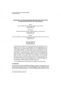
VNMR software from Varian. The chemical shifts reported in Table ALDH Signal Peptide IN VITRO IMPORT I are from spectra acquired a t 40 "C. Backbone protons were completely assigned for spectra acquired at ambient temperature. HowNative: ML-R-A-A-L-ST-A-R-R--P-A-L-SR-L-L[-sY-A-NH,] looX ever, the leucine and arginine side chain resonances could not be N-helix deletion: M-R-C:P-R-L-SR-L-L 0% adequatelyassigned because of poor TOCSY transfer at ambient temperature. Cheiix deletion: ML-R-A-A-L-sT-A-R-R"q 0% Minimization and Molecular Dynamics-A model structure for the Turn deletion: ML-R-A-A-L-sT-A-R-R-L-SR-L("A-NY1 110% linker-deleted pALDH peptide was built on a Silicon Graphics Iris N-C ShHt: MR-L-SR-L-L-R:G-P-L-R-A-A-L-sT-A-R 85% 4D/25S workstation utilizing University of California San Francisco MIDAS Plus software (39). Minimization and molecular dynamics were performed using a modified version of AMBER 4 (40) which Cytochrome Oxidase Subunit IV ( E n d o e tal., 1989) contains a flat-well pseudopotential to constrain the peptide with the M-L-SL-R-Q-Si-R-F-F-K-P-A-T-R-T-L~SSR-[-L-L-NH,] 1ooX NOE distances(41). In addition the peptidewas built using pseudoa- Natlve: tom residues (42) toavoid the need to assign stereospecifically many FIG.2. Summary of i n vitro import results for precursor of the amino acid residue side chain protons. A total of 45 interresidue distance constraints derived from peak volumes or intensities were protein. Various ALDH and cytochrome oxidase subunit IV native and mutant precursors. The signal peptides studied by NMR are at 40 "C. NOES were obtainedfrom a 50-msNOESYspectrum indicated with the C-terminal amide section in brockets. The helical medium (2categorized by their peak intensityss strong (>7, T > 300),mf (medium-fast, 300 > T > 401,f (fast, 40 > T > 01,and uf (very fast, NH not observed). In the case of Arg-10 spectral overlap prevented determining its half-life.
12, Ser-13, Leu-15, and Leu-16. These experiments allowed us to build a map of the approximate exchange rates of the amide protons. The relative NH exchange rate of each amino acid residue is shown in Fig. 9. The observed results indicate
that almost all of the amide protonsareprotected from exchange to some degree with the residues on the hydrophobic face of the peptide exchanging much more slowly.
It has been well established that mitochondrial leader sequences are capable of forming amphiphilic helices (3-15). Prior to the NMR studies of cytochrome c oxidase IV (20), ALDH (21), and most recently, the P-subunit of F1ATPase (23), there was no detailed structural analysis of the peptides. Both ALDH signal peptide in a micelle and F1 signal peptide in 50% trifluoroethanol (membranemimicking environments) showed evidence of a linker region separating two helical domains. However, at lower temperature ( 5 "C) F1 signal peptide appears to be a rigid helix throughout the sequence. Cytochrome c oxidase IV wascomposedof an N-terminal helical region followed by an essentially structureless region. Based upon our earlier NMR analysis (21), we had proposed that therole of the second helix in pALDH is to facilitate the binding of the N-terminal helix which is necessary for its import function. We further proposed that any alteration of the signal peptide whichwould destabilize the N-terminal helix would impair import. An example could be the introduction of glycines into the N-terminal domain or the deletion of a middle section of the precursor of ornithine transcarbamylase (27). Here we present data (Fig. 1) to show that the linker region is not necessary for import since the continuous helix that is formed in itsabsence still maintains the necessary N-terminal portion. Unexpectedly, it was found that the linker-deleted signal peptide was not cleaved after import. NMR and NH Exchange Rates of Linker-deleted PeptideOne important advantage of NMR spectroscopy over other biophysical probes of peptide conformation (such as CD spectroscopy) is that we can obtain details on not only the overall conformation but also on local conformational features and dynamics. The two-dimensional NOESY experiment for the signal peptide bound to a micelleallows us to distinguish segments of helical structure readily. The amide exchange rates provide information on the relative stabilityand dynamics of the secondary structure. Thus NH exchange for a micelle-bound peptide could require that hydroxide penetrate the hydrophobic interior of the micelle to exchange those amide hydrogens of the hydrophobic residues in the amphiphilic helix which are buried within the micelle. Alternatively exchange could occur via local disruption of the helical portions of the signal peptide either at the surface or interior of the micelle or total dissociation of the peptide into the aqueous solution. Because the peptide has little structure in purely aqueous solution (21), NH exchange of the totally dissociated or random coil part of the partially dissociated peptide would be rapid. As shown for many soluble globular proteins, under conditions in which protein is largely folded, the rate of amide exchange (keJ is given by k,, = K,,k, where kc is the ratelimiting intrinsic rateof exchange of the unfolded protein and Kop (= k,Jkf) is the unfolding equilibrium constant between stable, folded protein and the unfolded state (50-52). This exchange-limited unfolding mechanism occurs under conditions in which the folded state is stable and kc is relatively slow. Sykes and co-workers (53,541 have shown that therate of NH exchange of the M13 coat protein bound to a micelle is interpretable in termsof this mechanism. These conditions are most certainly true for the signal peptide bound to the micelle as well. Thus the observed rate of exchange of the micelle bound signal peptide will reflect both the intrinsic rate of exchange of the freely exposed amide to the solution, as well as the fractional population of the locally unfolded state exposed to solvent. Molday et al. (51; see also 52) have shown that most amide protons of random coil peptides exchange ( k c )within a
19912
Linker-deleted Mitochondrial Signal Peptide
relatively narrow range of values (generally less than 10-fold), depending upon the amino acid, nearest neighbor effects, and proximity to theN andC termini. Indeed simple solubilization of a hydrophobic tripeptide into a detergent micelle does not alter the minimum rate of peptide backbone NH exchange (55). Two-dimensional NOESY and amide exchange experiments of the ALDH signal peptide suggested that thepeptide contained two helical regions, both of which are amphiphilic, separated by a flexible linker region. The helix located closer to the C terminus was found to undergo NH exchange at a much slower rate thandid the helix located at theN terminus. This indicates that the hydrophobic face of the C-terminal helix is buried deeper within or more stably bound to the hydrophobic region of the micelle than the N-terminal helix (21). As shown byCD and NMR, the micelle bound, linkerdeleted ALDH contains a single a-helical segment that extends nearly over the entire length of the peptide. The amide protons in the helix located closer to the C terminus were found to undergo NH exchange at a much slower rate than those located toward the N-terminal portion of the helix. In addition, the rate of exchange of residues on the hydrophobic face was much slower than those of the polar face of the peptide. This suggests that the entirehydrophobic face of the linker-deleted signal peptide is also buried within the micelle and therefore protected from exchange with the solvent. The slow exchange is not attributable to hydrophobicity effects alone since Ser-13 is also observed to be slowexchanging. The C-terminal half of the helix appears to be more stable and/or buried deeper as shown by the very slow exchange for residues Leu-12, Ser-13, Leu-15, and Leu-16. These amide protons exchange unusually slowly for a small linearpeptide. Analysis of nearest neighbor effects (51) allows a prediction of the relative exchange rates for an unstructured peptide. The predicted order for base catalyzed exchange (witha relative rate difference of 4 0 ) is Ala-5, Leu-6, Leu-16 < Arg3, Arg-10, Ala-19 < Tyr-18 < Ala-4, Ala-9, Leu-12, Leu-15 < Arg-11, Arg-14 < Ser-7, Ser-13, Ser-17 < Thr-8 100) is Leu-12, Leu-15, Leu-16 < Ser-13 < Ala9 < Ala-5, Leu-6, Thr-8, Arg-11 < Ala-4, Ser-7, Arg-14, Ser17, Tyr-18,Ala-19 < Arg-3. Some amide protons of the signal peptide remain even after 21 h indicating that they are well sequestered from the solvent and that this portion of the peptide rarely dissociates from the surface. It should be noted that shifts in the pH at which exchange is at a minimum (normally about pH 3.0) can also affect the relative exchange rates (52), and no correction has been made for this effect. The differential NH exchange rates are thus interpreted in terms of local unfolding of the peptide, exposing NHs to solvent. Exchange rates of amide protons inthe linker-deleted signal peptide arealtered significantly from those inthe native sequence. Residues Arg-3, Arg-14, Ser-17, Tyr-18, and Ala-19 of the linker-deleted ALDH peptide exchange faster, whereas residues Leu-6, Thr-8, Ala-9, Arg-11, Leu-18, andSer-13 exchange more slowly than their corresponding residues in the native sequence. The exchange rates of residues Ala-4, Ala-5, Ser-7, Leu-15, and Leu-16 are approximately the same as those observed for the corresponding residues in the native ALDH signal peptide. These results indicate that deletion of the 3-residue linker has resulted in greater protection of the interior amide hydrogens. Generally we find that themagnitude of the observed NOE cross-peak volumes for the micelle bound peptide does not
depend on thelocation of the residues in the peptide sequence. This indicates that in contrast to thatreported for the native ALDH signal peptide (21) the a-helical character is uniform throughout the a-helix. Furthermore, ascan be seen in Fig. 5 almost all of the a-protons areshifted upfield relative to the random coil and typical of an a-helix(47). In the native signal peptide, however, a bimodal distribution of a-protons was observed with maxima centered near the middle of each of the a-helical regions separated by the R-G-Plinker. Deletion of the R-G-Plinker in native ALDH also appears to result in greater stabilizationof the N-terminal helix. To understand further why some NH exchange rates increased while others decreased we have utilized NMR data and the molecular modeling program MIDAS (39) to model build the structure of the linker-deleted ALDH. Using the AMBER (40) molecular mechanics/dynamics program, we first calculated a minimum energy structure for the peptide consistent with the NMR results for a single helix. NOESY distance restraints (for the helix) were included as an additional flat-well pseudoenergy penalty term in the force field. After restrained molecular dynamics, the structures obtained were as expected a-helical. The helix extended from residue 5 to 19 with all slowly exchanging residues on the same face of the peptide. From this model it could be seen that Arg-14, Ser-17, and Tyr-18 are now on the hydrophilic face of the helix and therefore would be expected to exchange faster (as observed). The structure accurately fits the helix wheel representation and indicates that deletion of the 3-residue R-GP linker hasresulted in a shift in the phase of the amphiphilic peptide in such a way as to place Arg-14, Ser-17, and Tyr-18 on the hydrophilic face of the peptide (Fig. 9). Note that at the C terminus, residues 17-19 (NH exchange rates, fast) are part of the mature protein. The decrease in the NHexchange rate for residues Leu-6, Thr-8, and Ala-9 is most likely attributed to an increased stabilization of the N-terminal portion of the helix (Fig. 9). Structural and Dynamical Requirements for Import of Mitochondriat Precursor Proteins-It has been suggested that the presequence for a proteindestined to be imported into the mitochondria must first bindto themitochondrial membrane and that the association is made possible by hydrophobic interaction of the membrane with an amphiphilic a-helix (315). Membrane potential effects favoring association of the positively charged presequence with the mitochondrial membrane also play an important role (3, 22, 56). Although never unequivocally proved, support for the hydrophobic interactions comes from the observation that many synthetic signal peptides will form helical structures when in an hydrophobic environment (19-21,23). However, based upon various helical prediction models and supported by mutational and spectroscopic conformational studies, the entire presequence does not have to be helical for efficient import. Deleting as much as one-half or even two-thirds of the residues in the presequence often has little effect on import. von Heijne et al. (1)proposed the existence of two domains in mitochondrial signal peptides that have different amphiphilic properties based on Chou-Fasmanand hydrophobic moment calculations. They suggested that these two domains might be involved in two steps of signal peptide cleavage after import. Our earlier NMR study (21) on the ALDH signal peptide and that by Endo et al. (20) on cytochrome c oxidase IV was consistent with a multidomain hypothesis. The ALDH signal peptide conformed to a two-helical domain, helixlinker-helix model. The linker could be described equally well as a flexible linker or that of a type Ior I1 turn. Because NOE cross-peaks between the two helices were not observed, it is
Linker-deleted Mitochondrial Signal Peptide unlikely that these helices are folded upon one another when bound to themicelle into asingle stable structure.Cytochrome c oxidase IV signal peptide appeared to be an N-terminal amphiphilic helix connected to a random coil C-terminal domain. Our previous NMR studyled us to propose a helix-"linker"helix model (21) for ALDH import (Fig. 10). In this model the presequence is largely random coil in solution (Fig. 1OA). The presequence portion of the precursor protein is initially anchored to the membrane via a C-terminal amphiphilic ahelix (Fig. loll). The N terminus canalso fold into an a-helix when membrane bound, and both segments can contribute to binding of the presequence to the membrane (Fig. lOC). By anchoring the signal peptide to the membrane, the protein can diffuse across the surface of the membrane until it meets an appropriate receptor. Diffusion upon the surface of the membrane would allow the precursor protein to search more quickly in two-dimensional space rather than the three-dimensional space of the cytosol. The receptor then recognizes the presequence (Fig. 10B). It is quite possible that too strong of an association with the membrane could be deleterious to thereceptor binding to the presequence. Deletion studies have determined that the first 9 residues of another signal sequence are of particular importance for efficient transport (26). Most presequences have been proposed to contain at least a single amphiphilic helix (1).After the receptor and protein form a complex, the protein can be transported. In this paper, our NMRresults with the linker-deleted peptide show that thepeptide is composed of one long continuous helix, similar to theF1signal peptide at low temperature (23). The import results summarized in Fig. 2 show that the linker is not required for import. However, deletion of either the C-terminal or N-terminal helix produces protein that is not imported (32). These results suggest that precursor protein import simply requires a sufficiently long amphiphilic helix (or two separate helices) to bind stablyto themembrane. Note the N and C helices of native ALDH signal peptide are
A h
4 .
wC
MEMBRANE
B
1
E 11
FIG. 10. Model for binding of precursor proteins to membrane and precursor protein translocation. Presequence portion of native or linker-deleted ALDH precursor protein shown with the RGP linker presequence shown in parentheses. A , random coil state and membrane. B, initial anchoring of the C-terminal amphiphilic ahelix of the precursor protein to the membrane. Diffusion of the protein to thereceptor is then followed by insertion of the N-terminal helix into thetranslocator protein(cylinder),leading to translocation. C, both N-terminal and C-terminal a-helical regions bound to membrane.
19913
only about 6-8 residues long; this represents only about two turns of a helix, and either helix on its own does not provide enough stabilization to ensure folding and binding to the membrane (57). The linker-deleted ALDH peptide contains a single helix of 12-14 residues which is long enough to provide a hydrophobic surface that can interact stably with the hydrophobic interior of the membrane (Fig. 1OC). In pALDH the turn can be removed; even the order of the two helices is not important (Fig. 2; N-C shift sequence). The function of the C helix in the native signal peptide is therefore to enhance the stabilityand binding of the N-terminal signal to the membrane. As shown in Fig. loll, partial dissociation of the N terminus from the membrane surface may facilitate binding of the precursor protein to thetranslocator protein. This model is consistent with other import data as well (Fig. 2; cf. 18, 20, 24-29). Possibly the most important structuralrequirement for efficient import of a mitochondrial precursor protein is a minimal hydrophobic surface provided by the amphiphilic a-helical presequence (21, 28, 29). The cytochrome c oxidase IV signal peptide also appears to have one long helix of about 12 residues (3+ turns) which is sufficient for import (20). F, signal peptide also has one or two relatively long helices (23), although comparison with the micelle data for ALDH and cytochrome c oxidase IV and the trifluoroethanol data for F1 makes comparison more difficult. In contrast topALDH signal peptide, the N-terminal helix is more stable incytochrome c oxidase IV and F1(in cytochrome c oxidase IV a stable C-terminal helix is not even observed andcan,as expected therefore be deleted). It appears to generally be the case that theC terminusof the signal peptide is not that important for import. Our data support the proposal that a necessary and sufficient condition for import is a sufficiently stable amphiphilic helix. If, as in pALDH, one of the helical domains is not stable enough and only weakly binds to the membrane, then additional amphiphilic helical stabilization is needed. A flexible linker or none at all can connect these two helical domains. Processing-Quite to our surprise, unlike native ALDH precursor protein, the linker-deleted precursor sequence is not proteolytically cleaved once this mutant precursor protein is imported. The R-G-P deletion is far removed from the cleavage site, suggesting that orientation or relative stability of the amphiphilic a-helix itself may play an important role in proper processing. Little is known about the specificity for cleavage of the precursor sequence from the precursor protein. It has been suggested that thecleavage of mitochondrial signal peptides is not only dependent upon the local sequence around the cleavage sites but also the remote portion of the signal peptide (30). The NMR data clearly show that the linkerdeleted peptide kept the same helical conformation around the cleavage site as that of the native pALDH signal peptide. However, the NHexchange rates of the last 3 residues that immediately follow the cleavage site changed dramatically after deletion of the linker. These exchange rates are slow in the linker-deleted the native peptide (21) butarefastin peptide. As explained by the modeling of the a-helix, the faster NH exchange rate is possibly related to thephase shift of the amphiphilic helix. One interpretation of these data suggests that processing of the precursor protein requires that the first several residues of the mature protein be part of the hydrophobic membrane associated face of the helix. By moving them toward the hydrophilic face, the proteolytic signal processing protein is perhaps unable to recognize these resi-' dues. Alternatively, the NH exchange data show that the linker-deleted single helix is more stably bound to the membrane thanthe C-terminal helix of the helix-linker-helix
Linker-deleted Mitochondrial Signal Peptide
19914
native sequence. Perhaps the single helix ofthe linker-deleted signal peptide is too stable. Thus import and processing may require a trade-off in the relative stability and dynamics of varioussegmentsof the precursor sequence. These results demonstrate the importance of coupling structural data with the interpretation of biological function. REFERENCES 1. von Heijne, G., Steppuhn, J., and Herrmann, R. 0. (1989) Eur. J. Biochem. 180,535-545 2. Watson, M. E. E. (1984) Nucleic Acids Res. 12,5145-5164 3. von Hei’ne G (1986) E M f O J. 6, 1335-1342 4. Tamm, K . i1991) Biochrm. Biophys. Acta 1071, 123-148 5. Swanson, S. T., and Roise, D. (1992) Biochemistry 31,5746-5751 6. Brigga, M. S., Gierasch, L. M., Zlotnick, A., Lear, J. D., and DeGrado, W. F. (1985) Science 228,1096-1099 7. Furuya, S., Mihara, K., Aimoto, S., and Omura, T. (1991) EMBO J. 10, 1759-1766 8. Glaser, S., and Cumsky, M. (1990) J. Biol. Chem. 266,8817-8822 9. Kuchinka, E., and Seelig, J. (1989) Biochemistry 28,4216-4221 10. Ono, H., and Tuboi, S. (1988) J. BioL Chem. 263,3188-3193 11. Roise, D. (1992) Proc. Natl. Acad. Sci. U.S. A. 89, 608-612 12. Tamm, L. K. (1986) Biochemistry 26,7470-7476 13. Epand, R., Hui, S., Argan, C., Gillespie, L., and Shore, G. (1986) J. Eiol. Chem. 261,10017-10020 14. Hovt. D.W..Cvr.D.M.. Gierasch. L. M.. and Douelas. M.G. (1991) , , J. Biol. Chek. 266,21693-21699 ’ 15. Tamm, L. K., and Bartoldus, I. (1990) FEBS Lett. 272,29-33 16. Horwich. A. L.. Kalousek, F.. Mellman. I., and Rosenberg. -. L. E. (1985) EMBO J. 4,1129-1135. . 17. Hurt, E. C., Muller, U., and Schatz, G. (1985) EMBO J. 4, 3509-3518 18. Pilgrim, D., and Young, E. T. (1986) Mol. Cell Biol. 7, 294-304 19. Roise, D., Theiler, F., Horvath, S. J., Tomich, J. M., Richards, J. H., Allison, D. S., and Schatz, G. (1988) EMBO J. 7,649-653 20. Endo, T., Shimada, I., Roise, D., and Inagaki, F. (1989) J. Biochem. 106, 396-400 21. Karslake, C., Piotto, M. E., Pak, Y. M., Weiner, H., and Gorenstein, D. G. (1990) Biochemistry 29,9872-9878 22. Rome, D., Horwlth, S., Tomich, M., Richards, J. H., and Schatz, G. (1986) EMBO J. 6, 1327-1334 23. Bruch, M. D., and Hoyt, D. W. (1992) Biochim. Biophys. Acta 1169, 81-
i.
92
24. Hurt, E. C., Pesold-Hurt, B., Suda, K., Oppliger, W., and Schatz, G. (1985) EMBO J. 4,2061-2068 25. Nishi, T., Nagashima, F., Tanase, S., Shimada, K., Ushio, Y., and Morino, Y. (1989) J. Biol. Chem. 264,6944-6051 26. Haldi, M., and Guarente, L. (1989) J. Bwl. Chem. 264,17107-17112
27. Horwich, A. L., Kalousek, F., Fenton, W. A., Pollock, R. A., and Rosenherg, L. E. (1986) Cell 44,451-459 28. Bedwell, D. M., Strobel, S. A,, Yun, K., Jongeward, G. D., and Emr, S. D. (1989) Mol. CeU. Biol. 9,1014-1025 29. Bedwell, D. M., Klionsky, D. J., and Emr, S. D. (1987) Mol. CeU. Biol. 7, 4038-4047 30. Hurt, E., Allison, D., Miiller, U., and Schatz,G. (1987) J. Biol. Chem. 262, 1420-1424 31. Pak, Y. K., and Weiner, H. (1990) J. Biol. Chem. 266,14298-14307 32. Wang, Y., and Weiner, H. (1993) J. Biol. Chem. 268,4759-4765 33. Wang, T. T. Y., Farres, J., andWeiner, H. (1989) Arch. Biochem. Biophys. 272,440-449 34. States, D. J., Haherkorn, R. A., and Ruehen, D. J. (1982) J. Magn. Reson. 48,286-292 35. Bax, A., and Davis, D. G. (1985) J. Magn. Reson. 66, 355-360 36. Braunschweiler, L., and Ernst, R. R. (1983) J. Magn. Reson. 63,521-528 37. Piantini, U., Sbrenson, 0. W., and Emst, R. R. (1982) J. Am. Chem. SOC. 104,6800-6801 38. Rance, M., Sbrensen, 0. W., Bodenhausen, G., Wagner, G., Ernst, R. R., and Wuthrich, K. (1983) Biochem. Biophys. Res. Commun. 117, 479ARK
39. LakGidge, R., and Ferrin, T. E. (1984) J. Mol. Graphics 2,56 40. Weiner, P. K., and Kollman, P. A. (1981) J. Com ut Chem. 2,287-303 41. Nikonowicz, E. P., Roongta, V., Jones, C. R., anfcorenstein, D.G. (1989) Biochemistry 28,8714-8725 42. Wuthrich, K., Billeter, M., and Braun, W. (1983) J. Mol. Biol. 169, 949961 43. Braun, W., Wider, G., Lee, K. H., and Wuthrich, K. (1983) J. Mol. Eiol. 169,921-948 44. PfacpefLN., Pfaller, R., and Neupert, W. (1988) Trends Biochem. Sci. 13, lb9-16’{
45. Yang, J. T., Wu, C. C., and Martinek, H. M. (1986) Methods Enzymol. 130,208-269 46. Wuthrich, K. (1986) NMR of Proteins and Nucleic Acids, John Wiley &
Sons, New York
47. Wishart, D. S., Sykes, B. D., and Richards, F. M. (1992) Biochemistry 31, 1647-1651 48. Roongta, V., Powers, R., Jones, C., Beakage, M. J., Shields, J. E., and Gorenstein, D. G. (1989) Biochemistry 28,1048-1054 49. Hahn, U., and Ruterjans, H. (1985) Eur. J. Bmchem. 162,481-491 50. Englander, S. W., and Kallenbach, N. R. (1984) Q.Reu. Bmphys. 19, 521655 51. Molday,R. S., Englander, S. W., and Kallen, R. G. (1972) Biochemistry 11,150-158 52. Roder, H.,Wagner, G., and Wuthrich, K. (1985) Biochemistry 24, 73967407 53. Henry, G. D., and S kes, B. D. (1990) Biochemistry 29,6303-6313 and Sykes, B. (1987) Biochemistry 26,3619-3626 54. Henry, G., Weiner, 55. ONeil, J. D. J., and Sykes B. D. (1989) Biochemistry 28,699-707 56. Skerjanc, I., Shore, G., and Silvins, J. (1987) EMBO J. 6,3117-3123 57. McLean, L. R., Hagaman, K. A., Owen, T. J., and Krstenansky,J. L. (1991) Biochemistry 30,31-37
j.,