Papers in Press. First published December 18, 2003 as doi:10.1373/clinchem.2003.022426
Clinical Chemistry 50:2 000 – 000 (2004)
Cancer Diagnostics
Improved Accuracy of Detection of Nasopharyngeal Carcinoma by Combined Application of Circulating Epstein–Barr Virus DNA and Anti-Epstein–Barr Viral Capsid Antigen IgA Antibody Sing-fai Leung,1* John S. Tam,2 Anthony T.C. Chan,1 Benny Zee,1 Lisa Y.S. Chan,4 Dolly P. Huang,1 Andrew Van Hasselt,5 Philip J. Johnson,1 and Y.M. Dennis Lo3
Background: Circulating Epstein–Barr viral (EBV) DNA and anti-EBV capsid antigen IgA (IgA VCA) represent two of the most sensitive peripheral blood markers of nasopharyngeal carcinoma (NPC), but direct comparative studies of these two markers are lacking. Methods: The sensitivities and specificities of IgA-VCA and EBV DNA for diagnosis of NPC were determined in 139 new cases of NPC and 178 healthy individuals, respectively. EBV DNA was also assessed in 36 healthy family members identified to have false-positive IgAVCA results at a screening clinic. EBV DNA was measured by a real-time quantitative PCR assay with a detection limit of 60 copies/mL. IgA-VCA was measured by semiquantitative indirect immunofluorescent method; a titer >1/10 was taken as positive. Results: The sensitivities of EBV DNA and IgA-VCA for diagnosis of NPC were 95% (95% confidence interval, 91–98%) and 81% (73– 87%), respectively. The combined marker panel had an overall sensitivity (positive result by either marker) of 99%. The concentrations of both markers showed dependence on cancer stage. The specificities of EBV DNA and IgA-VCA were 98% (96 –99%) and 96% (91–98%), respectively. Among 36 healthy family members with false-positive IgA-VCA results, three-fourths had undetectable EBV DNA,
whereas the others had increased EBV DNA concentrations that were significantly lower than in NPC patients. Conclusions: For diagnosis of NPC, EBV DNA identifies almost all false-negative IgA-VCA cases and gives a 99% diagnostic sensitivity when combined with IgAVCA. In the screening setting, EBV DNA identifies three-fourths of false-positive IgA-VCA cases. The selective application of EBV DNA in an IgA-VCA-based screening protocol could improve screening accuracy with only moderate increases in cost. © 2004 American Association for Clinical Chemistry
The association between specific serologic responses to Epstein–Barr virus (EBV)5 and nasopharyngeal carcinoma (NPC) has been exploited to develop serologic tumor markers for this cancer (1 ). The anti-viral capsid antigen IgA antibody (IgA-VCA), measured by indirect immunofluorescence (1 ) or ELISA (2 ), is one of the most widely used antibody markers used for assisting in diagnosis (1, 3 ) and for screening (4 –9 ). Its sensitivity in the diagnosis of WHO type II and III NPC in areas endemic (1 ) and nonendemic (3 ) for the disease has been generally reported to be ⬃85–90% (1, 3, 4, 10 –14 ), although a broader range had been reported in some studies. A false-positive rate of 2.36 – 6.71% had been found in screening studies in areas with endemic NPC (5 ), whereas in case– control studies a false-positive rate of 9 –18% has been reported (3 ). In attempts to improve the sensitivity and specificity of
Departments of 1 Clinical Oncology, 2 Microbiology, 3 Chemical Pathology, and 4 Surgery, The Chinese University of Hong Kong, Hong Kong SAR, Peoples Republic of China. *Address correspondence to this author at: Department of Clinical Oncology, Prince of Wales Hospital, Shatin, Hong Kong. Fax 852-2636-4346; e-mail
[email protected]. Received May 28, 2003; accepted November 26, 2003. Previously published online at DOI: 10.1373/clinchem.2003.022426
5 Nonstandard abbreviations: EBV, Epstein-Barr virus; NPC, nasopharyngeal carcinoma; IgA-VCA, anti-viral capsid antigen IgA antibody; and CI, confidence interval.
1 Copyright (C) 2003 by The American Association for Clinical Chemistry
2
Leung et al.: EBV DNA vs EBV IgA-VCA in NPC
a peripheral blood-based marker for NPC, several alternative markers had been developed. These were based on antibodies to different antigenic components of the EBV (9, 15–29 ). Attempts to use panels of antibody-based markers have generally been useful in improving the accuracy of NPC detection (13, 15, 20, 21, 29 –32 ). Plasma/ serum cell-free EBV DNA is a more recently developed peripheral blood-based molecular marker for NPC. It is conceptually distinct from other peripheral blood markers in that it directly measures tumor-derived EBV genomic material rather than an antibody response to genomic or peptidic components of the EBV. Although initial studies based on qualitative measurement systems showed that plasma EBV DNA has a sensitivity of only 59 –75% for diagnosis of NPC (33–35 ), its sensitivity in quantitative assays was as high as 90% (36 ). Such a diagnostic sensitivity appears to be at least as good as that of IgA-VCA, although direct comparative studies of the two markers have yet to be reported. Of further interest is the observation that plasma EBV DNA was found to be rarely detectable in patients who had complete eradication of cancer (37 ) and in individuals without cancer (34, 36, 37 ). These observations suggest that the marker may have a role in the cancer screening setting. In the present study, we aimed to directly compare the sensitivities and specificities of the two markers in NPC patients and individuals without cancer and to explore whether the markers play complementary roles in the diagnosis of and screening for NPC.
Materials and Methods sample size The sample size of the present study was based on estimation of sensitivity and specificity rates postulated at ⬃80 –90%. The estimate is considered accurate within a variation of 10%. To have 80% power to test the postulated rates at 5% significance with a one-sided test, we need to enter ⬃90 patients. The same number of patients is adequate to draw a 95% confidence interval around the sensitivity or the specificity with a range of 10%.
cancer cases for determination of sensitivity We recruited 139 patients with newly diagnosed NPC at our institution (group A) and collected data prospectively. Consecutive consenting patients were recruited at the oncology clinic in the period May 1998 to December 2000. Histologic details were available in all cases and were confirmed to be WHO type II/III carcinoma in 138 cases and WHO type I in 1 case. Tumor staging was according to the International Union Against Cancer 1997 stage classifications (38 ). Staging work up included in all cases clinical examination; computed tomography scans of the nasopharynx, skull base, and neck; chest radiography; and serum alkaline phosphatase measurements. Table 1 details the gender, age, and tumor stage distribution of the patients. Serum IgA-VCA and plasma EBV DNA
Table 1. Characteristics of study participants.
n Gender, % male Age, years Median Mean (SD)
Cancer patients (group A)
Healthy controls (group B)
139 74
178 26
48 49 (12)
48 52 (12)
Family members with positive IgA-VCA (group C)
36 39 39 39 (10)
were assessed before commencement of oncologic therapy.
non-cancer cases (controls) for determination of specificity
We recruited 178 healthy individuals ⱖ30 years at a community center (group B). The age and gender distribution of the cohort are detailed in Table 1. The age selection served to enhance the comparability with the cancer patient group, in consideration of the uncommon occurrence of NPC below the age of 30. To further address the clinical question of whether selective analysis for EBV DNA in IgA-VCA-positive individuals could reduce false positives in the screening setting, another cohort of 36 family members of NPC patients with known positive IgA-VCA results (titer ⱖ1/10) were recruited at a NPC screening clinic during a follow-up visit (group C). After blood sampling, the participants were followed up at 6-month to 1-year intervals and had a minimum follow-up time of 1 year. Their noncancer status was defined by the absence of clinical evidence of disease at 1 year or more after the EBV DNA assessment time point. The age and gender distribution of the non-cancer cohorts are detailed in Table 1. The higher proportion of females reflects the gender distribution of individuals attending the screening clinic.
tumor marker assay Plasma EBV DNA was measured by real-time quantitative PCR as described previously (36 ). DNA was extracted from plasma samples with a QIAamp Blood Kit (Qiagen) according to the “blood and body fluid protocol” as recommended by the manufacturer. A total of 400 – 800 L of the plasma samples was used for DNA extraction per column. The exact amount was documented for calculation of the target DNA concentration. A final volume of 50 L was used to elute the DNA from the extraction column, and 5 L of the eluted DNA was used per PCR. Circulating EBV DNA concentrations were measured by a real-time quantitative PCR system that amplified a DNA segment in the BamHI-W fragment region of the EBV genome. The principles of real-time quantitative PCR and the reaction set-up procedures were as described previously. Data were collected with an ABI Prism 7700 Sequence Detector and analyzed with the Sequence Detection System software (Ver. 1.6.3) devel-
3
Clinical Chemistry 50, No. 2, 2004
oped by PE Biosystems. Results are expressed as EBV genome copies/mL of plasma. Endogenous -globin was used as an “amplification control”, and all plasma DNA samples were also subjected to real-time PCR analysis for the -globin gene, which gave a positive signal on all tested samples, thus demonstrating the quality of the extracted DNA. Multiple negative water blanks were included in every analysis. Results were expressed as DNA copies/mL of plasma. The lower limit of reliable quantification was 5 copies/assay, although occasionally 1 copy/assay was also detectable (the latter corresponding to 12 copies of EBV DNA/mL of plasma). The precision of the EBV DNA assay near the lower limit of detection was determined by 20 replicate extractions of pooled NPC plasma samples with a mean EBV DNA concentration of 100 copies/mL. Because no recovery experiments had been performed, the results, expressed in copies/mL and assuming 100% recovery, represent the minimum estimates of the concentrations. Each of these replicate extractions was then subjected to realtime EBV DNA PCR. The CV at this low concentration of circulating EBV DNA was 58%. EBV IgA-VCA was measured by a semiquantitative immunofluorescence method (1 ). Briefly, B95.8 cell preparations were air-dried and fixed in cold acetone in wells on glass slides, and dilutions of patient sera were applied. After incubation the slides were washed with phosphatebuffered saline, and fluorescein isothiocyanate-conjugated anti-human IgA was applied to each well at appropriate concentrations. After incubation and washing, the slides were mounted with buffered glycerol and read with a fluorescence microscope. The antibody titer of the test serum was read as the reciprocal of the highest dilution showing definite apple-green fluorescence in 20% of the cells. A titer ⱖ1/10 was taken as positive. This cutoff titer was commonly adopted in previous studies on the marker. The study was approved by our institution, and written informed consent was obtained from all study participants.
Table 2. Comparison of sensitivities of markers by stage (group A). NPC stage
n
All stages 95% CI Stages I ⫹ II 95% CI Stages III ⫹ IV 95% CI
139 50 89
Sensitivity of IgA-VCA
Sensitivity of EBV DNA
81% (112/139) 73–87% 72% (36/50) 59–84% 85% (76/89) 78–92%
95% (132/139) 91–98% 90% (45/50) 82–98% 98% (87/89) 95–100%
results by either marker, the panel had a sensitivity of 99%.
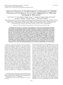
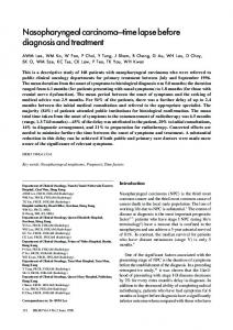
cancer cases (group a): relationship between marker concentration and cancer stage The distributions of EBV DNA concentrations and IgAVCA titers according to individual cancer stage are shown in Figs. 1 and 2, respectively. To assess the relationship between EBV DNA concentration and IgA-VCA titer and cancer stage, we tested the variance by first transforming EBV DNA values to a log scale and IgA-VCA titers to a linear scale. We found a significant relationship for each of the two markers with cancer stage (P ⬍0.01 for EBV DNA; P ⫽ 0.04 for IgA-VCA). Multiple group comparisons were also performed for marker concentrations among different cancer stages. We found that EBV DNA concentrations were significantly different between stage I and each of the other stages and between stages II and
Results cancer cases (group a): sensitivity of markers When we used detectable (ⱖ60 copies/mL of plasma) circulating EBV DNA concentrations and an IgA-VCA titer ⱖ1/10 to define positive results, the sensitivity of EBV DNA [95% confidence interval (CI), 91–98%] was higher than that of IgA-VCA (81%; 95% CI, 73– 87%; Table 2). We observed a difference in sensitivities among both early-stage and advanced-stage disease (Table 2). The 7 cases that were false negative for EBV DNA (stage I, 2 cases; stage II, 3 cases; stage III, 0 cases; stage IV, 2 cases) did not overlap with the 27 cases that were false negative for IgA-VCA (stage I, 5 cases; stage II, 9 cases; stage III, 8 cases; stage IV, 5 cases) except for 2 cases. When EBV DNA and IgA-VCA were combined as a marker panel and the sensitivity of the panel was defined as positive
Fig. 1. Scattergram of distribution of EBV DNA according to cancer stage. x axis, cancer stage; y axis, EBV DNA concentrations in log scale.
4
Leung et al.: EBV DNA vs EBV IgA-VCA in NPC
Fig. 2. Distribution of EBV IgA-VCA titers according to cancer stage.
IV. The IgA-VCA titers were significantly different only between stages I and IV. In other words, the concentrations of both markers exhibited a certain degree of cancer stage dependence, which was more significant for EBV DNA.
controls (group b): specificity of markers The specificity for EBV DNA was 98% (174 of 178) with a 95% CI of 96 –99%, and the specificity for IgA-VCA was 96% (170 of 178) with a 95% CI of 91–98%. There were four false-positive cases for EBV DNA and eight false-positive cases for IgA-VCA, but these cases did not overlap except for one case. Among the eight individuals with falsepositive IgA-VCA results, seven had a titer of 1/20 and one had a titer of 1/40. The EBV DNA concentrations in the four individuals with false-positive EBV DNA results were 72, 164, 239, and 3287 copies/mL, respectively, which were significantly lower than the concentrations in NPC (group A) patients (P ⬍0.01, Wilcoxon rank-sum test). For the only individual with false-positive results for both markers, the EBV DNA concentration was 164 copies/mL and the IgA-VCA titer was 1/10.
healthy family members of cancer cases with increased IgA-VCA titers (group c) Among the 36 healthy family members who had increased IgA-VCA titers (ⱖ1/10) identified at a screening clinic and who did not have evidence of cancer at followup, 27 had undetectable EBV DNA. For the other nine individuals, all had EBV DNA concentrations ⱕ344 copies/mL, apart from one individual with a concentration of 4840 copies/mL. For these nine individuals with falsepositive results for both markers, EBV DNA concentrations were significantly lower than those of true-positive
cases (i.e., group A NPC patients; P ⬍0.01, Wilcoxon rank-sum test).
Discussion The anti-VCA IgA antibody was chosen as the reference marker for this study because of its high sensitivity for diagnosis of NPC and the relative abundance of data related to this marker with its long track record in both case– control studies and population screening studies. Plasma/serum cell-free EBV DNA represents a direct measure of EBV genomic material and is conceptually distinct from other antibody-based markers for NPC. There is much clinical interest in comparing EBV DNA and IgA-VCA, first because the sensitivities of both markers for diagnosis of NPC are among the highest reported for peripheral blood markers of NPC, and second because their different mechanisms of production may allow minimization of false-positive cases when used in conjunction, which is an important issue in the cancer screening setting. The present study shows that the two markers have complementary roles in the diagnosis of and screening for NPC. There is almost no overlap of false-negative cases for these two markers and only limited overlap of the false-positive cases. This allows a potential diagnostic sensitivity of 99% when the two markers are used in a panel. The clinical interpretation is that if a patient is strongly suspected to have NPC, a negative result for both markers would make the diagnosis of NPC extremely improbable and should lead to consideration of alternative diagnoses. Such a high sensitivity has been reported for only a very limited number of marker panels (20 ). Although the reasons for false negativity of the markers is not certain, we observed that 5 of the 7 cases that were
5
Clinical Chemistry 50, No. 2, 2004
false negative for EBV DNA had stage I-II tumors, whereas the distribution of the 27 false-negative IgA-VCA cases was relatively even between early- and advancedstage tumors. In other words, the two markers appear to exhibit different degrees of cancer stage dependency. Although improvement in sensitivity is commonly paralleled by a reduction of specificity, the present study on healthy family members of NPC patients showed that use of EBV DNA can eliminate approximately threefourths of the false-positive IgA-VCA results. Although the cost of the EBV DNA test (approximately US $75.00 per test) is appreciably higher than that of the IgA-VCA test (less than US $10 per test), the latter can be used as the first step in a screening protocol, followed by the EBV DNA test in cases with positive IgA-VCA results. Such selective application of the EBV DNA test in a IgA-VCAbased screening protocol may lead to a significant improvement in accuracy of screening with only a limited increase in cost. Although the widely used anti-early antigen IgA antibody marker may serve a similar purpose of eliminating an appreciable proportion of false-positive IgA-VCA results, its sensitivity in detecting NPC is only ⬃70% (1, 10, 12 ). Thus, its combination with IgA-VCA in the screening setting may lead to missing an appreciable proportion of true cancers. This limitation is overcome by EBV DNA, which has a 90% sensitivity in detecting early-stage cancer and 95% overall detection rate for NPC. The reason for the presence of detectable EBV DNA and IgA-VCA in 2– 4% of healthy individuals without cancer is not clear, although the EBV DNA concentrations in false-positive cases were much lower than in NPC patients. Previous studies had shown that EBV DNA and EBER (small EBV-encoded RNA) are detectable only in tumor cells and not in nasopharyngeal tissue of healthy individuals (39 ). However, there have also been reports documenting EBV DNA in nonmalignant tissue (40 – 45 ). Some individuals with increased IgA-VCA had been shown to have nasopharyngeal lymphoid hyperplasia, in which EBV DNA and EBER were detectable (46 ). The alternative explanation for the higher rate of EBV DNA positivity in apparently healthy individuals with increased IgA-VCA titers is that they actually harbor occult NPC that was not clinically apparent (44, 47 ). An early study on healthy family members of NPC patients had also reported an increased incidence of IgA-VCA seropositivity in family members, and it was postulated that an autosomal recessive gene might be involved in the IgAVCA response (48 ). Continued follow-up surveillance of healthy individuals with increased IgA-VCA titers, especially in the family member group, is worthwhile to exclude emergence of cancer (4 ). These individuals may also serve as a target group for tissue sampling by noninvasive methods, such as nasopharyngeal brushing for EBV DNA (49 –51 ), which has been reported to have a high accuracy in diagnosis of NPC.
In conclusion, the strategy of combined marker panels has been increasingly explored in NPC with encouraging results (13, 30, 31 ). In the cancer screening setting, the sensitivity, specificity, quality control, and costs are relevant considerations. To date, only a few markers or marker panels had been reported to have both a sensitivity and specificity ⬎90% for detection of NPC (14, 18, 27, 36 ). The sensitivity of 99% of the combined IgA-VCA/EBV DNA marker panel and the specificity of 96 –98% of the respective markers in the present study warrant further studies in the screening setting. Because plasma/serum EBV DNA concentrations are based on direct measurement of genomic material in the peripheral blood, this marker is conceptually advantageous in terms of quality control and has potential to serve as a reference marker for comparison of results obtained with different EBV antibody-based markers, which are linked to different EBV source antigens.
This study was supported in part by the Hong Kong Research Grants Council (Central Allocation Research Projects), the Institute of Molecular Oncology of The Chinese University of Hong Kong, and the Kadoorie Charitable Foundations, Hong Kong. We acknowledge Frankie Mo and Maria Lai (Department of Clinical Oncology, The Chinese University of Hong Kong) for statistical support and database management for the study, and Katherine Chow (Department of Chemical Pathology, The Chinese University of Hong Kong) for experiments with the EBV DNA assay.
References 1. Henle G, Henle W. Epstein-Barr virus-specific IgA serum antibodies as an outstanding feature of nasopharyngeal carcinoma. Int J Cancer 1976;17:1–7. 2. Uen WC, Luka J, Pearson GR. Development of an enzyme-linked immunosorbent assay (ELISA) for detecting IgA antibodies to the Epstein-Barr virus. Int J Cancer 1988;41:479 – 82. 3. Neel HB 3rd, Pearson GR, Taylor WF. Antibodies to Epstein-Barr virus in patients with nasopharyngeal carcinoma and in comparison groups. Ann Otol Rhinol Laryngol 1984;93:477– 82. 4. Chien YC, Chen JY, Liu MY, Yang HI, Hsu MM, Chen CJ, et al. Serologic markers of Epstein-Barr virus infection and nasopharyngeal carcinoma in Taiwanese men. N Engl J Med 2001;345:1877– 82. 5. Zeng Y, Zhang LG, Li HY, Jan MG, Zhang Q, Wu YC, et al. Serological mass survey for early detection of nasopharyngeal carcinoma in Wuzhou City, China. Int J Cancer 1982;29:139 – 41. 6. Deng H, Zhao Z, Zhang Z. [Serologic screening on nasopharyngeal cancer in 338,868 persons in 21 cities and counties of Guangxi Region, China] Zhonghua Yu Fang Yi Xue Za Zhi 1995;29:342–3. 7. Zeng Y, Zhong JM, Li LY, Wang PZ, Tang H, Ma YR, et al. Follow-up studies on Epstein-Barr virus IgA/VCA antibody-positive persons in Zangwu County, China. Intervirology 1983;20:190 – 4. 8. Zong YS, Sham JS, Ng MH, Ou XT, Guo YQ, Zheng SA, et al. Immunoglobulin A against viral capsid antigen of Epstein-Barr virus and indirect mirror examination of the nasopharynx in the detection of asymptomatic nasopharyngeal carcinoma. Cancer 1992; 69:3–7.
6
Leung et al.: EBV DNA vs EBV IgA-VCA in NPC
9. Chen JY, Chen CJ, Liu MY, Cho SM, Hsu MM, Lynn TC, et al. Antibody to Epstein-Barr virus-specific DNase as a marker for field survey of patients with nasopharyngeal carcinoma in Taiwan. J Med Virol 1989;27:269 –73. 10. Henle W, Ho HC, Henle G, Kwan HC. Antibodies to Epstein-Barr virus-related antigens in nasopharyngeal carcinoma. Comparison of active cases with long-term survivors. J Natl Cancer Inst 1973;51:361–9. 11. Neel HB 3rd, Pearson GR, Weiland LH, Taylor WF, Goepfert HH, Pilch BZ, et al. Application of Epstein-Barr virus serology to the diagnosis and staging of North American patients with nasopharyngeal carcinoma. Otolaryngol Head Neck Surg 1983;91:255– 62. 12. Lynn TC, Hsieh RP, Chuang CY, Huang SC, Hsieh T, Tu SM. Epstein-Barr virus-associated antibodies and serum biochemistry in nasopharyngeal carcinoma. Laryngoscope 1984;94:1485– 8. 13. Cai WM, Li YW, Wu B, Liu YY, Hu YH, Gu XZ, et al. A double-blind study of four EB virus antibodies with evaluation by sequential discrimination. Int J Radiat Oncol Biol Phys 1983;9:1763– 8. 14. Low WK, Leong JL, Goh YH, Fong KW. Diagnostic value of Epstein-Barr viral serology in nasopharyngeal carcinoma. Otolaryngol Head Neck Surg 2000;123:505–7. 15. Chen MR, Liu MY, Hsu SM, Fong CC, Chen CJ, Chen IH, et al. Use of bacterially expressed EBNA-1 protein cloned from a nasopharyngeal carcinoma (NPC) biopsy as a screening test for NPC patients. J Med Virol 2001;64:51–7. 16. Hsu MM, Hsu WC, Sheen TS, Kao CL. Specific IgA antibodies to recombinant early and nuclear antigens of Epstein-Barr virus in nasopharyngeal carcinoma. Clin Otolaryngol 2001;26:334 – 8. 17. Connolly Y, Littler E, Sun N, Chen X, Huang PC, Stacey SN, et al. Antibodies to Epstein-Barr virus thymidine kinase: a characteristic marker for the serological detection of nasopharyngeal carcinoma. Int J Cancer 2001;91:692–7. 18. Dardari R, Khyatti M, Benider A, Jouhadi H, Kahlain A, Cochet C, et al. Antibodies to the Epstein-Barr virus transactivator protein (ZEBRA) as a valuable biomarker in young patients with nasopharyngeal carcinoma. Clin Cancer Res 2000;6:1046 –51. 19 Zhu XX, Zeng Y, Wolf H. Detection of IgG and IgA antibodies to Epstein-Barr virus membrane antigen in sera from patients with nasopharyngeal carcinoma and from normal individuals. Int J Cancer 1986;37:689 –91. 20. Chow KC, Ma J, Lin LS, Chi KH, Yen SH, Liu SM, et al. Serum responses to the combination of Epstein-Barr virus antigens from both latent and acute phases in nasopharyngeal carcinoma: complementary test of EBNA-1 with EA-D. Cancer Epidemiol Biomarkers Prev 1997;6:363– 8. 21. Cheng HM, Foong YT, Mathew A, Sam CK, Dillner J, Prasad U. Screening for nasopharyngeal carcinoma with an ELISA using the Epstein-Barr virus nuclear antigen, EBNA 1: a complementary test to the IgA/VCA immunofluorescence assay. J Virol Methods 1993;42:45–51. 22. Baylis SA, Purifoy DJ, Littler E. High-level expression of the Epstein-Barr virus alkaline deoxyribonuclease using a recombinant baculovirus: application to the diagnosis of nasopharyngeal carcinoma. Virology 1991;181:390 – 4. 23. Joab I, Nicolas JC, Schwaab G, de-The G, Clausse B, Perricaudet M, et al. Detection of anti-Epstein-Barr-virus transactivator (ZEBRA) antibodies in sera from patients with nasopharyngeal carcinoma. Int J Cancer 1991;48:647–9. 24. Hsu TY, Pai CY, Shieh SM, Cho SM, Liu MY, Chen JY, et al. Use of antigen expressed in bacteria for detection of EBV-specific thymidine kinase antibodies in sera from patients with nasopharyngeal carcinoma. J Med Virol 1992;38:214 –9. 25. Mathew A, Cheng HM, Sam CK, Joab I, Prasad U, Cochet C. A high incidence of serum IgG antibodies to the Epstein-Barr virus
26.
27.
28.
29.
30.
31.
32.
33.
34.
35.
36.
37.
38. 39.
40.
41.
42.
43.
replication activator protein in nasopharyngeal carcinoma. Cancer Immunol Immunother 1994;38:68 –70. Fones-Tan A, Chan SH, Tsao SY, Gan LH, Tan WH, Li B, et al. Enzyme-linked immunosorbent assay (ELISA) for IgA and IgG antibodies to Epstein-Barr-virus ribonucleotide reductase in patients with nasopharyngeal carcinoma. Int J Cancer, 1994;59: 739 – 42. Huang D. [The usefulness of serum antibody to Epstein-Barr virus-specific DNAase (EDAb) in early detection of nasopharyngeal carcinoma]. Zhonghua Zhong Liu Za Zhi 1993;15:289 –91. Ginsburg M. Antibodies against the large subunit of the EBVencoded ribonucleotide reductase in patients with nasopharyngeal carcinoma. Int J Cancer 1990;45:1048 –53. Liu MY, Shih YY, Chou SP, Chen CJ, Sheen TS, Yang CS, et al. Antibody against the Epstein-Barr virus BHRF1 protein, a homologue of Bcl-2, in patients with nasopharyngeal carcinoma. J Med Virol 1998;56:179 – 85. Cheng WM, Chan KH, Chen HL, Luo RX, Ng SP, Luk W, et al. Assessing the risk of nasopharyngeal carcinoma on the basis of EBV antibody spectrum. Int J Cancer 2002;97:489 –92. Liu MY, Chang YL, Ma J, Yang HL, Hsu MM, Chen CJ, et al. Evaluation of multiple antibodies to Epstein-Barr virus as markers for detecting patients with nasopharyngeal carcinoma. J Med Virol 1997;52:262–9. Dardari R, Hinderer W, Lang D, Benider A, El Gueddari B, Joab I, et al. Antibody responses to recombinant Epstein-Barr virus antigens in nasopharyngeal carcinoma patients: complementary test of ZEBRA protein and early antigens p54 and p138. J Clin Microbiol 2001;39:3164 –70. Mutirangura A, Pornthanakasem W, Theamboonlers A, Sriuranpong V, Lertsanguansinchi P, Yenrudi S, et al. Epstein-Barr viral DNA in serum of patients with nasopharyngeal carcinoma. Clin Cancer Res 1998;4:665–9. Shotelersuk K, Khorprasert C, Sakdikul S, Pornthanakasem W, Voravud N, Mutirangura A. Epstein-Barr virus DNA in serum/ plasma as a tumor marker for nasopharyngeal cancer. Clin Cancer Res 2000;3:1046 –51. Hsiao JR, Jin YT, Tsai ST. Detection of cell free Epstein-Barr virus DNA in sera from patients with nasopharyngeal carcinoma. Cancer 2002;94:723–9. Lo YMD, Chan LYS, Lo KW, Leung SF, Zhang J, Chan ATC, et al. Quantitative analysis of cell-free Epstein-Barr virus DNA in plasma of patients with nasopharyngeal carcinoma. Cancer Res 1999;59: 1188 –91. Lo YMD, Chan LYS, Chan ATC, Leung SF, Lo KW, Zhang J, et al. Quantitative and temporal correlation between circulating cell-free Epstein-Barr virus DNA and tumor recurrence in nasopharyngeal carcinoma. Cancer Res 1999;59:5452–5. American Joint Committee on Cancer. Manual for staging of cancer, 5th ed. Philadelphia: JB Lippincott, 1997. Sam CK, Brooks LA, Niedobitek G, Young LS, Prasad U, Rickinson AB. Analysis of Epstein-Barr virus infection in nasopharyngeal biopsies from a group at high risk of nasopharyngeal carcinoma. Int J Cancer 1993;53:957– 62. Gan YJ, Sullivan JL, Sixbey JW. Detection of cell-free Epstein-Barr virus DNA in serum during acute infectious mononucleosis. J Infect Dis 1994;170:436 –9. Sixbey JW, Vesterinen EH, Nedrud JG, Raab-Traub N, Walton LA, Pagano JS. Replication of Epstein-Barr virus in human epithelial cells infected in vitro. Nature 1983;306:480 –3. Tsai ST, Jin YT, Mann RB, Ambinder RF. Epstein-Barr virus detection in nasopharyngeal tissues of patients with suspected nasopharyngeal carcinoma. Cancer 1998;82:1449 –53. Zhang HY, Qu G, Deng ZW, Yao TH, Glaser R. Epstein-Barr virus DNA in nasopharyngeal biopsies. Virus Res 1989;12:53–9.
Clinical Chemistry 50, No. 2, 2004
44. Desgranges C, Bornkamm GW, Zeng Y, Wang PC, Zhu JS, Shang M, et al. Detection of Epstein-Barr viral DNA internal repeats in the nasopharyngeal mucosa of Chinese with IgA/EBV-specific antibodies. Int J Cancer 1982;29:87–91. 45. Desgranges C, Pi GH, Bornkamm GW, Legrand C, Zeng Y, de-The G. Presence of EBV-DNA sequences in nasopharyngeal cells of individuals without IgA-VCA antibodies. Int J Cancer 1983;32: 543–5. 46. Zong Y, Zhang J, Li Z, Chen G, Rong Z, Wu W. Epstein-Barr virus infection in nasopharyngeal lymphoid hyperplasia. Chin Med J (Engl) 1999;112:845–9. 47. Lanier AP, Henle W, Bender TR, Henle G, Talbot ML. Epstein-Barr virus-specific antibody titers in seven Alaskan natives before and after diagnosis of nasopharyngeal carcinoma. Int J Cancer 1980; 2:133–7.
7
48. Ho HC, Ng MH, Kwan HC. Factors affecting serum IgA antibody to Epstein-Barr viral capsid antigens in nasopharyngeal carcinoma. Br J Cancer 1978;37:356 – 62. 49. Sheen TS, Ko JY, Chang YL, Chang YS, Huang YT, Chang Y, et al. Nasopharyngeal swab and PCR for the screening of nasopharyngeal carcinoma in the endemic area: a good supplement to the serologic screening. Head Neck 1998;20:732– 8. 50. Tune CE, Liavaag PG, Freeman JL, van den Brekel MW, Shpitzer T, Kerrebijn JD, et al. Nasopharyngeal brush biopsies and detection of nasopharyngeal cancer in a high-risk population. J Natl Cancer Inst 1999;91:796 – 800. 51. Lin SY, Tsang NM, Kao SC, Hsieh YL, Chen YP, Tsai CS, et al. Presence of Epstein-Barr virus latent membrane protein 1 gene in the nasopharyngeal swabs from patients with nasopharyngeal carcinoma. Head Neck 2001;23:194 –200.