Improved Detection of Staphylococcus intermedius Group in a Routine Diagnostic Laboratory John Lee,a Aimee Murray,b Richard Bendall,a William Gaze,b Lihong Zhang,b Michiel Vosb Clinical Microbiology, Royal Cornwall Hospital, Truro, Cornwall, United Kingdoma; European Centre for Environment and Human Health, University of Exeter Medical School, University of Exeter, Exeter, United Kingdomb
The Staphylococcus intermedius group (SIG) includes zoonotic pathogens traditionally associated with dog bites. We describe a simple scheme for improved detection of SIG using routine laboratory methods, report its effect on isolation rates, and use sequencing to confirm that, apart from one atypical SIG strain, most isolates are Staphylococcus pseudintermedius.
S
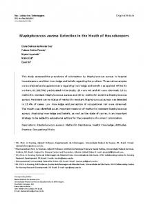
taphylococcus intermedius sensu lato is a veterinary pathogen occasionally reported from infected-animal (particularly dog) bites in humans (1, 2). Originally thought to be a single species, molecular characterization has resulted in its reclassification as Staphylococcus intermedius group (SIG), which includes S. intermedius, S. pseudintermedius, and S. delphini (3, 4). Non-bite-associated human isolates were reported in 1994 (5), but little has been documented since. Our aim was to improve the detection and identification of SIG from human samples using a simple phenotypic algorithm. This initial identification was confirmed by sequencing fragments of two housekeeping genes which were also used to determine diversity within SIG isolates. Initially, one of us (J. Lee) undertook a training session for laboratory staff to raise awareness of SIG and their colonial morphology and explain the algorithm. This was supplemented by posters in the laboratory and regular feedback. Following this, wound swabs (principally from skin and soft-tissue infections) were screened for SIG over the period from October 2010 to September 2013 at the Department of Clinical Microbiology, Royal Cornwall Hospital, Cornwall, United Kingdom. The specimens had been submitted for routine investigation from local hospitals and primary care physicians. Clinical details were taken from laboratory request forms accompanying the specimens from which SIG bacteria were isolated. No extra information was sought from requesting clinicians. Swabs were inoculated onto horse blood agar (Oxoid) and examined after overnight incubation in 5% CO2, our standard laboratory procedure. Bacterial colonies typical of SIG—white, entire, convex, glistening colonies 5 to 6 mm in diameter (see Fig. S1 in the supplemental material)—were selected for further characterization using a simple algorithm (Fig. 1). Briefly, suspect colonies were tested with a commercial latex reagent for clumping factor/protein A detection (Prolab StaphXtra) and for DNase production using commercial medium (Oxoid). Isolates that were latex negative and produced DNase were processed by the VITEK2 (bioMérieux) automated identification/susceptibility system using GP/AST cards. Strains identified by VITEK2 as “Staphylococcus intermedius” underwent PCR amplification of fragments of the housekeeping genes hsp60 and sodA using published primer pairs (6, 7). Sequences were trimmed, aligned, and analyzed with MEGA5 (8). Sensitivity testing was performed by using the VITEK2 system and the AST-P578 panel, interpreted by EUCAST guidelines. Sensitivity results were compared with those
March 2015 Volume 53 Number 3
FIG 1 Phenotypic algorithm for the detection of SIG isolates.
of 40 consecutive community-acquired Staphylococcus aureus isolates from our laboratory. No SIG isolates were recovered in our laboratory in the year preceding the study. SIG bacteria were isolated on 40 occasions from 39 patients during the study period. Sites of isolation included wounds (57%), ears (19%), diabetic ulcers (12%), dog bites (7%), and cutaneous ulcers (5%). Clinical details included
Received 11 September 2014 Returned for modification 5 October 2014 Accepted 22 November 2014 Accepted manuscript posted online 10 December 2014 Citation Lee J, Murray A, Bendall R, Gaze W, Zhang L, Vos M. 2015. Improved detection of Staphylococcus intermedius group in a routine diagnostic laboratory. J Clin Microbiol 53:961–963. doi:10.1128/JCM.02474-14. Editor: B. W. Fenwick Address correspondence to John Lee,
[email protected]. Supplemental material for this article may be found at http://dx.doi.org/10.1128 /JCM.02474-14. Copyright © 2015, American Society for Microbiology. All Rights Reserved. doi:10.1128/JCM.02474-14
Journal of Clinical Microbiology
jcm.asm.org
961
Lee et al.
FIG 2 Maximum likelihood tree based on the concatenated hsp60 and sodA gene fragments (1,000 bootstrap replicates). The collapsed branch contains all S. pseudintermedius strains in this study. Selected SIG strains isolated from different hosts were used for reference, and S. aureus was used as an outgroup.
impetigo, cellulitis, erythema, pain, purulent exudate, inflammation, and postsurgical infection. A history of animal contact was given in 4 of 40 requests; 2 of these were identified as bite wounds. The algorithm was fully operational between May 2012 and September 2013. During this 16-month period, SIG and S. aureus were isolated 32 and 10,777 times, respectively, from 39,380 specimens. Sequencing confirmed that all but one SIG isolate identified by the algorithm belonged to S. pseudintermedius. Analysis of sodA and hsp60 sequences revealed that the S. pseudintermedius strains, all isolated from local residents, were closely related (Fig. 2). One divergent strain, NW1, which came from a visitor to Cornwall, United Kingdom, could not be assigned to a recognized SIG species (Fig. 2). The sodA sequence of this strain was identical to a “staphylococcal species” described previously by Slettemeås and colleagues (9). The hsp60 sequence from this strain had 95% sequence similarity to an S. intermedius strain isolated from a pigeon (10) (see Fig. S2 in the supplemental material). The antibiotic susceptibilities of SIG isolates resembled those of local community-acquired S. aureus bacteria with universal susceptibility to oxacillin, erythromycin, fusidic acid, chloramphenicol, ciprofloxacin, clindamycin, gentamicin, linezolid, and rifampin. Tetracycline resistance was more common (15% versus 4.7%) in SIG isolates than in S. aureus. Polymyxin MICs were higher (range, 8 to 24 mg/liter) for S. pseudintermedius strains than for strain NW1 (4 mg/liter). Our results show that it is possible to identify SIG isolates using standard laboratory procedures. Recognition of the colonial morphology of SIG is essential for successful detection. Where this is followed by a simple algorithm, SIG can be identified reliably in clinical specimens. Before this method was introduced in our laboratory, SIG isolates were undetected and almost certainly underreported. Our study was limited in scope and did not attempt to identify colonies that were morphologically atypical, did not seek SIG among “latex-positive” strains, and did not perform genotyping on staphylococcal isolates which VITEK2 identified as nonSIG strains. It is therefore likely that some SIG strains were missed and that the true prevalence of these organisms is higher than our results suggest. In our population, S. pseudintermedius is readily detected, unlike S. intermedius sensu stricto. This confirms recent findings that “S. intermedius infections” described in earlier reports were likely to be S. pseudintermedius infections (11). We also isolated a SIG isolate belonging to a distinct, uncharacterized lineage that has
962
jcm.asm.org
been reported only once before (9). This isolate, NW1, had reduced susceptibility to polymyxin, which is a feature of S. intermedius sensu stricto and S. delphini (4). It may be a new species in the SIG group (9). The clinical details given by requesting clinicians were of little use in predicting SIG isolation; very few noted animal contact. This probably reflects the limited information supplied by clinicians rather than an absence of zoonotic risk. The pattern of samples yielding SIG resembled that for S. aureus, which supports a pathogenic role for S. pseudintermedius in humans. A number of SIG isolates came from diabetic patients, an association which warrants further study. Susceptibility to antistaphylococcal antibiotics was similar to that of community-acquired S. aureus isolates but with decreased susceptibility to tetracycline; this may reflect veterinary prescribing practice (12). In conclusion, SIG bacteria are underreported and often found where there is no stated history of animal contact to guide investigations. A simple modification of routine laboratory methods results in better detection of these organisms. Reporting of clinical isolates as “Staphylococcus intermedius group” when identified by phenotype is accurate and should be adopted by clinical laboratories. ACKNOWLEDGMENTS We thank staff at the Clinical Microbiology Unit of the Royal Cornwall Hospital for help with isolation and the Cornish Microbiological Society (Cowethas Corbryvyas Kernow).
REFERENCES 1. Kelesidis T, Tsiodras S. 2010. Staphylococcus intermedius is not only a zoonotic pathogen, but may also cause skin abscesses in humans after exposure to saliva. Int J Infect Dis 14:e838 – e841. http://dx.doi.org/10 .1016/j.ijid.2010.02.2249. 2. Talan DA, Goldstein E, Staatz D, Overturf GD. 1989. Staphylococcus intermedius: clinical presentation of a new human dog bite pathogen. Ann Emerg Med 18:410 – 413. http://dx.doi.org/10.1016/S0196-0644(89) 80582-7. 3. Devriese LA, Vancanneyt M, Baele M, Vaneechoutte M, De Graef E, Snauwaert C, Cleenwerck I, Dawyndt P, Swings J, Decostere A, Haesebrouck F. 2005. Staphylococcus pseudintermedius sp. nov., a coagulase-positive species from animals. Int J Syst Evol Microbiol 55:1569 – 1573. http://dx.doi.org/10.1099/ijs.0.63413-0. 4. Sasaki T, Kikuchi K, Tanaka Y, Takahashi N, Kamata S, Hiramatsu K. 2007. Reclassification of phenotypically identified Staphylococcus intermedius strains. J Clin Microbiol 45:2770 –2778. http://dx.doi.org/10.1128 /JCM.00360-07. 5. Lee J. 1994. Staphylococcus intermedius isolated from dog-bite wounds. J Infect 29:105. http://dx.doi.org/10.1016/S0163-4453(94)95276-0.
Journal of Clinical Microbiology
March 2015 Volume 53 Number 3
Staphylococcus intermedius Group
6. Sakamoto M, Suzuki N, Benno Y. 2010. hsp60 and 16S rRNA gene sequence relationships among species of the genus Bacteroides with the finding that Bacteroides suis and Bacteroides tectus are heterotypic synonyms of Bacteroides pyogenes. Int J Syst Evol Microbiol 60:2984 –2990. http://dx.doi.org/10.1099/ijs.0.021154-0. 7. Poyart C, Quesne G, Boumaila C, Trieu-Cuot P. 2001. Rapid and accurate species-level identification of coagulase-negative staphylococci by using the sodA gene as a target. J Clin Microbiol 39:4296 – 4301. http: //dx.doi.org/10.1128/JCM.39.12.4296-4301.2001. 8. Tamura K, Peterson D, Peterson N, Stecher G, Nei M, Kumar S. 2011. MEGA5: Molecular Evolutionary Genetics Analysis using maximum likelihood, evolutionary distance, and maximum parsimony methods. Mol Biol Evol 28:2731–2739. http://dx.doi.org/10.1093/molbev/msr121. 9. Slettemeås JS, Mikalsen J, Sunde M. 2010. Further diversity of the Staphy-
March 2015 Volume 53 Number 3
lococcus intermedius group and heterogeneity in the MboI restriction site used for Staphylococcus pseudintermedius species identification. J Vet Diagn Invest 22:756 –759. http://dx.doi.org/10.1177/104063871002200517. 10. Decristophoris P, Fasola A, Benagli C, Tonolla M, Petrini O. 2011. Identification of Staphylococcus intermedius group by MALDI-TOF MS. Syst Appl Microbiol 34:45–51. http://dx.doi.org/10.1016/j.syapm.2010.11 .004. 11. Weese JS. 2013. Phenotypic identification of Staphylococcus intermedius may be inaccurate. J Clin Microbiol 51:734. http://dx.doi.org/10.1128/JCM .02736-12. (Reply, 51:735, http://dx.doi.org/10.1128/JCM.02767-12.) 12. Blunt CA, Van Vuuren M, Picard J. 2013. Antimicrobial susceptibility profiles of Staphylococcus intermedius isolates from clinical cases of canine pyoderma in South Africa. J S Afr Vet Assoc 84:E1–E6. http://dx.doi .org/10.4102/jsava.v84i1.276.
Journal of Clinical Microbiology
jcm.asm.org
963