(IJCSE) International Journal on Computer Science and Engineering. Vol. ... degrees of membership. But the major ... Fuzzy C means is a method of clustering which allows one .... Electronics and Communication Engineering in the year. 2007 ...
P. Vasuda et. al. / (IJCSE) International Journal on Computer Science and Engineering Vol. 02, No. 05, 2010, 1713-1715
Improved Fuzzy C-Means Algorithm for MR Brain Image Segmentation P.Vasuda M.Tech Student, Dept. of ECE G.Narayanamma Institute of Technology and Science Hyderabad, India.
Abstract— Segmentation is an important aspect of medical image processing, where Clustering approach is widely used in biomedical applications particularly for brain tumor detection in abnormal Magnetic Resonance Images (MRI). Fuzzy clustering using Fuzzy C- Means (FCM) algorithm proved to be superior over the other clustering approaches in terms of segmentation efficiency. But the major drawback of the FCM algorithm is the huge computational time required for convergence. The effectiveness of the FCM algorithm in terms of computational rate is improved by modifying the cluster center and membership value updation criterion. In this paper, convergence rate is compared between the conventional FCM and the Improved FCM. Keywords- Medical Imaging, Segmentation, Fuzzy C-Means, Improved C-Means Algorithm
S.Satheesh Assistant Professor, Dept of ECE G.Narayanamma Institute of Technology and Science Hyderabad, India. besides being susceptible to human errors. Several automated techniques have been developed which removes the drawbacks of manual segmentation. Clustering is one of the widely used image segmentation techniques which classify patterns in such a way that samples of the same group are more similar to one another than samples belonging to different groups [3,9,10]. There has been considerable interest recently in the use of fuzzy clustering methods, which retain more information from the original image than hard clustering methods. Fuzzy C-means algorithm is widely preferred because of its additional flexibility which allows pixels to belong to multiple classes with varying degrees of membership. But the major operational complaint is that the FCM technique is time consuming.The drawback of the FCM is improved by the improved FCM algorithm.
I. INTRODUCTION
II. FUZZY C- MEANS CLUSTERING
The field known as biomedical analysis has evolved considerably over the last couple of decades. The widespread availability of suitable detectors has aided the rapid development of new technologies for the monitoring and diagnosis, as well as treatment, of patients. Over the last century technology has advanced from the discovery of xrays to a variety of imaging tools such as MRI, Computed Tomography (CT), Positron Emission Tomography (PET) and ultrasonography [2]. Three-dimensional (3-D) processing and visualization of medical images is a rapidly growing area of research and MRI has provided a means for imaging tissue at very high resolutions providing the desired information for use in fields like reparative surgery, radiotherapy treatment planning, stereotactic neurosurgery, and others [6,7].Furthermore, new techniques are helping to advance fundamental biomedical research. Image segmentation plays a major role in the field of biomedical applications. The segmentation technique is widely used by the radiologists to segment the input medical image into meaningful regions [1, 4, 5, 8]. The specific application of this technique is to detect the tumor region by segmenting the abnormal MR input image. The size of the tumor region can be tracked using these techniques which aid the radiologists in treatment planning. The primitive techniques are based on manual segmentation which is a time consuming process
Fuzzy C means is a method of clustering which allows one pixel to belong to one or more clusters[9,10]. The FCM algorithm attempts to partition a finite collection of pixels into a collection of “C” fuzzy clusters with respect to some given criteria [3]. Fuzzy C-means Algorithm is based on minimization of the following objective function
ISSN : 0975-3397
c
c
n
J (U , c1 , c2 ,..., cc ) J i uijm d ij2 i 1
i 1 j 1
uij is between 0 and 1; Ci is the centroids of cluster I;dij is the Euclidean distance between ith centroids and jth data point; mЄ [1,∞] is a weighting function. Fuzzy portioning of known data sample is carried out through an iterative optimization of the objective function n
u ij
1 d ij k 1 d kj c
2
m 1
c ij
j1 n
u imj x
j1
This iteration will stop when max ij uij where
k 1
j
u imj
uij
k
,
is a termination criterion between 0 and 1, whereas
1713
P. Vasuda et. al. / (IJCSE) International Journal on Computer Science and Engineering Vol. 02, No. 05, 2010, 1713-1715 IV. RESULTS AND DISCUSSIONS
k are the iteration steps. This procedure converges to a local minimum or a saddle point of Jm. The algorithm is composed of the following steps: 1. Initialize U=[uij] matrix, U(0) 2. At k-step: calculate the centers vectors C(k)=[cj] with U(k) N
cj
u i 1 N
m ij i
u
m ij
i 1
1.
x
(a)
Update U(k) , U(k+1)
uij
(b)
1 2
|| xi c j || m 1 k 1 || xi ck || c
2.
If || U(k+1) - U(k)||< then STOP; otherwise return to step 2. III. IMPROVED FUZZY C MEANS ALGORITHM
n
ci
u j 1 n
m ij
u j 1
yj uij m ij
Where dij y j ci y = Reduced Dataset
ISSN : 0975-3397
1 dij k 1 kj c
d
(d)
(e) 250
200
C P U tim e
The improved FCM algorithm is based on the concept of data compression where the dimensionality of the input is highly reduced. The data compression includes two steps: quantization and aggregation [3]. The quantization of the feature space is performed by masking the lower 'm' bits of the feature value. The quantized output will result in the common intensity values for more than one feature vector. In the process of aggregation, feature vectors which share common intensity values are grouped together. A representative feature vector is chosen from each group and they are given as input for the conventional FCM algorithm. Once the clustering is complete, the representative feature vector membership values are distributed identically to all members of the quantization level. Since the modified FCM algorithm uses a reduced dataset, the convergence rate is highly improved when compared with the conventional FCM. The improved FCM algorithm uses the same steps of conventional FCM except for the change in the cluster updation and membership value updation criterions. The modified criterions are showed below
(c)
150
100
50
2 m 1 0
1
2
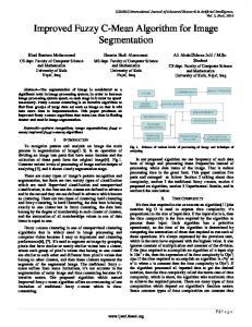
(f) Figure: a) Original Gray Level Brain Image b) Original Colormap Image c) Color Space Translated Image d) Truly Segmented Image using C-Means e) Truly segmented image using Modified C-Means f) FCM Vs Improved FCM CPU Time
1714
P. Vasuda et. al. / (IJCSE) International Journal on Computer Science and Engineering Vol. 02, No. 05, 2010, 1713-1715 In this work, a comparative analysis is performed on the techniques based on the performance measures.The clustered output reveals the same detection of the tumor for both techniques. Convergence rate is the time period required for the system to reach the stabilized condition.Significant improvement is achieved in Improved FCM over FCM in terms of convergence rate.Better results may achieved if more than two bits are changed in the bit mask. V. CONCLUSIONS The results show that FCM and MFCM method can successively segment a tumor provided the parameters are chosen properly. The visualization and detective valuations of the results of the segmentation show the success of the approaches.The modified FCM algorithm yields superior convergence rate.The tumor identification and the investigation are carried out for the potential use of MRI data for improving the tumor shape and 2D visualization of the surgical planning. VI. FUTURE SCOPE Future research in MRI segmentation should strive toward improving the accuracy, precision, and computation speed of the segmentation algorithms, while reducing the amount of manual interactions needed. This is particularly important as MR imaging is becoming a routine diagnostic procedure in clinical practice. It is also important that any practical segmentation algorithm should deal with 3D volume segmentation instead of 2D slice by slice segmentation, since MRI data is 3D in nature. Volume segmentation ensures continuity of the 3D boundaries of the segmented images whereas slice by slice segmentation does not guarantee continuation of the boundaries of the tissue regions between slices.
[7] Michael Bomans,Karl-Heinz Hohne.Ulf Tiede and Martin Riemer,“3-D Segmentation of MR Images of the Head for 3-D Display”,Page Number 177-183, IEEE Transactions on Medical Imaging,Vol.9,No. 2,July 1990. [8] Rafael C. Gonzalez, Richard E.Woods, “ Digital Image processing”, published by Pearson Education,Inc.,2002. [9] Sorin Istrail, “An Overview of Clustering Methods”, With Applications to Bioinformatics. [10] Steven Eschrich, Jingwei Ke, Lawrence O. Hall and Dmitry B. Goldgof Fast “Accurate Fuzzy Clustering through Data Reduction”, Page Number 1-18,November 13 IEEE 2002.
P.Vasuda received B.Tech degree from G.Narayanamma Institute of Technology and Science, Hyderabad, India in Electronics and Communication Engineering in the year 2007.Currently; she is pursuing her M.Tech degree from G.Narayanamma Institute of Technology and Science, Hyderabad, India in Digital Electronics and Communication Engineering. Her area of interest includes Image Processing and Digital Communications. S.Satheesh received the B.Tech degree in Electronics and Communication Engineering from Nagarjuna University, Guntur, India, in 2001, and the M.E degree in Electronics and Communication Engineering from Osmania University, Hyderabad, India, in 2005.He is currently pursuing the Ph.D degree in Electronics and Communication Engineering at Jawaharlal Nehru Technological University Hyderabad, AP, India. His research interests are in the area of Medical Image Processing and Signal Processing.
REFERENCES [1] Anton Bardera, Jaume Rigau, Imma Boada, Miquel Feixas, and Mateu Sbert, “Image Segmentation Using Information Bottleneck Method”,Page Number 1601-1612, IEEE Transactions on Image Processing, Vol. 18, No. 7, July 2009. [2] J.Jaya and K.Thanushkodi, “Segmentation of MR Brain tumor using Parallel ACO”,Page Number 150-153, (IJCNS) International Journal of Computer and Network Security,Vol. 2, No. 6, June 2010. [3] Jude hemanth.D, D.Selvathi and J.Anitha,“Effective Fuzzy Clustering Algorithm for Abnormal MR Brain Image Segmentation”,Page Number 609-614, International/Advance Computing Conference (IACC 2009),IEEE,2009. [4] Jian Wu, Feng Ye, Jian-Lin Ma, Xiao-Ping Sun, Jing Xu, Zhi-Ming, “ The Segmentation and Visualization of Human Organs Based on Adaptive Region Growing Method” ,Page Number-439-443, IEEE 8th International Conference on Computer and Information Technology Workshops978-0-7695-3242-4/08,IEEE,2008. [5] Marcus karnan and T.logeswari, “An Improved Implementation of Brain Tumor Detection using Soft Computing”,Page Number 6-10, (IJCNS) International Journal of Computer and Network Security,Vol. 2, No. 1, January 2010 [6] Marroquin J.L , B. C. Vemuri, S. Botello, F. Calderon, and A. FernandezBouzas, “ An Accurate and Efficient Bayesian Method for Automatic Segmentation of Brain MRI”,Page Number 934-945, IEEE Transactions on Medical Imaging, Vol. 21, No. 8, AUGUST 2002.
ISSN : 0975-3397
1715