Folia Microbiol. 50 (1), 31–39 (2005)
http://www.biomed.cas.cz/mbu/folia/
Improved Method of Detection and Molecular Typing of Borrelia burgdorferi sensu lato in Clinical Samples by Polymerase Chain Reaction without DNA Purification N. RUDENKOa,b, M. GOLOVCHENKOa,b, J. NĚMECa, J. VOLKAERTc, N. MALLÁTOVÁd, L. GRUBHOFFERa,b aFaculty of Biological Sciences, University of South Bohemia, 370 05 České Budějovice, Czechia bInstitute of Parasitology, Academy of Sciences of the Czech Republic, 370 05 České Budějovice, Czechia
fax +420 385 310 388 e-mail
[email protected] cDepartment Industriele Wetenschappen en Technologie, Karel de Grote-Hogeschool, Antwerpen, Belgium dDepartment of Clinical Parasitology and Mycology, Regional Hospital, České Budějovice, Czechia
Received 6 October 2003 Revised version 22 June 2004
ABSTRACT. A simple assay by polymerase chain reaction was used for the of detection of Borrelia burgdorferi, causative agent of Lyme borreliosis (LB). It involves no DNA purification and is based on the amplification of a specific region of ospA gene of B. burgdorferi, followed by direct detection of the PCR product with SYBR Green I by agarose gel electrophoresis. The method was used to analyze samples from patients with LB diagnosis, with presumable infection with the LB spirochete, those with unclear clinical symptoms and after the course of an antibiotic treatment. Spirochetal DNA was detected by PCR even in contaminated samples in which B. burgdorferi was overgrown by fungi and other bacteria. Spirochetal DNA was detected and borrelia species was identified in cerebrospinal fluid of two patients hospitalized with the diagnosis “fever of unknown origin”. Western blot and ELISA were negative in both cases. Total analysis of 94 samples from the hospital in České Budějovice (South Bohemia, Czechia) showed infection with B. burgdorferi sensu stricto in 11 % and B. garinii in 15 % of cases. The highest prevalence was found for B. afzelii (43 %). Co-infection was confirmed in 24 % of the analyzed symplex; 7 % of samples that were B. burgdorferi sensu lato positive gave no results in DNA amplification with B. burgdorferi sensu stricto-, B. garinii- and B. afzelii-specific primers. The proposed reliable, rapid, unexpensive and specific technique could form the basis of laboratory tests for routine detection and identification of Lyme-disease spirochete in different samples.
Lyme borreliosis (LB) is a disease of epidemiological, clinical as well as social importance, especially in Europe and in the USA (Burgdorfer et al. 2000, 2001; Weber 2001). It is caused by infection with the spirochete Borrelia burgdorferi sensu lato. During the past several years, various molecular approaches have been developed and successfully used for the identification and typing of LB-related spirochetes (Tenover et al. 1997; Schwarzová and Čižnár 2004a,b). Application of molecular typing methods for the classification of B. burgdorferi sensu lato provides the framework for the systematic approach to characterizing the differences in infectivity as well as in pathogeneicity between strains. Sensitive molecular typing techniques do not require large amounts of material or cultivation of spirochete and thus play an increasingly important role in elucidation of the pathogenic potential of different B. burgdorferi genotypes (Elias et al. 2002; Stoitsova et al. 2003). Ultimately, the development of suitable animal models for the investigation of the tissue tropisms of different B. burgdorferi sensu lato species provides more direct evidence for the correlation between Borrelia species and clinical symptoms of LB (Balmelli and Piffaretti 1995; Wang et al. 1999). Routine diagnosis is done by serological detection of Borrelia-specific antibodies but the sensitivity of this method during early infection is low and antibody concentrations decrease slowly only after therapy (Craft et al. 1984; Kmety 2000). Antibody titers are usually not detectable before 4–6 weeks post infection and may never become positive. In contrast, positive Lyme serology may just indicate a past borrelia infection without any proof of actual disease. Cultivation of B. burgdorferi from body fluids is slow and inefficient, indicating a need for new diagnostic methods. Since PCR has proved to be a sensitive and fast method for the diagnosis of microorganisms that are difficult to culture, this new technology has been used for the diagnosis of LB (Rosa and Schwan 1989; von Stedingk et al. 1995). Under experimental conditions, the sensitivity of PCR is extremely high and DNA of a single gene copy can be detected. Here we aimed to determine the diagnostic value of PCR with the samples that were
32
N. RUDENKO et al.
Vol. 50
not specially collected and suitable for PCR (human serum) but obtained in the hospital from patients with reactive serology for antibodies against causative agent of LB. Valuable information was also provided by comparison of the assay results. MATERIALS AND METHODS Borrelia strains (controls) and culturing. Reference strains of B. burgdorferi sensu lato were used as positive controls in the detection and identification of the pathogen in samples using PCR technique: Borrelia burgdorferi strain B31 (obtained from Dr. J.F. Anderson, Connecticut Agricultural Experiment Station, New Haven, USA, and successfully cultivated in our laboratory), Borrelia garinii strain NE 426 and Borrelia afzelii strain NE 632 (kindly supplied by Dr. L. Gern, University of Neuchatel, Switzerland; both strains were cultivated in our laboratory). Borrelia cultures were maintained in BSK-H complete medium at 34 °C. The growth was checked once a week by dark-field microscopy. The bacteria were subcultured when the titer achieved ≈108 spirochetes per mL. Observation of clinical manifestation. The group under investigation was rather heterogeneous (age, sex, symptoms) and included patients currently hospitalized, those under investigation during the post-hospitalization period, and outpatients with a light form of borreliosis or unclear diagnosis. The samples for PCR analysis of spirochete were taken from the patients with the diagnosis “Lyme borreliosis” and from those with nonspecific symptoms like headache, weakness, fatigue, inflammatory affliction of joints and muscle pain. Patients and clinical samples. The samples were collected at the Department of Infectious Diseases of the Regional Hospital in České Budějovice (Czechia). In total, 94 clinical samples (sera and cerebrospinal fluids; CSF), both serologically positive and negative for B. burgdorferi antibodies, were examined. The samples were collected in two periods – April–September 2001 and February–May 2002. All selected samples were examined by ELISA (VIR-ELISA anti-Borrelia IgM/IgG (Viro-Immun Labor, Germany) and Western Blot (ID Blot Borrelia IgM and ID Blot Borrelia IgG® (DPC, Germany) for Lyme immunoglobulin M (IgM)- and immunoglobulin G (IgG)-antibody in the laboratory of the Department of Clinical Parasitology of the Regional Hospital. The supplied samples and the results of serology were used as references. The clinical diagnosis based on the results of ELISA and Western blot was obtained from the same laboratory. Sample preparation with Chelex® 100 resin. The sample preparation involved no DNA purification. The most suitable way appeared to be a treatment of the serum or CSF by Chelex® 100 resin (BioRad Laboratories, USA). The resin binds all possible inhibitors of the PCR reaction, which may be present in the samples. A 5 % Chelex suspension in TE buffer (10 mmol/L Tris-HCl, pH 7.2; 1 mmol/L EDTA, pH 8.0) was used. The samples (sera, CSF) were diluted 1 : 5 (V/V) with phosphate-buffered saline (PBS; in mmol/L: NaCl 137, Na2HPO4 10, KCl 2.7; KH2PO4 2), vortexed vigorously and centrifuged (12 000 g, 45 min, 4 °C). The supernatant was discarded and 200 µL of 5 % Chelex 100 in TE buffer was added. The tube was vigorously vortexed to resuspend the pellet and incubated overnight at 56 °C. (The overnight incubation was used for the sake of convenience but it could be reduced to 15–30 min if necessary; cf. Vignoli et al. 1995; Schmidt et al. 1996.) The samples were then incubated for 15 min at 98–100 °C, vortexed for 10 s and chilled on ice. Supernatant was used directly in PCR. The samples were stable at 4 °C. Primer selection. Seventeen primers for PCR designated by different authors and directed on different regions of the borrelia genome for pathogen detection in ticks were checked (Marconi and Garon 1992; Sparagano et al. 1999). The best results were obtained with the following: primers based on B. burgdorferi sensu lato ospA gene sequences that have been designed for use in the PCR to type all (sensu lato – SL, primers set) or each (GI, GII and GIII primer sets) of the B. burgdorferi sensu lato genospecies involved in Lyme disease. The primers were synthesized by Generi-Biotech (Czechia). The sequences of oligonucleotide primers were designed and described by Demaerschaltck et al. (1995). Analysis of primer specificity. To analyze the specificity of the PCR primers, genomic DNA from B. burgdorferi, B. garinii and B. afzelii were purified using the Wizard genomic DNA purification kit (Promega, USA) and used as a template in “rapid multiplex PCR” in an amount of 30 ng per reaction. Control DNA amplification was run under the following conditions: 95 °C 5 min, 94 °C 1 s, 55 °C 1 s, 72 °C 10 s, 30 cycles. GI, GII and GIII primer sets were added to the reaction mixture simultaneously in a final concentration of 20 pmol each. Reaction conditions and composition of reagents used for the control and the clinical group of samples were identical. PCR products were resolved on 1 % agarose gel, SYBR Green I being used for DNA visualization. Detection of spirochete DNA in spiked positive control. To prove the effect of Chelex treatment of clinical samples on the increasing sensitivity of the PCR reaction, the following experiment was conducted:
2005
DETECTION OF B. burgdorferi IN CLINICAL SAMPLES BY PCR WITHOUT DNA PURIFICATION
33
purified genomic DNA of B. burgdorferi B31 (8, 80, and 800 pg, 8, 80, and 800 ng) was mixed with human serum (200 µL), as a simulation of infected human samples. All samples were divided into 3 parts, from which the first one was used directly for PCR without any treatment, the second part was once treated with Chelex and the last part was twice treated with the Chelex. DNA amplification. Twenty-five µL of supernatant from a Chelex-treated sample were used as a template in 30 cycles of amplification under the following conditions: 95 °C 30 s, 55 °C 30 s, 72 °C 1 min. The reaction buffer (concentration of components after dilution 1 : 10 in 50 µL of reaction volume; 50 mmol/L KCl, 10 mmol/L Tris-HCl pH 9.0, 1.5 mmol/L MgCl2, 0.1 % Triton X-100) was supplemented with Taq DNA polymerase (Promega). Due to the high A/T content in B. burgdorferi genome, the dNTPs mixture (all 10 mmol/L, in µL ATP 26,dTTP 26, dCTP 14, dGTP 14) was used in a final concentration of 100 µmol/L. Twenty pmol of each primer and 0.5 µL of Taq DNA polymerase (5 U/µL) were used. PCR products were resolved on 1 % agarose electrophoresis gel with subsequent visualization under UV light. Controls for PCR primer specificity. To confirm the Borrelia-specificity of the primers used and to eliminate the possible appearance of false-positive results of cross-reaction, 2 control samples with clinically significant titers were obtained from the Department of Infectious Diseases of the Regional Hospital in České Budějovice: (a) 4 serovars of leptospira (L. sejroe 1 : 3200, L. istrica 1 : 3200, L. sorex-jalna 1 : 800, L. canicola 1 : 800), and (b) syphilis-causing spirochete Treponema pallidum. Genomic DNAs from the samples were purified using the Wizard Genomic DNA purification kit (Promega, USA) and used as a template in PCR in an amount of 30 ng per reaction. Conditions of preparation, PCR reaction and analysis of positive control (B. burgdorferi), negative controls (leptospiras and T. pallidum) and clinical samples were identical. To increase the sensitivity of control PCR two subsequent reactions were conducted with B. burgdorferi DNA, leptospira and T. pallidum DNA. Primers 23SN1 and 23SC1 with annealing temperature 52 °C were used for amplification of the spacer region between 5S and 23S rRNA genes, producing a 380-bp PCR product. Amplified intergenic region was used as a template in subsequent nested PCR with 23SN2 and 5SCB primers with annealing temperature of 55 °C, providing a 225-bp PCR product. Reagent composition was the same as for Borrelia detection. Primers for spacer and nested PCR were designed by Rijpkema et al. (1995). Direct sequencing of PCR produkt. To ensure the specificity of the reaction, direct sequencing of the PCR product was done with 2 randomly picked SL1/SL2 positive samples. The spacer PCR of both samples was conducted under conditions described above. The products were cut out of the gel, purified using QIAquick Gel extraction kit (Qiagen, Germany) and sequenced in both directions with 23SN1 and 23SC1 primers. CEQ 2000 dye terminator cycle sequencing kit (Beckman Coulter) was used for direct sequencing of the PCR product. Sequences were analyzed on the ABI Prism 877 ITC automated DNA sequencer (Beckman) followed by analysis with DNAStar software (DNAStar Ltd., UK) and compared with GenBank Database searches used in the BLAST programs of the National Center for Biotechnology Information (Bethesda, USA). RESULTS AND DISCUSSION The diagnosis of LB has recently been made by clinical, epidemiological and serologic techniques, however, all these disease confirmations can be unreliable and problematic. Antigen detection by monoclonal antibodies did not exceed the experimental level, has a low yield and cannot be used as a routine test. Serologic tests are subject to false-positive results at a rate of 20–40 % (Agger and Case 1997). Silverstaining technique can identify other spirochetes, besides B. burgdorferi, further increasing the number of false-positive results. Enzyme-linked immunosorbent assay (ELISA), currently used for diagnosis, appeared to yield some false-positive and false-negative results; moreover, it does not distinguish different genospecies involved in LB. Western blotting improves the specificity of ELISA, but still it does not solve the problem of completely confirming only 30–50 % of suspected cases. Even direct detection of spirochetes through cultivation or dark-field microscopy cannot be considered reliable because of a low pathogen concentration in body fluids. We detected B. burgdorferi in patient samples by PCR. The specificity of PCR for B. burgdorferi as tested by using DNA from 4 serovars of leptospiras and T. pallidum as templates showed no cross-reactivity; also nonspecific PCR products were not detected (Fig. 1). Many different methods of sample treatment for obtaining better results in PCR reactions had already been described but all of them included a DNApurification step for its further use as a template in PCR. The large number of laboratories for daily use adopted the method for purification of nucleic acids described by Boom et al. (1990); however, in spite of all positive features, the purification leads to a loss of DNA. Since the nucleic acids are usually present in
34
N. RUDENKO et al.
Vol. 50
relatively impure and highly variable clinical specimens, such as serum or urine, and in rather low amounts any loss of DNA during purification can bring undesirable complications. The lysis of cells releases the nucleic acids which can be bound to silica particles (or membrane) and subsequently purified but the recovery of single-stranded, double-stranded, covalently closed or relaxed circular DNA is usually only ≈50 % (Boom at al. 1990). The recovery of pg amounts of DNA is ≈40 %. Addition of carrier DNA (500 ng) to the samples increased the yield to 40–53 %. However, it was already demonstrated that the presence of ≥500 ng of nontarget DNA can completely inhibit the PCR reactions (Alvarez et al. 1994; Cogswell et al. 1996). The yields for DNA fragments ≤4 kb were estimated to be ≈50 %. Lower recovery of intact fragments of >4 kb is presumably due to shearing. Significant shearing and approximately the same recovery were observed in the case of DNA ≈48 kb in size. Purification of genomic DNA very often resulted in the formation of very tight silica-beads–DNA complexes which could no longer be re-disolved. Approxi-
Fig. 1. Specificity analysis of PCR primers used for Borrelia burgdorferi detection in clinical samples. Genomic DNA from Treponema pallidum (1), 4 serovars of leptospira (L. sejroe 1 : 3200, L. istrica 1 : 3200, L. sorex-jalna 1 : 800, L. canicola 1 : 800) (2) and B. burgdorferi (3) were used as template in PCR; 23SN1/23SC1 primer set was used for amplification of the spacer (s) region between 5S and 23S rRNA genes, producing the 380-bp PCR product; amplified intergenic region was used as template in the following nested (n) PCR with 23SN2/5SCB primers, providing the 225-bp PCR product; 4 – negative control (no DNA in the reaction).
mately 8 % of DNA has not been bound to silica particles, a similar 8 % was lost during the washing step, and 30 % could not be eluted. Numerous data have been published comparing purification techniques based on different strategies of DNA extraction and differing in cost, labor and time consumption (Wilson 1997; McOrist et al. 2002; Angelini et al. 2003; Exner and Lewinski 2003; Gomez-Couso et al. 2004; Kuhad et al. 2004; Simmon et al. 2004). The classical 4-step process with the use of commercially available kits provided better results than the standard phenol–chloroform procedure. The spin-column technique is a 3-step process having an advantage in that it does not require any chemical reagents for extraction. Microwave irradiation or resin-binding is an alternative approach which by-passes the need to perform the DNA extraction step. The anion-binding resin method (GeneFizz) is a single-tube single-step process without any centrifugation; the lysis and the DNA extraction are accomplished directly in the amplification tube. An autoclave method preparing bacterial DNA for PCR eliminates, on the other hand, the use of detergents, organic solvents and mechanical cellular disruption approaches, thereby significantly reducing processing time and cost. All the above techniques represent relatively rapid, reliable, simple, cost-saving methods (starting from 0.12 EUR per test), reducing the time, the loss of material, and the risk of cross contamination (Angelini et al. 2003). All of them, however, have a similar limiting factor – the potential reduction of Taq-polymerase activity due to residual inhibitory substances which may interfere with enzyme reactions during PCR. Complete failure or false-negative amplification reactions have been reported (Wilson 1997); this makes the methods less suitable for treating multiple clinical samples in routine detection of pathogen DNA. Recently, the detection of pathogens in clinical samples (cerebrospinal fluid, synovial fluid, blood or urine) is conducted by a wide range of specifically developed techniques (Rosa and Schwan 1989; Marconi and Garon 1992; Demaerschaltck et al. 1995; Rijpkema et al. 1995; Vignoli et al. 1995; Figueroa et al. 1996; Priem et al. 1997; Brettschneider et al. 1998; Angelini et al. 2003; Exner et al. 2003; Lodin 2003; Táborský et al. 2003). Some of them use also the Chelex® 100 resin (Schmidt et al. 1996; Mercier et al. 1997; Moravcová et al. 2000). We developed a technique that requires no DNA purification but is based on the removal or, at least, lowering of PCR inhibiting substances in the samples and provides pathogen genetic material suitable for PCR. Chelex® 100 resin binds the remaining inhibitors of the PCR reaction and helps with the stabilization of dsDNA (Walsh et al. 1991). The general advantages of the Chelex technique are the simplicity, versatility
2005
DETECTION OF B. burgdorferi IN CLINICAL SAMPLES BY PCR WITHOUT DNA PURIFICATION
35
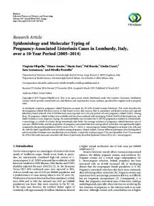
of application, sparing the genetic material in the sample, reproducibility, and the lowest costs among all the current techniques available on the market. We improved the use of Chelex to make it more effective even with samples that have been known as unsuitable for PCR pathogen detection in clinical (i.e. serum) or natural (hard ticks) samples without the need of purification of the genetic material (Danielova et al. 2004). We optimized the PCR cycle to minimize the duration of the reaction for amplification of short Borrelia-specific sequences. The amount of amplified pathogen DNA was then sufficient to be visualized on the agarose gel without further re-amplification of the PCR product. The use of SYBR Green I further increased the sensitivity of DNA detection on the gel. A total of 94 clinical samples, analyzed by PCR for the presence of B. burgdorferi DNA (14 % of clinical samples were CSF, 86 % blood serum) were treated with Chelex once. The round of PCR with SL1/SL2 primers (a 307-bp PCR product) detected the presence of spirochete from B. burgdorferi sensu lato group. Direct sequence of 2 PCR products (spacer) from randomly picked SL1/SL2 positive samples showed 96 % homology (identity 216–224 bp) to B. afzelii genotype D-3 5S-23S ribosome RNA intergenic spacer region (gi|20530690|AF497982.1). The genospecies were subsequently identified from SL-positive samples by the 2nd round of PCR with species-specific GI, GII, GIII primer sets for detection of B. burgdorferi sensu stricto (544 bp), B. garinii (345 bp) and B. afzelii (190 bp), respectively. The use of control sample (purified DNA + serum) in the reaction showed the increasing of efficiency of PCR after the Chelex treatment – no PCR products were obtained from untreated spiked samples regardless of the amount of spirochetal DNA used as a template, 10 ng of borrelia DNA was detected in samples treated once with the Chelex, and 10 pg of pathogen DNA was detected in the same serum samples treated twice with Chelex (Fig. 2); it demonstrated the presence of PCR inhibitors in human serum. The samples that were SL negative were eliminated by the repeated treatment. The comparison of PCR results with those obtained from the
Fig. 2. Effect of Chelex® 100 treatment on increasing of sensitivity of PCR reaction. Different amounts of purified genomic DNA of B. burgdorferi were mixed with human serum and used as template in PCR directly (1–6), after single (1´–6´) or double (1´´–6´´) Chelex treatment. Concentration of DNA in reaction: 1 – 1 pg , 2 – 10 pg, 3 – 100 pg, 4 – 1 ng , 5 – 10 ng, 6 – 100 ng; arrow – the SL1/SL2 specific 307-bp PCR product; M – DNA size marker (GeneRuler DNA Ladder Mix; MBI Fermentas).
serological tests conducted in the local hospital showed that the ELISA analysis detected 72 samples (68 %) to be positive while the repeated use of PCR revealed the presence of B. burgdorferi sensu lato DNA only in 46 cases (49 %). In 42 cases (45 %) PCR gave similar results with serology, 27 % of them were similar in ELISA, western blot and PCR. Positive results similar in PCR and both ELISA or Western-blot analysis were obtained in 53 %; the remaining 20 % were only PCR-positive. Comparison of ELISA and Westernblot analysis revealed similarity in 40 % of cases, the rest of the samples showed different results (Table I). Equivocal values of ELISA test were considered to be negative regardless of the previous positive result (negative results of serology do not mean the absence of the infection, as the titer of antibodies could be, in some cases, very low or even zero). On the other hand, scant data can be found in the literature describing the speed of clearance of Borrelia DNA from the organism; unfortunately, our results of Western-blot analysis were often confusing. Out of the group of samples with available Western-blot results (34 of 94 samples, i.e. 37 %) all revealed the presence of Borrelia DNA by PCR, 14 patients from this group were after or in the process of antibiotic treatment.
36
N. RUDENKO et al.
Vol. 50
Table I. Clinical symptoms and laboratory resultsa of a selected group of patients with Lyme borreliosis Patient
ELISA
Western blot
PCR
sampleb
sex, agec
tick bite
clinical symptoms
IgM
IgG
IgM
IgG
GI
GII
GIII
S
F28
yes
erythema migrans
–
+
+/–
+
–
–
+
S
M26
no
fatigue, weakness, myalgia
–
+/–
–
–
–
–
+
S
F63
yes
fatigue, weakness, headache
–
+
–
+
–
–
+
S
F17
yes
erythema migrans
+/–
–
+
–
–
–
+
S
M12
yes
fatigue, weakness
+/–
–
+
–
–
–
+
S
F48
yes
erythema migrans, fatigue, weakness, arthralgia
–
+
+/–
+/–
–
–
+
S
F45
no
fatigue, weakness, arthralgia
+
–
+/–
–
–
–
+
S
M39
no
fatigue, weakness, headache
–
+/–
–
+
–
+
–
CSF
M39
no
fatigue, weakness, headache
–
–
–
–
–
+
–
S
M53
no
fatigue, weakness, headache, arthralgia
–
+/–
–
+
+
+
–
S
M53
yes
no complains
–
+
+
+
–
+
+
S
F50
no
arthralgia
–
+
–
+
+
+
S
F67
yes
fatigue, weakness, paralysis
+
+
+/–
+/–
+
+
+
S
M53
no
–
+/–
+/–
+
+
+
+
CSF
M53
no
fatigue, weakness, myalgia headache, arthralgia fatigue, weakness, myalgia headache, arthralgia
–
–
–
–
+
+
+
a(+) – positive, (–) – negative, (+/–) – equivocal. cF – female, M – male (both age in years).
bS – serum, CSF – cerebrospinal fluid.
The next step of PCR with the species-specific primers showed the following distribution of different Borrelia species: the highest prevalence was found for B. afzelii – 20 cases (43 %). Co-infection, i.e. the presence of more than one Borrelia species, was found in 11 cases (24 %). B. garinii represented 7 cases (15 %), DNA of B. burgdorferi sensu stricto was detected in only 5 cases (11 %). Three samples (7 %) that exhibited the presence of B. burgdorferi sensu lato DNA showed no result in the subsequent reaction with GI, GII and GIII primers, supporting the presence of species different from B. burgdorferi sensu stricto, B. afzelii or B. garinii (Table II). The data are in good agreement with our results on the distribution of Borrelia species among infected ticks I. ricinus in South Bohemia in 2000 and subsequent years. In spite of the year and number of infected ticks, the prevalence of B. afzelii was always dominant, followed by the coinfection and infection with B. garinii. B. burgdorferi sensu stricto was present in a rather small number of ticks. Spirochete species other than the above 3 were detected by PCR with SL1/SL2 primers in 10 % of ticks (Danielova et al. 2004). Out of 46 samples positive for Borrelia DNA we selected (with ELISA, Western blot and PCR results) a relatively small group (15 cases) of patients in an early stage of the disease, who had not been treated with any antibiotics, but displayed the clinical manifestations of the disease (Table I). All 15 cases had the stated diagnosis of LB. Among this group only 47 % of patients (7 cases) recalled the tick bite. The cases, in which the presence of more than one Borrelia species was detected, covered 40 % (6 cases). B. afzelii was detected in 47 % (7 cases) of patients and B. garinii in 13 % (2 cases), which corresponds to the species distribution determined in the whole group of 94 clinical samples. Surprisingly, B. burgdorferi sensu stricto never appeared as a single infection in samples of this group, but was present in 5 of 6 cases of coinfection, always accompanying B. afzelii and/or B. garinii. The occurrence of specific strains of Borrelia in the positive analyzed samples mostly corresponded to the clinical symptoms of relevant patients. On the other hand, unambiguous assignment “syndrome genospecies” was in most cases not possible due to the presence of more than one strain of Borrelia in one sample and, often, a considerable nonspecificity of the observed symptoms. Long-lasting headache in 5 of 6 cases
2005
DETECTION OF B. burgdorferi IN CLINICAL SAMPLES BY PCR WITHOUT DNA PURIFICATION
37
was associated with B. garinii, fatigue and weakness – with B. garinii and B. afzelii, affliction of the motoric apparatus – with B. burgdorferi sensu stricto. In three cases with the occurrence of erythema migrans, PCR reaction revealed the presence of B. afzelii. Table II. Analysis of DNA amplification by PCR Resulta Cases
numberb %
SL1/SL2, (–)
SL1/SL2, (+)
GI, (+)
GII, (+)
GIII, (+)
Co-infection
Other
48 51
46 49
5 11
7 15
20 43
11 24
3 7
a(+) – positive, (–) – negative; SL1/SL2 – presence of B. burgdorferi sensu lato spirochete, GI – presence of B. burgdorferi sensu
stricto (Bb ss), GII – presence of B. garinii (Bg), GIII – presence of B. afzelii (Ba); co-infection – presence of >1 species of spirobTotal 94. chete in the sample; other – samples that were SL positive but contained spirochete other than Bb ss, Bg, Ba.
A total of 7 of 15 patients confirmed the tick bite, although erythema migrans was observed only in 3 cases; in these cases the development of erythema migrans was connected with the occurrence of B. afzelii. Only 1 of the group of patients under the investigation developed paralysis of the lower limbs; in this case the PCR analysis detected the presence of 3 strains of borrelia in 1 sample (B. afzelii, B. garinii and B. burgdorferi sensu stricto). All other patients exhibited only nonspecific symptoms. Long-lasting fatigue and weakness was observed in 11 cases, 6 of them being linked with the occurrence of B. garinii. Headaches were registered in 6 cases, out of which 5 proved to have B. garinii in the serum samples. Out of 8 patients with B. garinii, 6 cases were characterized by a mixed infection, with at least one of the other strains of the B. burgdorferi complex. Further symptoms were an inflammatory affliction of joints and muscle pain; out of 7 patients with such syndromes, 4 had co-infection and 3 revealed the presence of B. afzelii. In 2 samples of cerebrospinal fluid, negative according to ELISA and Western blot for B. burgdorferi antibodies, PCR revealed the presence of B. burgdorferi DNA. Both patients were at that moment hospitalized at the Infectious Department with the diagnosis of “fever of unknown origin”; both were complaining of tiredness and headache, indicating an affliction of the nervous system. Further detection of B. burgdorferi strain indicated the presence of B. garinii in 1 sample and B. garinii accompanied by B. afzelii and B. burgdorferi sensu stricto in the other, which correlates with the symptoms of neuroborreliosis. These cases confirm a higher sensitivity and specificity of PCR compared to ELISA and Western blot, and further demonstrate the general usefulness of PCR in diagnostics. The Center for Disease Control and Prevention (CDC; Atlanta, USA) recommends a two-test approach for good results, obtained by an enzyme-immunoassay testing, followed by a western immunoblotting of the ELISA-positive specimens (Center for Disease Control and Prevention 2000). False-positive results and cross reactions explain the number of negative samples in our study. Unfortunately, neither ELISA nor western blot alone can produce reliable results that might be successfully used as a basis for diagnostic determination. At the same time, the correct treatment and recovery of the sick patients relies on the accuracy of the diagnosis, i.e. on the accuracy and reliability of the techniques used. As a possible conclusion we could say that PCR can greatly contribute to the accurate diagnosis of LB and to the further understanding of human disorders associated with Borrelia infection. The use of PCR as a reliable, sensitive, specific, fast and inexpensive method alone or together with ELISA or Western blot could improve the diagnostics of LB, reducing drastically false and equivocal results. Further advantage could be the possibility to determine which or even how many species are present in cases of co-infection of Borrelia species, and the link between species and the clinical picture. The test can be used without some interference of possible inhibitors in human sera or cerebrospinal fluids after an easy treatment with Chelex® 100 resin. We thank the colleagues from the Department of Infectious Diseases of the Regional Hospital in České Budějovice (Czechia) for their collaboration and for generously providing the clinical samples. This work was supported by the grant 524/03/1326 of the Grant Agency of the Czech Republic and grant MSM 123 100 003 of the Ministry of Education, Youth and Sport of the Czech Republic. REFERENCES AGGER W.A., CASE K.L.: Clinical comparison of borreliacidal-antibody test with indirect immunofluorescence and enzyme-linked immunosorbent assays for diagnosis of Lyme disease. Mayo Clin.Proc. 72, 510–514 (1997). ALVAREZ A.J., BUTTNER M.P., TORANZOS G.A., DVORSKY E.A., TORO A., HAIKES T.B., MERTIKAS-PIFER L.E., STETZENBACH L.D.: Use of solid phase PCR for enhanced detection of airborne microorganisms. Appl.Environ.Microbiol. 60, 374–376 (1994).
38
N. RUDENKO et al.
Vol. 50
ANGELINI A., DI FEBBO C., BACCANTE G., DI NISIO M., DI ILIO C., CUCCURULLO F., PORRECA E.: Identification of three genetic risk factors for venous thrombosis using multiplex allele-specific PCR assay: comparison of conventional and new alternative methods for the preparation of DNA from clinical samples. J.Thromb.Thrombolysis 16, 189–193 (2003). BALMELLI T., PIFFARETTI J.C.: Association between different clinical manifestations of Lyme disease and different species of Borrelia burgdorferi sensu lato. Res.Microbiol. 146, 329–340 (1995). BOOM R., SOL C.J.A., SALIMANS M.M.M., JANSEN C.L., WERTHEIM-VAN DILLEN P.M.E., VAN DAE NOORDAA J.: Rapid and simple method for purification of nucleic acids. J.Clin.Microbiol. 28, 495–503 (1990). BRETTSCHNEIDER S., BRUCKBAUER H., KLUGBAUER N., HOFFMAN H.: Diagnostic value of PCR for detection of Borrelia burgdorferi in skin biopsy and urine samples from patients with skin borreliosis. J.Clin.Microbiol. 36, 2658–2665 (1998). BURGDORFER W.: Arthropod-borne spirochetosis: a historical perspective. Eur.J.Clin.Microbiol.Dis. 20, 1–5 (2001). BURGDORFER W., BARBOUR A.G., HAYES S.F., BENACH C.J.L., GRUNDWALD D.E., DAVIS S.J.P.: Lyme disease: a tick-born spirochetosis? Science 216, 1317–1319 (1982). Center for Disease Control and Prevention. Lyme disease – United States. J.Am.Med.Assoc. 287, 1259–1260 (2000). COGSWELL F.B., BANTAR C.E., HUGHES T.G., GU Y., PHILLIP M.T.: Host DNA can interfere with detection of Borrelia burgdorferi in skin biopsy specimens by PCR. J.Clin.Microbiol. 34, 980–982 (1996). CRAFT J.E., GRODZICKI R.L., STEERE A.C.: Antibody response in Lyme disease: evaluation of diagnostic tests. J.Infect.Dis. 149, 789– 795 (1984). DANIELOVÁ V., DANIEL M., RUDENKO N., GOLOVCHENKO M.: Prevalence of Borrelia burgdorferi sensu lato genospecies in host-seeking Ixodes ricinus ticks in selected South Bohemian locations (Czech Republic). Central Eur.J.Public Health 12, in press (2004). DEMAERSCHALTCK I., MESSAOUD A.B., KESEL M.D., HOYOIS B., LOBET Y., HOET P., BIGAIGNON G., BOLLEN A., GODFROID E.: Simultaneous presence of different Borrelia burgdorferi sensu lato genospecies in biological fluids of Lyme disease patients. J.Clin.Microbiol. 33, 602–608 (1995). ELIAS A.F., STEWART P.E., GRIMM D., CAIMANO M.J., EGGERS C.H., TILLY K., BONO L.J., AKINS D.R., RADOLF J.D., SCHWAN T.G., ROSA P.: Clonal polymorphism of Borrelia burgdorferi strain B31 MI: implications for mutagenesis in an infectious strain background. Infect.Immun. 70, 2139–2150 (2002). EXNER M.M., LEWINSKI M.A.: Isolation and detection of Borrelia burgdorferi DNA from cerebral spinal fluid, synovial fluid, blood, urine, and ticks using the Roche MagNA Pure system and real-time PCR. Diagn.Microbiol.Infect.Dis. 46, 235–240 (2003). FIGUEROA R., BRACERO L.A., AGUERO-ROSENFELD M., BENECK D., COLEMAN J., SCHWARTZ I.: Confirmation of Borrelia burgdorferi spirochetes by polymerase chain reaction in placentas of women with reactive serology for Lyme antibodies. Gynecol. Obstet.Invest. 41, 240–243 (1996). GOMEZ-COUSO H., FREIRE-SANTOS F., AMAR C.F.L., GRANT K.A., WILLIAMSON K., ARES-MAZÁS M.E., MCLAUGHLIN J.: Detection of Cryptosporidium and Giardia in molluscan shellfish by multiplex nested-PCR. Internat.J.Food Microbiol. 91, 279–288 (2004). KMETY E.: Dynamics of antibodies in Borrelia burgdorferi sensu lato infections. Bratisl.Lek.Listy 101, 105–107 (2000). KUHAD R.C., KAPOOR R.K., LAL R.: Improving the yield and quality of DNA isolated from white-rot fungi. Folia Microbiol. 49, 112– 116 (2004). LODIN Z.: Inflammatory and autoimmune diseases of the nervous system; possibilities of laboratory diagnostic methods in cerebrospinal fluid. Folia Microbiol. 48, 839–848 (2003). MARCONI R.T., GARON C.F.: Development of polymerase chain reaction primer sets for diagnosis of Lyme disease and for speciesspecific identification of Lyme disease isolates by 16S rRNA signature nucleotide analysis. J.Clin.Microbiol. 30, 2830–2834 (1992). MCORIST A.L., JACKSON M., BIRD A.R.: A comparison of five methods for extraction of bacterial DNA from human fecal samples. J.Microbiol.Meth. 50, 131–139 (2002). MERCIER G., BURCKEL A., LUCCOTE G.: Detection of Borrelia burgdorferi DNA by polymerase chain reaction in urine specimens of patients with erythema migrans lesions. Mol.Cell.Probes 11, 89–94 (1997). MORAVCOVÁ L., LASIKOVÁ Š., PICHA D., ŽĎÁRSKÝ E.: Determination of specific DNA of Borrelia burgdorferi in urine of patients with Lyme borreliosis. (In Czech) Klin.Mikrobiol.Inf.Lék. 6, 221–224 (2000). PRIEM S., RITTIG M.G., KAMRADT T., BURMESTER G.R., KRAUSE A.: An optimized PCR leads to rapid and highly sensitive detection of Borrelia burgdorferi in patients with Lyme borreliosis. J.Clin.Microbiol. 35, 685–690 (1997). RIJPKEMA S.G.T., MOLKENBOER M.J.C.H., SCHOULS L.M., JONGEJAN F., SCHELLEKENS J.F.P.: Simultaneous detection and genotyping of three genomic groups of Borrelia burgdorferi sensu lato in Dutch Ixodes ricinus ticks by characterization of the amplified intergenic spacer region between 5S and 23S rRNA genes. J.Clin.Microbiol. 33, 3091–3095 (1995). ROSA P.A., SCHWAN T.G.: A specific and sensitive assay for the Lyme disease spirochete Borrelia burgdorferi using the polymerase chain reaction. J.Infect.Dis. 160, 1018–1029 (1989). SCHMIDT B., MUELLEGGER R.R., STOCKENHUBER C., SOYER H.P., HOEDL S., LUGER A., KERL H.: Detection of Borrelia burgdorferispecific DNA in urine specimens from patients with erythema migrans before and after antibiotic therapy. J.Clin.Microbiol. 34, 1359–1363 (1996). SCHWARZOVÁ K., ČIŽNÁR I.: Combined infection of Ixodes ricinus with three Borrelia burgdorferi sensu lato genotypes. Folia Microbiol. 49, 297–300 (2004a). SCHWARZOVÁ K., ČIŽNÁR I.: Immunochemical analysis of lipopolysaccharide-like component extracted from Borrelia burgdorferi sensu lato. Folia Microbiol. 49, 625–629 (2004b). SIMMON K.E., STEADMAN D.D., DURKIN S., BALDWIN A., JEFFREY W.H., SHERIDAN P., HORTON R., SHIELDS M.S.: Autoclave method for rapid preparation of bacterial PCR-template DNA. J.Microbiol.Meth. 56, 143–149 (2004). SPARAGANO O.A, ALLSOPP M.T., MANK R.A., RIJPKEMA S.G., FIGUEROA J.V., JONGEJAN F.: Molecular detection of pathogen DNA in ticks (Acari: Ixodidae): a review. Exp.Appl.Acarol. 23, 929–960 (1999). VON STEDINGK L.V., OLSSON I., HANSON H.S., ASBRINK E., HOVMARK A.: Polymerase chain reaction for detection of Borrelia burgdorferi DNA in skin lesions of early and late Lyme borreliosis. Eur.J.Clin.Microbiol.Infect.Dis. 14, 1–5 (1995).
2005
DETECTION OF B. burgdorferi IN CLINICAL SAMPLES BY PCR WITHOUT DNA PURIFICATION
39
STOITSOVA S.R., GRUBHOFFER L., NEBESÁŘOVÁ J.: Exposed and hidden lectin-binding epitopes at the surface of Borrelia burgdorferi. Folia Microbiol. 48, 654–658 (2003). TÁBORSKÝ L., ADAM P., SOBEK O., DOSTÁL M., DVOŘÁKOVÁ J., DUBSKÁ L.: Levels of apolipoprotein A-II in cerebrospinal fluid in patients with neuroborreliosis are associated with lipophagocytosis. Folia Microbiol. 48, 849–856 (2003). TENOVER F.C., ARBEIT R.D., GOERING R.V., MICKELSEN P.A., MURRAY B.E., PERSING D.H., SWAMINATHAN B.: Interpreting chromosomal DNA restriction patterns produced by pulsed-field gel electrophoresis: criteria for bacterial strain typing. J.Clin. Microbiol. 33, 2233–2239 (1997). VIGNOLI C., DE LAMBALLERIE X., ZANDOTTI C., TAMALET C., DE MICCO P.: Advantage of a rapid extraction of HIV1 DNA suitable for polymerase chain reaction. Res.Virol. 146, 159–162 (1995). WALSH P.S., METZGER D.A., HIGUCHI R.: Chelex 100 as a medium for simple extraction of DNA for PCR-based typing from forensic material. Biotechniques 10, 506–513 (1991). WANG G., VAN DAM A.P., SHWARTZ I., DANKERT J.: Molecular typing of Borrelia burdorferi sensu lato: taxonomic, epidemiological and clinical implications. Clin.Microbiol.Rev. 12, 633–653 (1999). WEBER K.: Aspects of Lyme borreliosis in Europe. Eur.J.Clin.Microbiol.Dis. 20, 6–13 (2001). WILSON I.G.: Inhibition and facilitation of nucleic acid amplification. Appl.Env.Microbiol. 63, 3741–3751 (1997).