The alternate fast tuning and detuning of fiducial markers during sampling is analyzed regarding an improvement of the tracking of interventional devices.
Improved Tracking of Resonant Circuits based on Rapid Optical Switching H. Eggers
', S. Weiss ', P. Boemert I , P. Boesiger *
' Philips Research, Sector Technical Systems, Hamburg, Germany 'Institute of Biomedical Engineering, University of Zurich and Swiss Federal Institute of Technology Zurich, Zurich, Switzerland
The alternate fast tuning and detuning of fiducial markers during sampling is analyzed regarding a n improvement of the tracking of interventional devices. Sampling with twice the usual frequency and simultaneously switching the local signal enhancement of resonant circuits is shown to enable a robust detection of the markers' positions in difference images without prolonging measurement times. Results of initial experiments with a spiral acquisition underline the potential of this approach. Introduction Small resonant circuits as fiducial markers have lately attracted considerable attention in interventional magnetic resonance imaging [1,2]. In combination with optical means to rapidly detune them, they allow a robust localization irrespective of the background signal and facilitate the circumvention of safety hazards associated with long conducting wires. Previous work showed that an incremental difference reconstruction enables a fast image-based tracking of such fiducial markers [3]. Infomation on their position is derived from the difference of two images, which are reconstructed from the sets of data acquired while the resonant circuit was tuned and detuned respectively. The optical switching of the signal enhancement bas so far been performed in between two read-out intervals only. In order to preserve the temporal resolution of the imaging sequence, both sets of data could not be sampled fully. The resulting subsampling artifacts caused the detection of the resonant circuit's position to be occasionally unreliable, particularly when spiral instead of radial acquisitions were used due to inferior properties of their point-spread functions in case of subsampling. This work takes up an approach originally proposed for field-inhomogeneity catheters [41. Instead of modulating the currents flowing through the wires of such catheters with exactly half the sampling frequency, it proposes to switch resonant circuits at this rate during read-out intervals. A doubling of the sampling and the switching frequency then allows to avoid subsampling artifacts in both sets of data without prolonging measurement times. Results of initial experiments based on this approach demonstrate its potential to render the tracking of tuned fiducial markers more robust. Materials a n d Methods For the data acquisition, a fast gradient echo sequence, which samples kspace on interleaved spiral trajectories, was employed. By suitably increasing the sampling frequency and accordingly adjusting the filter settings, a net oversampling by a factor of two along each interleaf was achieved. The resonant circuit was either tuned or detuned for the excitations and then switched before the acquisition of every sample. The two interleaved data sets were separated and reconstructed individually, taking into account the actual location of the respective samples in k-space. The difference of the resulting two images finally formed the basis for determining the resonant circuit's position. To accurately switch the signal enhancement of the fiducial marker, the acquisition software was extended to trigger a function generator immediately before the activation of the two readout gradients. The function generator drove the laser diode at one end of the optical fiber with a rectangular waveform of defined frequency throughout the subsequent sampling. Experiments were performed on a 1.5 T Gyroscan ACS-NT system (Philips Medical Systems, Best, The Netherlands) using an optically detunable resonant circuit 121, that was integrated into the tip of a clinical catheter and immersed in a tracking phantom.
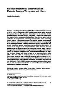
Imaging was successfully performed with the described set-up at different sampling and switching rates, demonstrating the feasibility of the
© Proc. Intl. Soc. Mag. Reson. Med. 10 (2002)
proposed approach. In Fig. I, results obtained at a sampling frequency of 33.6 kHz and a switching frequency of 16.8 kHz, which means that the local signal enhancement of the resonant circuit was toggled for the acquisition of every sample, are shown. The measurement of a 256' matrix was segmented into 64 interleaves in this case, each with a read-out interval of 60 ms. All images were deblurred using a conjugate phase reconstruction to correct off-resonance artifacts. Experiments at considerably higher frequencies no longer resulted in a reliably detectable signal in the difference images, since the resonant circuit's speed of tuning and detuning was insufficient. Compared to previously investigated approaches [3], the robustness of the localization depended less on the excitation angle
Fig. 1: Images of a resonant circuit, at the position of which the white arrow points, immersed in a tracking phantom. Shown are the results of a separate reconstruction of the two data sets and their difference.
Discussion a n d Conclusion As pointed out in [ 5 ] , the local signal enhancement of tuned fiducial markers results from a seeming amplification of both the irradiated signal during excitation as well as the emanated signal during relaxation. The approach proposed in this work, however, selectively exploits only the enhancement of the emanated signal to generate a detectable variation in images at the location of a resonant circuit. Although this difference in signal is consequently reduced in magnitude, the advantage of almost entirely avoiding the misinterpretation of artifacts far outweighs this drawback. In addition, finding an appropriate compromise between the requirements of the imaging and the tracking regarding the excitation angle is substantially simplified. The approach may directly be applied to other sampling schemes as well. Moreover, it lends itself to projection- instead of image-based tracking, as described in [2, 61, in which case it allows to halve the number of additional projection measurements inserted into the imaging sequence. The resonant circuit employed currently switches too slowly to be used in standard real-time imaging. The required acceleration by approximately one order of magnitude therefore necessitates an optimization of its speed of tuning and detuning. Further raising the sampling frequency while keeping the switching frequency constant would additionally allow to image transient states of resonant circuits in the same measurement time. In conclusion, the proposed approach allows a robust tracking of resonant circuits without prolonging measurement times or reducing the signd-tonoise ratio of the imaging. References I. Wong, E. Y., et al., JMugn Reson Imaging, 12, 632-638, 2000 2. Weiss, S., et al., In: Proc ISMRM, 1, 544, 2001. 3. Eggers, H., et al., In: Proc ISMRM, 3, 2162, 2001. 4. Glowinski, A,, et al., In: Proc ISMRM, 1,408,2000. 5. Burl, M., et al., Mugn Reson Med, 36,491-493, 1996. 6. Dumoulin, C.L., et.al., Mugn Reson Med, 29, 41 1-415, 1993.