But in this paper the fusion criterion is to minimize different error ... These methods are used according the .... conventional DWT using Debauchees 8 filters and.
ISSN 2250-0987 UNIASCIT, Vol 3 (2), 2013, 379-382
IMPROVING MEDICAL IMAGES USING WAVELET IMAGE FUSION TECHNIQUES Vivek Shrivastava Basant Dhakad Vikash Shrivastava Department of Informatin Technology ,Head of Department ,ITM College Bhilwara Rajsthan India * Department of Informatin Technology ,Student ,ITM College Bhilwara Rajsthan India Senior System Architect, I-Gate Global Solution Private Limited Noida, New Delhi,India *
#
Abstract: Today, image fusion as one kind of information integrated technology has played an important role in many fields. Most of previous image fusion methods aim at obtaining as many as information from the different images. But in this paper the fusion criterion is to minimize different error between the fused image and the input images. This paper presents the use of image fusion of medical images. Multi-sensor image fusion is the process of combining information from two or more images into a single image. The resulting image contains all the information of the input images. It is containing more information as compared to individual input images. we know that the image with higher contrast contain more edge-like features. In term of this view, we proposed a medical image fusion scheme based on an improved wavelet coefficient contrast. Image fusion techniques is very useful in medical application because in the medical application, CT (Computed Tomography) and MRI (Magnetic Resonance Imaging) images needs to be fused with high frequency for diagnosis purpose. In this paper, I have described methods for medical image fusion – simple fusion rule based on wavelet, contrast based image fusion, discrete packet based and maximum pixel replacement based on wavelet transform. These methods are used according the requirement of fused image and application.
Keyword-- Medical Image fusion, Wavelet transform, wavelet coefficient contrast, Medical diagnosis, edge preservation. 1. INTRODUCTION The main purpose of image fusion technique is to achieve useful corresponding information from multi images into a single image as much as possible. A number of algorithm are available for the image fusion. Most of the image fusion algorithm aims at obtaining as much as possible information in the fused image, with detecting the error of input images. In case of medical fusion, images contains more contrast and edge approximating information, which needs to preserve in fused image. The image fusion technology in such aspects as medicine, remote sensing, computer vision, weather forecast has also been widely applied. In the medical image fusion, images are taken by CT and MRI. CT (Computed tomography) is a specialized X-ray imaging technidsque. It may be performed plain or after the injection of a “Contrast
Agent”. CT creates the image by using an array of individual small X-Ray sensors and a computer. By spinning the X-Ray source and the sensor/detectors around the patient, data is collected from multiple angles. A computer then processes this information to create an image on the video screen. These images are called "sections" or "cuts" because they appear to resemble cross-sections of the body. This technique eliminates the problem of conventional X-rays, where all the shadows overlap. Magnetic Resonance Imaging (MRI), is a diagnostic technique that uses nuclear magnetic resonance to produce cross-sectional images of organs and other internal body structures. The patient lies inside a large, hollow cylinder containing a strong electromagnet, which causes the nuclei of certain atoms in the body (especially those of hydrogen) to align magnetically. The patient is then subjected to radio waves, which cause the aligned nuclei to flip. When the radio waves are withdrawn, the nuclei return to their original positions, emitting radio waves that are then detected by a receiver and translated into a two-dimensional picture by computer.[2][4] CT image offers high resolution in the visualization of bone structures, but its soft tissue contrast is poor. Conversely, MR imaging offers high contrast for the visualization of the soft tissue morphology, but it produces weak signal intensity in bone. Due to their complementary information, it is desired that both X-ray computed tomography (CT) and magnetic resonance imaging (MRI) are integrated. To spatially relate the two datasets, image fusion techniques are employed. Fused images are valuable in clinical diagnosis, in planning surgery, and in the image guided surgical interventions. Non-contrast computed tomography (CT) is the standard brain imaging study for the initial evaluation of patients with acute stroke symptoms. Magnetic resonance imaging (MRI) has been proposed as an alternative to CT in the emergency stroke setting. However, the accuracy of MRI relative to CT for the detection of hyper-acute intra-cerebral hemorrhage has not been demonstrated. The present work aims at selecting the optimal method for fusion of MRI and CT images. The fused image gives higher detection accuracy than either MRI or CT images [11].
379
ISSN 2250-0987 UNIASCIT, Vol 3 (2), 2013, 379-382
For medical diagnosis, CT provides the better information on denser tissue with less distortion, While MRI offers better information on soft tissue with more distortion. The goal of image fusion is to obtain useful complementary information from multimodality images. The simplest way to obtain a fused image from two or more medical images is to average them. Other application can also use this image fusion algorithm for preserving the contrast. The fused image can have complementary spatial and spectral resolution characteristics. But the standard image fusion techniques can distort the spectral information of the multispectral data, while merging.
but a feature at a time. A schematic diagram of the Laplacian Pyramid fusion methods is show in figure.
2.1 Multimodality medical image fusion Medical images are used by doctors to get information about the human body and to detect any disease. There are many tools available to captures the images of the human body (eg. X-rays, CT ,MRI etc). X-Rays is used to detect broken bones in the human body. CT (Computed tomography) is used to provide structure of bones in patient body. These techniques provide images, Which is used to provide us a detail information regarding body parts. MRI (magnetic resonance imaging) is used to get information about soft tissues, organs in human body. MRI technique does not use X-rays and radiations that harm to our body. Ultrasound is a safe and less harmful image processing, in which high frequency sound waves are used. Medical fusion is an very important field of research, diagnosis and treatment. Multimodality medical image fusion is a technique for the merge all the information of the images that are captured by different modalities in a single image. That techniques is very useful for the doctors[1][4]. 2.
Image Fusion Techniques
Image fusion methods can be classified into two groups – Spatial domain fusion [8] and Transform domain fusion. The image fusion methods such as averaging, Brovey method, principal component analysis (PCA) and HIS based methods fall under spatial domain approaches. The disadvantage of spatial domain fusion method is the high pass filtering based technique. Spatial distortion can be very well handled by frequency domain approaches on image fusion. The discrete wavelet transforms has become a very useful tool for fusion. Some other fusion methods are Lapacian pyramid based, Curvelet transform based etc. 3.1 Laplacian pyramid based Laplacian pyramid (LP) is derived from the gaussian pyramid (GP), which is a multiscale representation obtained through a recursive reduction. The Laplacian pyramid implements a “pattern selective” approach to image fusion, so that the composite image is constructed not a pixel at a time,
The Laplacian Pyramid representation was introduced by Burt and Adelson [2]. It is easy to implement and computationally efficient. The Laplacian Pyramid transform is specifically designed for capturing image details over multiple scales. Each band-pass level is sampled at precisely its Nyquist frequency making it less sensitive to noise. All these properties make the Laplacian pyramid transform a well-suited representation for the fusion task. Laplacian Pyramid implements a pattern selective approach to image fusion, so that the
380
ISSN 2250-0987 UNIASCIT, Vol 3 (2), 2013, 379-382 composite image is constructed not a pixel at a time, but a feature at a time. Given the image sequence {I1 , I 2, ....., I n } , the entire fusion algorithm is outlined by the following steps: 1. Generate a Laplacian pyramid images
Li for each of the
Ii .
2. Merge the pyramids
Li by taking the maximum at
each pixel of the pyramid, obtaining the Laplacian pyramid representation L of the fusion result. 3. Reconstruct the fusion result I from its Laplacian pyramid representation. 4. Normalize the dynamic range of the result so that it resides within the range of [0,1]. 3.2 Wavelet Transform The most common form of transform image fusion is wavelet fusion. The wavelet transform decomposes the image into low-high, high-low, high-high spatial frequency bands at different scales and the low-low band at the coarsest scale. The low-low band contain the average image information whereas the other bands contain directional information due to spatial orientation [4]. Wavelet transform fusion is more formally defined by considering the wavelet transforms ω of the two input images I1 ( χ, у ) and I2 ( χ, у ) together with fusion rule Φ [12]. The inverse wavelet transform ω-1 is computed and the fused image I ( χ, у ) is reconstructed: I ( χ, у ) = ω-1 (Φ(ω(I1 ( χ, у )), ω(I2 ( χ, у )))) An alternative to fusion using pyramid based multiresolution representations is fusion in the wavelet transform domain. The multiresolution wavelet representation is argued to be superior in several respects to that obtained with pyramidal methods [3]: 1. Spatial orientation is introduced in the wavelet decomposition process, unlike pyramidal representations which do not include directional information. 2. The wavelet transform can be tailored to extract highly salient textures/edges while suppressing noise through the choice of the mother wavelet and high- and low-pass filters. 3. The different scales in the wavelet decomposition have a higher degree of independence than those in the pyramidal representations, which are correlated with each other. The wavelet transform decomposes the image into lowhigh, high-low, and high-high spatial frequency bands at different scales and the low-low band at the coarsest scale. The L-L band contains the average image information whereas the other bands contain directional information due to spatial orientation. Higher absolute values of wavelet coefficients in the high bands correspond to salient features such as edges or lines. Since larger absolute transform coefficients correspond to sharper brightness changes, a good integration rule is to select, at every point in the transform domain, the coefficients whose absolute values are higher [4], [5].
3.3 Discrete Wavelet Transform The maximum pixel replacement fusion rule or absolute maximum (AM) detail coefficient fusion rule selects the detail coefficient in each sub – band with greatest magnitude. The input source images is decomposed using discrete wavelet transform. This is because as said approximation coefficients are interpreted as the mean intensity value of the source images with all salient features encapsulated by the detail coefficient sub bands at their various scales[5]. Wavelet transform has good spatial and frequency localization characteristics which shows itself mainly at three aspects: frequency feature compression (feature compression in the frequency domain), space compression feature and structure similarity of wavelet coefficients among different scales. Frequency compression feature means that the energy of original image concentrates at low frequency sub band. Space compression feature indicates that the energy of high frequency sub band mainly distributes at the
381
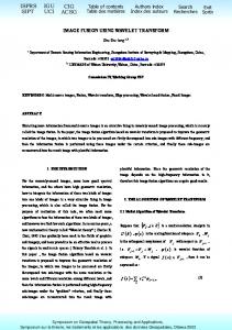
ISSN 2250-0987 UNIASCIT, Vol 3 (2), 2013, 379-382 corresponding positions of the edges of original image. Structure similarity of wavelet coefficients refers to the general consistence of the distributions of wavelet coefficients in high frequency sib bands of the same orientation. 3. Experiment Result Medical image fusion performance can be evaluated in term of doctor’s perception and quantitative criterions. In this section, by fusing CT and MRI images we try to compare the performances of proposed fusion scheme in the previous section to Laplacian pyramid gradient pyramid(GP), the original contrast pyramid (CP), the conventional DWT using Debauchees 8 filters and wavelet coefficient contrast pyramid for medical diagnosis, doctors usually observe the image manually and fuse them in mind. Figure shows the one fusion example[1][2].
. 4. CONCLUSION Image fusion techniques were utilized to facilitate detection of acute intra-cerebral hemorrhage by fusing MRI and CT images at the same level. Results have shown that Wavelet method is more suitable than other techniques for fusing medical images. In addition, the fused CT-MRI image contains more information than the source images. As a conclusion, fusion of CT and MRI images leads to higher diagnosis accuracy. Thus, image fusion can be considered as an assistant diagnostic tool especially when CT and MRI scans give different results[1]. In this paper we have used the image fusion scheme based on a new wavelet coefficient contrast for the medical domain. The visual experiments and the quantitative analysis demonstrate that the proposed medical image fusion method can preserve the important structure information such as edge of organs, outlines of tumors compared to other image fusion methods. This characteristic makes the proposed methods an efficient application in medical diagnosis. Further practical application will be investigated in our future work with more medical images. 5. REFERENCE [1] I. P. I. Pappas, M. Styner, P. Malik, L. Remonda, and M. Caversaccio, “Automatic Method to Assess Local CT-MR Imaging Registration Accuracy on Images of The Head,” AJNR: 26(1), pp. 137144, January 2005 [2] P. J. Burt., “The Pyramid as Structure for Efficient Computation,” In Multiresolution Image Processing and Analysis, pp. 6-35. Springer Verlag, 1984.
CT image
[3] J. J. Lewis, R. J. O’Callaghan, S. G. Nikolov, D. R. Bull, C. N. Canagarajah, “Region-Based Image Fusion Using Complex Wavelets,” report, The Centre for Communications Research, University of Bristol, 2004 [4] M. I. Smith, J. P. Heather, “Review of Image Fusion Technology in 2005,” Proceedings of the SPIE, Volume 5782, pp. 29-45, 2005 [5] Z. S. Long, “Image Fusion Using Wavelet Transform,” Symposium on Geospatial Theory, Processing and Applications, Ottawa 2002 [6] V. Petrović, C. Xydeas, “Computationally Efficient Pixel-level Image Fusion,” report, Manchester Avionics Research Center (MARC), University of Manchester, 2000, available at: http://imaging.utk.edu/~priya/GAweb/petrovic.doc
MRI image
[7] Y. Wang and B. Lohmann, “Multisensor Image Fusion: Concept, Method, and Applications,” Univ. Bremen, Bremen, Germany, Tech. Rep., 2000. [8] L. R. Liang, C. G. Looney, “Image Fusion with Spatial Frequency,” Computer Science Department, University of Nevada, USA, 2002 [9] F. Laliberté, L. Gagnon, and Y. Sheng, “Registration and Fusion of Retinal Images – An Evaluation Study,” IEEE Transactions on Medical Imaging, Vol. 22, No. 5, MAY 2003. [10] Z. Wang, D. Ziou, C. Armenakis, D. Li, and Q. Li, “A Comparative Analysis of Image Fusion Methods,” IEEE Transactions on Geoscience and Remote Sensing, Vol. 43, NO. 6, JUNE 2005 [11] C. S. Kidwell et al., “Comparison of MRI and CT for Detection of Acute Intra-cerebral Hemorrhage,” JAMA, Vol. 292, No. 15, pp. 18231830, 2004.
Fusion image
[12] Area level fusion of Multi-focused Images using multi-stationary Wavelet Packet Transform, International Journal of Computer Application (0975-8887), Volume 2 – No. 1, May 2010.
382