Clin Orthop Relat Res (2011) 469:1213–1215 DOI 10.1007/s11999-010-1537-8
IN BRIEF
In Brief Patellar Fractures John Scolaro MD, Joseph Bernstein MD, Jaimo Ahn MD, PhD
Published online: 26 August 2010 Ó The Association of Bone and Joint Surgeons1 2010
Introduction
Injury Considerations
Despite the patella’s small size, patellar fractures can lead to profound pain and impairment owing to their articulation with the distal femur and their critical function in the extensor mechanism of the lower extremity. Treatment often alleviates the impairment—knee extension is restored—but even well-executed treatment may be associated with poor outcomes.
Fractures of the patella may result from either direct or indirect mechanisms. The classic indirect mechanism is a fall on the feet in which the quadriceps eccentrically fire to decelerate the body. When the force of the fall overwhelms the resistance to knee flexion, the extensor mechanism fails. Depending on the velocity of the force (owing to viscoelastic properties, ie, time-dependent strain, of tissues) either the tendons will rupture or the bone will break. Indirect forces typically lead to transverse fractures [6] with, at times, substantial displacement of the fracture fragments. By contrast, a direct blow more likely results in comminution, articular injury, anterior soft tissue damage, and thus open injury [4]. Combinations of patterns are common: a direct impact accompanied by knee flexion and quadriceps contraction can cause marked fragment displacement and soft tissue injury [6].
Structure and Function The patella is a sesamoid bone in the extensor mechanism between the quadriceps tendon and the infrapatellar ligament (patellar tendon). It functions as a lever for knee extension, effectively augmenting the force of the quadriceps. This position and function has three important consequences: (1) the quadriceps is weaker without a patella; (2) there are large compressive forces at the patellofemoral joint; and (3) the patella is subject to high tension forces [7]. The combination of compressive and tensile forces has implications for both mechanisms of injury, methods of treatment, and ultimate outcomes.
Each author certifies that he has no commercial associations (eg, consultancies, stock ownership, equity interest, patent/licensing arrangements, etc) that might pose a conflict of interest in connection with the submitted article. J. Scolaro, J. Bernstein, J. Ahn (&) Department of Orthopaedic Surgery, University of Pennsylvania School of Medicine, 2 Silverstein Pavilion, Philadelphia, PA 19104, USA e-mail:
[email protected];
[email protected]
Diagnosis and Classification A patellar fracture must be suspected in all patients who have sustained a direct high-energy blow to the anterior knee or cannot actively extend their knee or perform a straight-leg raise after a flexion injury or fall. Anteroposterior and lateral plain radiographs must be obtained but often underestimate the degree of comminution and articular step-off, especially in the axial plane. An axial or sunrise view is helpful for evaluation but is difficult to obtain in a patient with an acute injury owing to discomfort. With major trauma but no fracture, images should be assessed carefully for evidence of quadriceps tendon or patellar ligament injury.
123
1214
Scolaro et al.
Fractures should be described by fracture orientation and position (eg, transverse, inferior pole), degree of comminution, and displacement (especially affecting the articular surface). Vertical fractures are possible but rare. A well-corticated fragment in the superolateral quadrant is more than likely part of a bipartite patella and not an acute fracture [1]. Avulsion fractures are most commonly located at the inferior pole of the patella and often encountered in the pediatric population as patellar sleeve fractures. In the adult population, avulsion fractures occur at the origin of the patellar tendon and often cause disruption of the extensor mechanism [3]. Consideration of soft tissue status is important as the patella is nearly subcutaneous. Foremost, an open fracture must be considered when there is a break of or blood on the skin. A full-thickness skin laceration in the absence of a fracture should be injected to test for a traumatic arthrotomy (which then should be addressed by de´bridement in the operating room) [9]. In addition, the state of the extensor mechanism must be assessed. If there is doubt whether the inability to extend the knee results from true disruption as opposed to pain-related guarding, infusing the knee with lidocaine can increase specificity.
Treatment The goals of treatment are a functional extensor mechanism, articular congruity, and full, painless motion of the knee. Nonoperative treatment may be reasonable in
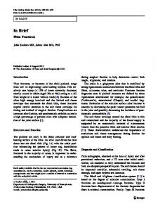
Fig. 1A–B (A) A lateral radiograph shows a transversely oriented patella fracture with wide displacement. (B) A lateral radiograph, obtained after open reduction and internal fixation using a plate and screws, shows the tension-band configuration with fracture reduction.
123
Clinical Orthopaedics and Related Research1
patients with an intact extensor mechanism and minimal intraarticular step-off. Surgery should be deferred in patients whose preoperative risk assessment is bleak, or in those who are not apt to regain function in any case (eg, patients with a prior failed extensor mechanism, joint ankylosis, or who are nonambulatory). Small fragments may be excised or repaired with suture or screw fixation if possible. Every effort should be made to preserve and stabilize as much of the patella as possible to maintain the moment arm and avoid loss of poor outcomes which can occur when greater than 40% of the patella is removed [2]. Removal of the entire patella may be contemplated for patients with massively comminuted fractures. Excision addresses the pain related to the fracture but decreases the mechanical advantage the patella provides to the extensor mechanism. The loss of quadriceps strength after total patellectomy can be as great as 49% and enough loss to preclude climbing stairs or getting out of a chair [13]. Therefore we believe total patellectomy should be considered only after all techniques for salvage have been exhausted. Transverse fractures (Fig. 1A) are best treated with a tension-band technique (traditionally two axial Kirschner wires with a figure-of-eight wire anteriorly) (Fig. 1B) which neutralizes tension forces anteriorly and converts them into stabilizing compressive forces at the articular surface [8]. This technique is stronger than a simple cerclage wire in transverse fractures [14] and can be combined with screw fixation in simple and in more comminuted
Volume 469, Number 4, April 2011
fractures to increase stability [5]. The reduction must be checked by palpating or observing the articular surface of the patella through a retinacular arthrotomy (surgically created if needed); fluoroscopy is not sufficiently accurate. If there are multiple fracture lines, it may be reasonable to first provisionally fix small fragments with screws to create a two-part transverse fracture, which then is addressed with a tension band, circumferential fixation, or both.
Patellar Fractures
1215
4.
It may be reasonable to fix (rather than excise) a comminuted fracture even with bad articular damage, as this can preserve bone stock for a knee replacement later.
5.
Tension-band wires ultimately will break. Use of suture, and not metal, will spare you a ‘broken wire xray’.
References Outcomes The typical outcome after surgery is union of the fracture and function inversely proportionate to the severity of the initial injury. Still, even seemingly mild injuries can be associated with development of patellofemoral arthrosis [12]. Prolonged immobilization after surgery also can lead to stiffness and loss of knee motion [11]. Early displacement after operative fixation can occur in 10% to 20% of patients following ORIF, often attributable to inadequate fixation [10, 11]. Overall rates of patellar nonunion are reportedly between 2.4% to 12.5% [6]. Hardware failure is not uncommon even with good healing (especially with wire banding), but removal is necessary only if prominent or symptomatic.
Five Pearls 1.
A patellar fragment, especially one in the superior lateral quadrant, may be a bipartite patella (often bilateral) and not a fracture.
2.
In the setting of injury but normal bony anatomy observed on radiographs, consider a quadriceps rupture or avulsion. (Failure of the infrapatellar ligament results in a high-riding patella.)
3.
Repair of the patella should be accompanied by access to the joint surface and retinacular repair.
1. Atesok K, Doral MN, Lowe J, Finsterbush A. Symptomatic bipartite patella: treatment alternatives. J Am Acad Orthop Surg. 2008;16:455–461. 2. Bo¨stman O, Kiviluoto O, Nirhamo J. Comminuted displaced fractures of the patella. Injury. 1981;13:196–202. 3. Bostro¨m A. Fracture of the patella: a study of 422 patellar fractures. Acta Orthop Scand Suppl. 1972;143:1–80. 4. Carpenter JE, Kasman R, Matthews LS. Fractures of the patella. Instr Course Lect. 1994;43:97–108. 5. Carpenter JE, Kasman RA, Patel N, Lee ML, Goldstein SA. Biomechanical evaluation of current patella fracture fixation techniques. J Orthop Trauma. 1997;11:351–356. 6. Harris RM, Fractures of the patella and injuries to the extensor mechanism. In: Bucholz, RW, Heckman, JD, Court-Brown CM, eds. Fractures in Adults. Ed 6. Philadelphia, PA: Lippincott Williams and Wilkins; 2006:1969–1998. 7. Huberti HH, Hayes WC, Stone JL, Shybut GT. Force ratios in the quadriceps tendon and ligamentum patellae. J Orthop Res. 1984;2:49–54. 8. Muller ME, Allgower M, Schneider R, Willeneger H. Manual of Internal Fixation: Techniques Recommended by the AO Group. Berlin, Germany: Springer; 1979:248–253. 9. Nord RM, Quach T, Walsh M, Pereira D, Tejwani NC. Detection of traumatic arthrotomy of the knee using the saline solution load test. J Bone Joint Surg Am. 2009;91:66–70. 10. Nummi J. Fracture of the patella: a clinical study of 707 patellar fractures. Ann Chir Gynaecol Fenn Suppl. 1971;179:1–85. 11. Smith ST, Cramer KE, Karges DE, Watson JT, Moed BR. Early complications in the operative treatment of patella fractures. J Orthop Trauma. 1997;11:183–187. 12. Sorensen KH. The late prognosis after fracture of the patella. Acta Orthop Scand. 1964;34:198–212. 13. Sutton FS Jr, Thompson CH, Lipke J, Kettelkamp DB. The effect of patellectomy on knee function. J Bone Joint Surg Am. 1976;58:537–540. 14. Weber MJ, Janecki CJ, McLeod P, Nelson CL, Thompson JA. Efficacy of various forms of fixation of transverse fractures of the patella. J Bone Joint Surg Am. 1980;62:215–220.
123