Original Article
Incidence and risk factors of aplastic anemia in Latin American countries: the LATIN case-control study Eliane Maluf,2 Nelson Hamerschlak,1 Alexandre Biasi Cavalcanti,1 Álvaro Avezum Júnior,1 José Eluf-Neto,3 Roberto Passetto Falcão,4 Irene G. Lorand-Metze,5 Daniel Goldenberg,6 Cézar Leite Santana,7 Daniela de Oliveira Werneck Rodrigues,8 Leny Nascimento da Motta Passos,9 Luis Gastão Mange Rosenfeld,1 Marimilia Pitta,13 Sandra Loggetto,13 Andreza A. Feitosa Ribeiro,1 Elvira Deolinda Velloso,1 Andrea Tiemi Kondo,1 Erika Oliveira de Miranda Coelho,10 Maria Carolina Tostes Pintão,4 Hélio Moraes de Souza,11 José Rafael Borbolla,12 and Ricardo Pasquini2 1
Hospital Israelita Albert Einstein/Instituto Israelita de Ensino e Pesquisa Albert Einstein, São Paulo, Brazil; 2Hospital das Clínicas da Universidade Federal do Paraná, Curitiba, Brazil; 3Faculdade de Medicina da Universidade de São Paulo, São Paulo, Brazil; 4 Faculdade de Medicina de Ribeirão Preto da Universidade de São Paulo, Ribeirão Preto, Brazil; 5Faculdade de Ciências Médicas da Universidade Estadual de Campinas, Campinas, Brazil; 6Hospital de Clínicas José de San Martín, Buenos Aires, Argentina; 7 Instituto de Hemoterapia de Goiânia, Goiânia, Brazil; 8Hemocentro Regional de Juiz de Fora, Juiz de Fora, Brazil; 9Fundação de Hematologia e Hemoterapia do Amazonas (HEMOAM), Manaus (AM), Brazil; 10Fundação Hemope, Recife, Brazil; 11Hemocentro Regional de Uberaba, Uberaba, Brazil, and 12Hospital San José Tec de Monterrey, Monterrey, México, Centro de Hematologia de São Paulo
ABSTRACT Background Associations between aplastic anemia and numerous drugs, pesticides and chemicals have been reported. However, at least 50% of the etiology of aplastic anemia remains unexplained. Design and Methods This was a case-control, multicenter, multinational study, designed to identify risk factors for agranulocytosis and aplastic anemia. The cases were patients with diagnosis of aplastic anemia confirmed through biopsy or bone marrow aspiration, selected through an active search of clinical laboratories, hematology clinics and medical records. The controls did not have either aplastic anemia or chronic diseases. A total of 224 patients with aplastic anemia were included in the study, each case was paired with four controls, according to sex, age group, and hospital where the case was first seen. Information was collected on demographic data, medical history, laboratory tests, medications, and other potential risk factors prior to diagnosis. Results The incidence of aplastic anemia was 1.6 cases per million per year. Higher rates of benzene exposure (≥30 exposures per year) were associated with a greater risk of aplastic anemia (odds ratio, OR: 4.2; 95% confidence interval, CI: 1.82-9.82). Individuals exposed to chloramphenicol in the previous year had an adjusted OR for aplastic anemia of 8.7 (CI: 0.87-87.93) and those exposed to azithromycin had an adjusted OR of 11.02 (CI 1.14108.02). Conclusions The incidence of aplastic anemia in Latin America countries is low. Although the research study centers had a high coverage of health services, the underreporting of cases of aplastic anemia in selected regions can be discussed. Frequent exposure to benzene-based products increases the risk for aplastic anemia. Few associations with specific drugs were found, and it is likely that some of these were due to chance alone. Key words: aplastic anemia, incidence, risk factors, benzene. Citation: Maluf E, Hamerschlak N, Cavalcanti AB, Avezum Júnior Á, Eluf-Neto J, Passetto Falcão R, Lorand-Metze IGH, Goldenberg D, Leite Santana C, de Oliveira Werneck Rodrigues D, Nascimento da Motta Passos L, Mange Rosenfeld LG, Pitta M, Loggetto S, Feitosa Ribeiro AA, Velloso ED, Kondo AT, de Miranda Coelho EO, Tostes Pintão MC, Moraes de Souza H, Borbolla JR, and Pasquini R. Incidence and risk factors of aplastic anemia in Latin American countries: the LATIN case-control study. Haematologica 2009;94:1220-1226. doi:10.3324/haematol.2008.002642
©2009 Ferrata Storti Foundation. This is an open-access paper. | 1220 |
haematologica | 2009; 94(9)
Funding: this study was supported by a grant from Sanofi-Aventis. The study sponsors were not involved at all in the study design, data collection, data analysis, data interpretation, or report writing. The corresponding author had full access to all the study data, and had ultimate responsibility for the decision to submit for publication. Acknowledgments: the authors want to thank Rodrigo Callado, Joan Laport and David Kaufman for their observations and suggestions on the text. Manuscript received on November 25, 2008. Revised version arrived on March 23, 2009. Manuscript accepted on April 10, 2009. Correspondence: Nelson Hamerschlak, Centro de Pesquisa Clínica, Instituto Israelita de Ensino e Pesquisa Albert Einstein Av. Albert Einstein, 627/701, Piso Chinuch, São Paulo (SP), Brazil, CEP 05651-901. E-mail:
[email protected]
Incidence and risk factors of aplastic anemia
Introduction Aplastic anemia (AA) is a hematologic condition characterized by bone marrow hypoplasia or aplasia resulting in pancytopenia. It is a severe disease and its etiology has been attributed to medications,1,2 chemicals,1-3 and environmental factors.4 Although bone marrow transplants have increased the survival rate of patients with AA, most people do not have access to this therapy, and fatality rates of the disease remain high.5 Several studies published to date disagree as to the etiology of AA, which may be partially explained by their different methodologies.2 Well-founded studies with good statistical power, assessing the association between AA and drugs and other risk factors are scarce.1 A recent review of the epidemiology of aplastic anemia shows that most cases of aplastic anemia appear to be secondary to the immunological destruction of the hematopoietic cells. The risk of development of autoimmune diseases has been linked to host genetics, and a few risk factors have been identified that affect the immune response and the susceptibility of the hematopoietic target cell.6 Methodologically well-founded studies have found an incidence of AA ranging from 1.4 to 14 cases per million people,7-9 with higher rates in Asian countries than in Western ones.9,10 A study conducted in Southern Brazil, from 1999 to 2000, reported an incidence of 2.4 cases per million per year.1 This wide variation in incidence among regions is generally thought to be due to environmental, rather than genetic factors.7,11-13 Based on data from several publications indicating that environmental factors play a major role in the development of AA2, the fact that the risk factors have not yet been well-described for our context, and because treatment is not widely available in developing countries, this study has been carried out since 2002,14 with the purpose of providing more information for prevention of the disease. The LATIN study is an international case-control study designed to identify risk factors for agranulocytosis and AA, including drugs, other diseases, and environmental factors, using a methodological approach similar to that used in previous studies.2,3 Its secondary objective was to estimate the incidence rates of both agranulocytosis and AA in some Latin American countries.14 This report focuses on the risk factors and incidence rate of AA.
in Mexico (Monterrey). In the pilot phase of the LATIN study, an active search for AA cases was carried out in the Brazilian states of Paraná, Minas Gerais, Goiás, Pernambuco and Amazonas, and in the city of Ribeirão Preto (state of São Paulo) and adjacent cities. The geographical area covered was extremely large. Following the pilot phase (after April 2003), the area covered by each study center was then restricted, including only regions with better medical systems, to avoid underreporting of cases. Therefore, instead of covering whole states, a small region formed by the cities adjacent to each study center was defined as their catchment area. The only exception was the center of Curitiba, for which the catchment area continued to be the whole state of Paraná. This reduced the study area in Brazil from 2.25 million km2 to 295 thousand km2. (Figure 1). Buenos Aires (Argentina) and Monterrey (Mexico) joined the study after the pilot phase. For all the sites, an active search of AA patients was carried out on a weekly basis, among hematology clinics in predefined areas. Records of death due to hematologic diseases were also obtained from government agencies on an annual basis. An informed consent was obtained from all the patients and controls. The local and national research ethics committees approved this study.
Participants Cases All the patients with acquired AA included in the study had been living in the area covered by the study site for more than three months, and had undergone peripheral blood testing and bone marrow study. All the patients were submitted to peripheral blood count, bone marrow aspiration, and/or bone marrow biopsy. All the patients under two years of age were submitted to a specific test to exclude Fanconi anemia. The eligible patients were those who met at least two of the three
Design and Methods Design and settings The LATIN study14 is an international, multi-center, case-control study. Due to the vast size of Brazil (covering an area of more than 8.5 million km2 and with a population in 2007 of nearly 190 million)15,16 and its high regional population diversity, it is not possible to assess the incidence of AA at a national level. Therefore, this study included seven sites in representative areas of six Brazilian regions, plus two additional study sites, one in Argentina (Buenos Aires) and one
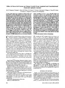
Figure 1. Location of Brazilian study sites.
haematologica | 2009; 94(9)
| 1221 |
E. Maluf et al.
peripheral blood count criteria below, together with a compatible bone marrow study:17 1. White cells < 3,5×10⁹/L; 2. Platelets < 50×109/L; 3. Hemoglobin < 10.0 g/dL or hematocrit < 30%. The bone marrow biopsy and/or myelogram had to show hypocellularity, but no fibrosis or leukemic, lymphomatous or carcinomatous infiltration. Cases with hypocellular myelodysplasia were also ruled out. Cases and controls were also excluded from the study if they had other severe hematologic diseases (neural tube defects, neoplasias, megaloblastic anemia) or systemic diseases usually associated with neutropenia or pancytopenia (such as lupus, HIV infection, and hypersplenism) or if they had previously undergone any organ transplantation, chemotherapy, radiotherapy or immunosuppressive therapy. Felty’s syndrome, Kostmann’s syndrome, Shwachman-Diamond syndrome, neutropenias of the autoimmune, isoimmune, chronic hypoplasic or cyclic types, reticular dysgenesis (dyskeratosis congenita) and dyskeratoses were also conditions for exclusion. To exclude alternative diagnoses, the final acceptance of cases for inclusion in the study required a characteristic hypocellular bone marrow biopsy (marrow cellularity