Unusual association of diseases/symptoms
CASE REPORT
Incidental pathologically proven pulmonary hamartoma in a patient with carcinoma tongue Aditya Jindal,1 Karan Madan,1 Raje Nijhawan,2 Navneet Singh1 1
Department of Pulmonary Medicine, Postgraduate Institute of Medical Education & Research (PGIMER), Chandigarh, India 2 Department of Cytology & Gynecological Pathology, Postgraduate Institute of Medical Education & Research (PGIMER), Chandigarh, India Correspondence to Dr Navneet Singh,
[email protected]
SUMMARY Pulmonary hamartomas are usually clinically silent and found incidentally on chest radiographs. They can lead to diagnostic confusion especially in patients who have been previously treated for primary cancers at other sites. This can lead to consideration of metastatic malignancy as the primary diagnostic possibility. In this case, evaluation of a solitary pulmonary nodule (SPN) in a patient with carcinoma of tongue led to the diagnosis of pulmonary chondroid hamartoma. This highlights the fact that a pulmonary nodule in a patient with progressive cancer at another site does not always indicate pulmonary metastasis. BACKGROUND Pulmonary hamartomas are the most common benign lung tumours and account for up to 6% of all solitary pulmonary nodule (SPN).1 They are usually clinically silent and found incidentally on chest radiographs. Their incidence peaks from the fourth to the sixth decade and they are more common in men (2:1 to 3:1).2 To the best of our knowledge, this is the second case report of a pulmonary chondroid hamartoma in a patient with squamous cell carcinoma of the tongue. Onizawa et al3 had previously reported a case of a 66-year-old woman with squamous cell carcinoma of the tongue with an SPN that was later proven to be a chondroid hamartoma after wedge resection.
CASE PRESENTATION A 50-year-old male patient presented with a history of painless lump on the left side of the neck of 6 weeks duration. There was a history of hoarseness of voice for the last 2 weeks. He was a chronic smoker with a 30-pack-year smoking history. On oral examination, there was an indurated lesion (2.5 cm × 1.5 cm) over the base of the tongue on
To cite: Jindal A, Madan K, Nijhawan R, et al. BMJ Case Rep Published online: [please include Day Month Year] doi:10.1136/bcr-2013008942
the left side. The lesion was not crossing the midline. Left-sided vocal cord palsy was observed. Two firm and mobile, left upper deep cervical lymph nodes (level II) were palpable. Biopsy from the tongue lesion showed a squamous cell carcinoma. A diagnosis of carcinoma base of tongue stage IV-A (T2N2bM0) was made. The patient refused surgical management and was referred to the radiotherapy department where he received external beam radiation therapy (50 G, 25 fractions and 4 weeks) during the next 1 month. On repeat clinical examination, the mass at the base of the tongue had resolved. The cervical lymph nodes persisted, albeit reduced in size. The patient was subsequently lost to follow-up. He presented 6 months later with increasing size of the neck mass. During work up for systemic metastases, a non-calcified SPN was observed in the right lower zone on the chest radiograph (figure 1, left and middle panel). No previous chest radiograph was available for comparison. CT of the thorax lung window sections showed evidence of a well-defined, non-calcified 2.8 cm × 2.6 cm sized nodule in the right lower lobe with normal surrounding lung parenchyma and without any enlarged hilar or mediastinal lymph nodes (figure 1, right panel). The patient was referred to the pulmonary medicine department for opinion regarding the nature of the SPN. In view of history of heavy smoking and presence of clinically active malignancy, a differential diagnosis of pulmonary metastasis from tongue cancer versus synchronous (primary) lung cancer was entertained. CT-guided fine needle aspiration cytology from the SPN (figure 2) showed clusters of as well as scattered benign epithelial and spindle-shaped cells, which were intimately mixed with adipose tissue and chondroid material, along with occasional foci of calcification. A diagnosis of pulmonary chondroid hamartoma was confirmed. Since the patient
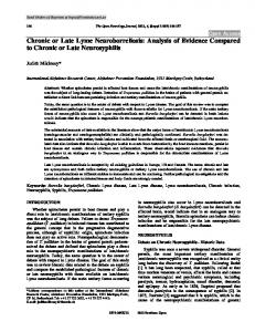
Figure 1 Chest radiograph showing presence of a solitary pulmonary nodule in the right lower zone on posteroanterior view (left panel) and right lateral view (middle panel). CT of the thorax showing a well-defined 2.8×2.6 cm lesion in the right lower lobe with smooth margins and normal surrounding parenchyma (right panel).
Jindal A, et al. BMJ Case Rep 2013. doi:10.1136/bcr-2013-008942
1
Unusual association of diseases/symptoms
Figure 2 Microphotograph of a fine needle aspiration cytology smear from a solitary pulmonary nodule showing benign bronchial epithelial cell cluster and mesenchymal element in the form of chondroid connective tissue (left panel H&E, ×200). A higher magnification of the chondroid tissue is depicted in the right hand panel (H&E, ×400). had no pulmonary symptoms and the lesion was benign, it was felt that no active intervention was needed for the same and he was again referred to otolaryngology and radiotherapy services for subsequent palliative treatment.
DISCUSSION Hamartomas are slow growing benign lung tumours. Most pulmonary hamartomas are parenchymal lesions though endobronchial hamartomas are also reported. More frequently, endobronchial hamartomas are symptomatic and may present with haemoptysis and/or central airway obstruction.4 Radiologically, the typical appearance is of a peripheral homogenous round lesion with smooth borders as seen in the index patient. The classical ‘popcorn calcification’ is present in only 10–30% of patients.5 CT is more sensitive in the detection of calcification as well as of fat in a pulmonary lesion. Most hamartomas demonstrate a predominant chondroid differentiation (80%) with the presence of a mesenchymal tissue (fibromyxoid stroma) and chondroid material, as was seen in the index case.6 Other patterns may include fibroblastic, fatty or osseous differentiation with the latter being the least common (3%). Asymptomatic hamartomas require no treatment. In symptomatic patients, the treatment of pulmonary hamartomas is primarily surgical and this is usually enucleation or wedge resection. Infrequently, lobectomy or pneumonectomy may be required. Treatment of endobronchial hamartomas may be performed by flexible or rigid bronchoscopy.4 Pulmonary hamartomas can sometimes give rise to diagnostic confusion especially in patients who have been previously treated for primary cancers at other sites and the same holds true for the index case also. This has been most frequently reported for breast and renal carcinomas.2 3 Considering the slow-growing nature of a pulmonary chondroid hamartoma, it is possible that the SPN on chest radiograph might have been present at the initial evaluation also. However, as a previous radiograph was not available for comparison and there was no documentation of the same in patient’s record, it is possible that this was a recent development. In summary, this case highlights the fact that in a patient with cancer with active/recurrent
2
disease, an asymptomatic solitary pulmonary lesion may not always indicate metastasis. A cytological/histopathological confirmation of the aetiology of such lesions may prove useful in establishing an accurate diagnosis and thus appropriately staging patients with cancer, which can ultimately influence management decisions.
Learning points ▸ Pulmonary hamartomas are often a cause of diagnostic confusion in patients who have been/are being treated for cancer at other sites. ▸ In a patient with active cancer, an asymptomatic solitary pulmonary nodule (SPN) may not always indicate metastasis. ▸ A pathological confirmation of the diagnosis of SPN in such settings may prove extremely useful in management decisions.
Competing interests None. Patient consent Obtained. Provenance and peer review Not commissioned; externally peer reviewed.
REFERENCES 1
2 3 4 5 6
Murray J, Kielkowski D, Leiman G. The prevalence and age distribution of peripheral pulmonary hamartomas in adult males. An autopsy-based study. S Afr Med J 1991;79:247–9. Sodhi KS, Virmani V, Jindal SK, et al. Pulmonary hamartoma. Ann Acad Med Singapore 2009;38:1110. Onizawa K, Furuya Y, Suzuki K, et al. Pulmonary chondroid hamartoma: an incidental finding in a case of tongue carcinoma. J Oral Maxillofac Surg 1998;56:898–900. Kim SA, Um SW, Song JU, et al. Bronchoscopic features and bronchoscopic intervention for endobronchial hamartoma. Respirology 2010;15:150–4. Briccoli A, Farinetti A, Del Prete P, et al. [Pulmonary hamartoma]. Minerva Chir 1993;48:813–19. Ganti S, Milton R, Davidson L, et al. Giant pulmonary hamartoma. J Cardiothorac Surg 2006;1:19.
Jindal A, et al. BMJ Case Rep 2013. doi:10.1136/bcr-2013-008942