Welkriana etJournal al. Indonesian of Biotechnology, December, 2011
Vol. 16, No. 2, I.J. pp.126-131 Biotech.
T-786c Polymorphism in nitric oxide synthase 3 gene and Nitrit Oxide Level of Diabetic Retinopathy in Javanese Population Putri Widelia Welkriana1, Sunarti2*, and Pramudji Hastuti2 1
Postgraduated Program of Basic and Biomedical Science, Faculty of Medicine, Universitas Gadjah Mada, Yogyakarta, Indonesia 2 Department of Biochemistry, Faculty of Medicine, Universitas Gadjah Mada, Yogyakarta, Indonesia
Abstract Complication of retinopathy in type 2 DM is caused of lower level of NO. Nitric oxide level is synthesized from L-arginin in reaction that catalyze Nitric oxide synthase (NOS) 3. The T-786C mutation in NOS 3 gene decreases the expression of nitric oxide synthase (NOS) 3 so decreases NO synthesis. To investigate the association between T-786C polymorphism in NOS 3 gene with NO level of diabetic retinopathy patients. This study was a case control study, consist of 40 patient of type 2 diabetic with DR (case group) and 40 patient of type 2 diabetic without DR (control group) of Javanese ethnic. The genotyping of T-786C polymorphism was performed by PCR-RLFP. Level of NO was measured by spectrophotometry. Chi square test and odd ratio were used to analyze the association of the T-786C polymorphism in NOS 3 gene with DR. Differences of NO level between TT and TC genotypes were analyzed using independent t test. The distribution of T-786C polymorphism in NOS 3 gene of DR subjects showed that frequency of TT genotype was 22.5% and TC genotype was 77.5%. Non DR subjects showed the frequency of TT genotype was 50% and TC genotype was 50%, (p= 0.011). Frequency of T allele in DR group was 61.25% and C allele was 38.75%, and frequency of T allele in non DR group was 75% and C allele was 25%, (p= 0.62). Odd ratio of TC genotype was 3.444(CI; 95% : 0.964-3.735) and C allele was 1.898 (CI; 95% : 1.310-9.058). The NO level of TC genotype was 1.43+0.126 and TT genotype was 11.27+5.87 (p=0.000). Level of NO between RD and non RD showed not different significantly (p=0.160) for retinopathy. The T-786C polymorphism of NOS 3 gene is risk factor for retinopathy in type 2 Diabetes Mellitus. Individual with TC genotype of NOS 3 gene has lower level of NO than TT genotype. Keywords : Diabetic Retinopathy, Polymorphism, Nitric Oxide, Nitric Oxide Synthase.
In Yogyakarta, the DM patients has reached approximately 150/10.000 citizens, therefore its prevalence is 1.6% (Depkes RI, 2008). The complications of DM is retinopathy, nephropathy, and peripheral neuropathy. Diabetic Retinopathy (DR) is a microvascular complication which causes blindness (Gustaviani, 2006). The prevalence of DR in the world is 40%, in Indonesia is about 4050% (Masduki et al., 2008). In Yogyakarta, the prevalence of DR in rural area is 8.7%, while in suburban is 7.7% (Gustaviani, 2006). Retinal defect caused by hyperglycemia involves some pathways, such as nonenzymatic glycation, protein kinase C activation, increase of oxidative stress, polyol
Introduction Diabetes Mellitus (DM) is a metabolic disease characterized by hyperglycemia. It is caused by defect on insulin secretion, insulin activity, or both (Gustaviani, 2006). Based on WHO database, the number of DM patients in the world has reached 171 million people (2.8% of world population). In 2007, the prevalence of DM in Indonesia was approximately 5.7% (Depkes RI, 2008). *Corresponding Author:
Sunarti Department of Biochemistry, Faculty of Medicine, Universitas Gadjah Mada, Yogyakarta, Indonesia, Email :
[email protected]
126
Welkriana et al.
I.J. Biotech.
Supariasa et al., 2001), did not suffer from dyslipidemia, systolic blood pressure < 180 mmHg and diastolic blood pressure < 120 mmHg (JNC 7, 2003), did not have any eye disease. Informed consent was obtained from each subject. Diagnosis of DM was established when the level of fasting plasma glucose ≥ 120 mg/ dL and 2-h plasma oral glucose tolerant ≥ 200 mg/dL (American Diabetes Association, 2010). The criterion of retinopathy was based on the photofundus examination that showed one of retinopathy symptoms, such as microaneurism, hemorrhage, exudates, or/and neovascularization. The data obtained from the subjects were age, sex, course of DM, BMI, and blood pressure. NO level was measured using spectrophotometry with Griess reaction. The measurements of GDP and G2PP levels used GOD-PAP, while triglyceride, HDL, and LDL HDL, and LDL levels were measured using CHOD-PAP levels were measured using CHOD- PAP. The data was analyzed with independent t test.
pathway, and decrease of NO. Chronic hyperglycemia increases free radicals, especially reactive oxygen species, in all of tissues causes oxidative stress. Oxidative stress cause change of the structural and functional retinal microvascular (Miyamoto et al., 2000) that is thickening of basal membrane and microvascular, loses the capillary form. Furthermore, structural changes may alter the function of pericyte cells and endothelial cells, such as decrease of blood stream, loss of intercellular junction, and increase of vessel permeability. The contractility of pericytes of retinal capillary is inhibited by high concentration of glucose. It is the early step of DR pathogenesis (Gillies and Su, 1993). Several studies reported that several genes influence the development of DR. One of those gene is endothelial nitric oxide synthase (eNOS) or NOS 3 gene. NOS 3 gene is located in chromosome 7 (7q36). This gene encodes NOS 3 which catalyzes the formation of NO by the conversion of L-arginin to L-citrullin (Mayer and Hemes, 1997). Ezzidi et al., 2008 investigated T-786C polymorphism in NOS 3 gene that located in -786 bp upstream from NOS 3 gene. This polymorphism causes the conversion of timine to cytocine which leads to the decrease of NO synthesis. Low level of NO causes vasoconstriction, platelet aggregation, and angiogenesis. The objective of this study was to give description about the relationship of T-786C polymorphism in NOS 3 gene as risk factor for DR in type 2 DM patients of Dr. Sardjito Hospital, Yogyakarta and level of NO.
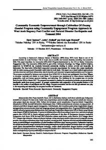
Genotyping of NOS 3 gene polymorphism The T-786C polymorphism in NOS 3 gene was determined with PCR-RLFP method. The PCR conditions were 94oC for 5s; followed by 40 cycles of 94oC for 1 min, 61oC for 1 min, 72oC for 1 min; and final extension at 72oC for 5 min. The primers used in this study were 5’-TGG AGA GTG CTG GTG TAC CCC A-3’ (forward primer) and 5’-GCC TCC ACC CCC ACC CTG TC3’ (reverse primer). From this amplification, PCR product of 180 bp was expected (Ezzidi et al., 2008). The enzyme for restriction was MspI restriction enzyme (New England Biolabs). The result of digestion for 16 h was then electrophorized with 2% agarose gel and visualized with ethidium bromide under UV light. Two fragments; 140 and 40 bp; represented TT genotype. Four fragments; 140, 90, 50, and 40 bp; represented TC genotype, while three fragments; 90, 50,
Materials and Methods This research was a case-control study with 40 subjects of type 2 DM patients with DR as the case group and 40 subjects of type 2 DM patients without DR as the control group. The patients were the outpatients of Dr. Sardjito Hospital and they are all Javanese. The inclusion criteria of the research subject were aged 35-65 years, have been diagnosed as type 2 DM patient minimally for 5 years, body mass index ≤ 25 kg/m (WHO, 2006;
127
Welkriana et al.
I.J. Biotech.
this study. The characteristics of the studied population were shown in Table 1. BMI of DR patients and non DR patients was significantly different, but the average value of both groups was in normal range (Table 1). The result of genotype analysis of T-786C polymorphism in NOS 3 gene using PCR-RFLP is shown in Figure 1. The genotype and allele distribution of T-786C polymorphism in NOS 3 gene is shown in Table 2 while the Hardy-Weinberg equilibrium is shown in (Table 3).
and 40 bp; represented CC genotype. The difference of genotype and allele frequencies between case and control groups were analyzed by chi-square test. The Odd Ratio test was used to analyze the extent to which various genotypes and alleles of T-786C polymorphism in NOS 3 gene was associated with DR. Results A total of 80 Javanese that consisted of 40 type 2 DM patients with DR and 40 type 2 DM patients without DR, was enrolled in
Table 1. Characteristics of the studied population Variables Number of subjects Sex : male (%) female (%) Age (year)# BMI (kg/m2)# Systolic blood pressure(mmHg)# Diastolic blood pressure(mmHg)# GDP (mg/dL)# Glucose 2 PP (mg/dL) Triglyceride (mg/dL)# Total cholesterol (mg/dL) HDL (mg/dL) LDL (mg/dL)#
Diabetic Retinopathy 40 13 (32.5%) 27 (67.5%) 55,58+7,91 21,93+2,83 124,25+11,07 79,50+9,04 167,18 +69,83 232,41+76.13 122,91+26,89 172.81+47,02 75,34+17,01 101,95+23,80
Non DR 40 12 (30%) 28 (70%) 56,03+8,26 23,37+1,84 123,00+12,03 80,25+8,00 142,26+66,23 206.29+91.80 137,02+33,12 171,02+26,65 72,19+ 12,95 101,41+19,30
Note : # calculated with Mann Whitney test
Figure 1. Genotype of TC and TT at -786 position NOS 3 gene
128
p
(95%CI)
0,797 0,018* 0,648 0,809 0,062 0,362 0,862 0,302 0,252 0,130
54,01-57,59 4,69-4,81 2,08-2,9 1,01-1,89 11,58-12,94 0,04-0,08 13,07-13,89 10,47-11,88 10,90-11,98 92,88-91,81
Welkriana et al.
I.J. Biotech.
Table 2. The genotype frequency of T-786C polymorphism in NOS 3 gene Genotype Type 2 DM TT TC DR 9 (22.5%) 31 (77.5%) non DR 20 (50%) 20 (50%)
CC 0 (0%) 0 (0%)
Table 5. The average of NO level between genotypes of T-786C polymorphism in NOS 3 gene in type 2 DM patients
p (CI=95%)
type 2 DM
0.011
DR and non DR
Pearson Chi-square analysis showed there was significantly different of genotype -786 NOS 3 gene in DR and non DR patients (p = 0.011).
Result
TT TC CC
Observed value 29 51 0
Expected value 37 35 8
7.99+5.25
1.23+1.71
0,000*
The average of NO level in individuals with TT genotype was higher than that in individuals with TC genotype (7.99+5.25 mg/dL vs 1.23+1.71 mg/dL) and this was statistically different (p=0.000). The average of NO level between genotypes in DR and without DR type 2 DM was shown in Table 6.
Table 3. The genotype frequency of T-786C polymorphism in NOS 3 gene between the observed and the expected values (Hardy-Weinberg equilibrium) Genotype
NO level (pg/mL) p TT genotype TC genotype (CI=95%) (n=60) (n=20)
p (95%CI)
Table 6. The average type 2 DM of NO level between genotypes in patients with DR and without DR
0.05
type 2 DM
With Hardy-Weinberg equilibrium, TT genotype was 37, TC genotype was 35, and CC genotype was 8. With Pearson chi-square analysis showed a significant difference between the observed value and the expected (HardyWeinberg equilibrium) (p= 0.05) (Table 3).
DR non DR
NO blood level (pg/mL) p TT genotype TC genotype (CI=95%) 11.27 +5.87 1.43+2.13 0.000* 6.52+4.32 0.93+0.65 0.000*
In type 2 DM patients with DR, the average of NO level in individuals with TC genotype was lower than that in individuals with TT genotype, and this was statistically different (p=0.000). Similar condition was also met in type 2 DM patients without DR.
Table 4. The odds ratio of T-786C genotype and allele in NOS 3 gene Type 2 DM 95% CI DR non DR OR (n=40) (n=40) Genotype TC 31 20 3.444 1.310-9.058 TT 9 20 Allele C 31 20 1.898 0.964-3.735 T 49 60 Variables
Discussion Genotyping of T-786C polymorphism in NOS 3 gene in this research only found 2 genotypes, TT and TC genotypes. This result was different with the result of Ezzidi et al.(2008) and Awata et al.(2004) That found TT, TC, and CC genotypes. Liew et al.(2009) stated that development of DR was influenced by environmental and genetic factors in population study. TC genotype and C allele in -786 NOS 3 gene was significant different between type 2 DM with DR and non DR. Individuals with TC genotype had 3.4 time increased risk for DR than individuals with TT genotype, while C allele carriers had 1.9 increased risk for DR. However, Ezzidi et al. (2008)
The TC genotype and C allele were risk factor for DR in type 2 DM patients with OR 3.444 and 1.898 respectively (Table 4). The probability to have complication was P = OR/(1+OR). The probability of individuals with TC genotype to suffer from DR was 77.49%, while the probability of C allele carriers to suffer from DR was 65.49%. The influence of TT and TC genotypes of NOS 3 gene toward NO level in type 2 DM was shown in Table 5. 129
Welkriana et al.
I.J. Biotech.
This study reported that lower level of NO occurred in individuals with TC genotype than those with TT genotype. Awata et al. (2004) reported the lower of NOS 3 activity in blood of type 2 DM patients with DR than type 2 DM patients without DR. The lower of NOS 3 activity cause the lower level of NO in type 2 DM patients with DR compared to type 2 DM patients without DR. A low NO level induce the proliferation of smooth muscle of retinal microvascular (Singh et al., 2009). That condition stimulate oxidative stress in retina that lead to change in the structural and functional of retina. Free radical from ROS induced DNA modifications. Reactive Oxygen Species (ROS) increased the vascular permeability and occlusions, decreased of blood flow, disturbed the retinal microvascular barrier, which eventually resulted in retinal neovascularization in DR patient (Cattaruza et al., 2004). In summary, the T-786C polymorphism in NOS 3 gene was a risk factor for DR in type 2 DM patient. Individuals with TC genotype had lower NO level than TT homozygous individuals. Further study on T-786C polymorphism in NOS 3 gene in different ethnics and its association with other variables, such as blood pressure.
reported that there was no statistically significant different of T-786C polymorphism between DR patients and non DR patients in Tunisia (p=0.069). Awata et al.(2004) also reported a similar results as Ezzidi et al.(2008) (p=0.367). The different of ethnic was one of the factors which influence the different in genotype frequency. Moreover, the different of environment in each population influence the development of DR (Liew et al., 2009). The T-786C polymorphism is located in basal region of NOS 3 gene promoter, that is an active functional element. Ezzidi et al. (2008) reported that allele differences in T-786C polymorphism influence the function and binding activity of nuclear factor that changes transcription level of NOS 3 gene. Decreased expression of NOS 3 gene identified in retinal endothelial cells occur in individuals with CC genotype (Awata et al., 2004). Shear stress stimulates expression of NOS 3 synthesis in endothelial cells, especially the expression of NOS 3 in basal membrane of individuals with TT genotype (Cattaruza et al., 2004). On the other hand, the expression of NOS 3 in individuals with CC genotype did not influence by shear stress. There was variance in the promoter region to interact with transcription factor which stimulated by shear stress. Besides that, the binding between variant in promoter region and transcription factor inhibit protein binding to DNA. In other word, the replication of A1 protein interacted with promoter with C allele. A1 protein as repressor protein (Miyamoto et al.,2000). The frequency of TT and TC genotypes of NOS 3 gene between type 2 DM patients with DR and without DR was statistically different (p=0,011). In this study, the Hardyweinberg equlibrium was not fulfilled. Perhaps it was caused by the small number of the studied population, which was not yet enough to fulfilled the equilibrium. Moreover, all of the subjects were type 2 DM patients in Dr. Sardjito Hospital, who were possibly did not represented the population in the community.
Acknowledgements The author would like to thank Dra. Tasmini, M.Kes, dr. Maliyah Madiyan for their help, dr. Haryo Yudono, Sp.M.,M.Sc. and dr.Elly as the advisor of photofundus at Dr. Sardjito Hospital Yogyakarta. The authors would also thank the Head and staffs of Biochemistry Laboratory of Faculty of Medicine Universitas Gadjah Mada; A. Cahyono, Agustina W. Djuma, and Jems K.R.Maay for the assistances. The author also very grateful to all of the type 2 DM patients in Dr. Sardjito Hospital, Yogyakarta who werewilling. References American Diabetes Association. 2010. Standar of Medical Care in Diabetes.Diabetes Care. 31(1), S12-S35.
130
Welkriana et al.
I.J. Biotech.
Awata, T., Neda, T., Iizuka, H., Kurihara, S., Ohkubo, T., Takata, N., et al. 2004. Endothelial nitric oxide synthase gene is associated with diabetic macular edema in type 2 diabetes. Diabetes Care 27, 2184–2190. Cattaruza, M., Guzik, T.J., Slowdowski, W. 2004. Shear stress insentivity of endothelial nitric oxide synthase expression as a genetic factor for coronary heart disease. Cir. Res.,95, 841-847. Departemen Kesehatan R.I. 2008. Laporan Riset Dasar Kesehatan Yogyakarta Tahun 2007. Departemen Kesehatan. Jakarta. Ezzidi, I., Mtiraoui, N., Mohamed, M.B.H., Mahjoub, T., Kacem, M., Almawi, W.Y. 2008. Endothelial nitric oxide synthase Glu298Asp, 4b/a, and T-786C polymorphisms in type 2 diabetic retinopathy. Clin.Endocrinol., 68, 542– 546. Gillies, M.C., Su, T. 1993. High glucose inhibits retinal capillary pericyte contractility in vitro. Ophthalmol., 34, 3396-3401. Gustaviani, R. 2006. Diagnosis dan Klasifikasi Diabetes Mellitus. Dalam Aru W.S., Bambang S., Idrus A., Marcellus S.K., Siti S.(Eds), Buku Ajar Ilmu Penyakit Dalam, Jakarta : Departemen Ilmu Penyakit Dalam FKUI. JNC 7. 2003. Seventh Report of Joint National Community on Prevention, Detection, Evaluation, and Treatment of High Blood Pressure American Hearth Association. Inc. Hipertension. 42, 1206-1252. Liew, G., Klein, R., Wong, T.Y. 2009. The role of genetics in susceptibility to diabetic retinopathy. Opthamol. Clin. 49(2):3552. Masduki, I., Agni, A.N., Hartono. 2008. Diabetic retinopathy diagnosis with opthalmoscope by trained doctor. Berkala Ilmu Kedokteran. 40(1),26-31. Mayer, B., Hemmens, B. 1997. Biosynthesis and action of nitric oxide in mammalian cells. Biochem. Jour., 22, 477-481.
Miyamoto, Y. Saito, Y., Nakayama, M. 2000. Replication protein A1 reduce transcription of the eNOS gene containing a-786T→C mutation associated with coronary spastic angina. Hum. Mol. Genet., 9, 2629-2637. Singh, P.P., Mahadi, F., Roy, A., Sharma , P. 2009. Reactive oxyden spesies, reactive nitrogen spesies and antioxidants in etiopathogenesis of diabetes mellitus type 2. Indian J. Biochem., 24(4), 324342. Supariasa IDN, Bakri B, Fajar I. 2001. Penilaian Status Gizi. Penerbit Buku Kedokteran EGC. Jakarta WHO. 2006. World Health Organization: definition, diagnosis and classification of diabetes mellitus and its complications: Report of a WHO Consultation.
131