Manuscript Number: HEP-13-1748.R2 Inflammatory and Metabolic Biomarkers and Risk of Liver and Bilary Tract Cancer
Krasimira Aleksandrova1, Heiner Boeing1, Ute Nöthlings2,3, Mazda Jenab4, Veronika Fedirko4,5,6, Rudolf Kaaks7, Annekatrin Lukanova7,8, Antonia Trichopoulou9,10, Dimitrios Trichopoulos10,11,12, Paolo Boffetta13, Elisabeth Trepo14, Sabine Westhpal15 , Talita DuarteSalles4, Magdalena Stepien4, Kim Overvad16, Anne Tjønneland17, Jytte Halkjær17, MarieChristine Boutron-Ruault18,19,20,
Laure
Dossus18,19,20,
Antoine Racine18,19,20, Pagona
Lagiou9,11,12, Christina Bamia9,10, Vassiliki Benetou9,10, Claudia Agnoli21, Domenico Palli22, Salvatore Panico23, Rosario Tumino24, Paolo Vineis25,26, Bas Bueno-de-Mesquita27, 28, Petra H. Peeters26,29, Inger Torhild Gram30, Eiliv Lund30, Elisabete Weiderpass30,31,32,33, J Ramón Quirós34,
Antonio
Agudo35,
María-José
Sánchez36,37,
Diana
Gavrila38,39,
Aurelio
Barricarte37,39, Miren Dorronsoro40, Bodil Ohlsson41, Björn Lindkvist42, Anders Johansson43, Malin Sund44, Kay-Tee Khaw45, Nicholas Wareham46, Ruth C Travis 47, Elio Riboli26, Tobias Pischon48
1
Department of Epidemiology, German Institute of Human Nutrition Potsdam-Rehbrücke,
Nuthetal, Germany; 2
Institute of Epidemiology, Christian-Albrechts University of Kiel, Kiel, Germany;
3
Nutritional Epidemiology Unit, Department of Nutritional and Food Science, Institut für
Ernährungs- und Lebensmittelwissenschaften, Rheinische Friedrich-Wilhelms-Universität Bonn, Germany; 4
International Agency for Research on Cancer (IARC-WHO), Lyon, France;
5
Department of Epidemiology, Rollins School of Public Health, Emory University, Atlanta
GA, USA; This article has been accepted for publication and undergone full peer review but has not been through the copyediting, typesetting, pagination and proofreading process which may lead to differences between this version and the Version of Record. Please cite this article as doi: 10.1002/hep.27016
Page 3 of 47
Hepatology
6
Winship Cancer Institute, Emory University, Atlanta, GA, USA;
7
Division of Cancer Epidemiology, German Cancer Research Center, Heidelberg, Germany;
8
Department of Medical Biosciences/Pathology, University of Umeå, Umeå, Sweden;
9
WHO Collaborating Center for Food and Nutrition Policies, Department of Hygiene,
Epidemiology and Medical Statistics, University of Athens Medical School, Athens, Greece; 10
Hellenic Health Foundation, Athens, Greece;
11
Department of Epidemiology, Harvard School of Public Health, Boston, USA;
12
Bureau of Epidemiologic Research, Academy of Athens, Athens, Greece;
13
Institute for Translational Epidemiology, Mount Sinai School of Medicine, New York,
USA; 14
Centre de Bioloqie Republique, Lyon, France
15
Institute of Clinical Chemistry, Otto-von-Guericke-University Magdeburg, Germany;
16
Section for Epidemiology, Department of Public Health, Aarhus University, Aarhus,
Denmark; 17
Diet, Genes and Environment Danish Cancer Society Research Center Copenhagen,
Denmark; 18
Inserm, Centre for research in Epidemiology and Population Health (CESP), U1018,
Nutrition, Hormones and Women's Health team,F-94805, Villejuif, France; 19
Univ Paris Sud, UMRS 1018, F-94805, Villejuif, France;
20
IGR, F-94805, Villejuif, France;
21
Nutritional Epidemiology Unit, Fondazione IRCCS Istituto Nazionale Tumori, Milano-Italy;
22
Molecular and Nutritional Epidemiology Unit, Cancer Research and Prevention Institute -
ISPO, Florence, Italy; 23
Department of clinical and experimental medicine-Federico II University,Naples, Italy;
24
Cancer Registry and Histopathology Unit, "M.P.Arezzo" Hospital, Ragusa, Italy;
25
HuGeF Foundation, Turin, Italy; Hepatology
Hepatology
26
Page 4 of 47
Division of Epidemiology, Public Health and Primary Care, Imperial College, London,
United Kingdom; 27
National Institute for Public Health and the Environment (RIVM), Bilthoven, The
Netherlands; 28
Department of Gastroenterology and Hepatology, University Medical Centre, Utrecht, The
Netherlands; 29
Julius Center for Health Sciences and Primary Care, University Medical Center, Utrecht, the
Netherlands; 30
Department of Community Medicine, Faculty of Health Sciences, University of Tromsø,
Norway 31
Department
32
Department of Medical Epidemiology and Biostatistics, Karolinska Institutet, Stockholm,
of
Research,
Cancer
Registry
of
Norway,
Oslo,
Norway;
Sweden; 33
Samfundet Folkhälsan, Helsinki, Finland;
34
Public Health Directorate, Asturias, Spain;
35
Unit of Nutrition, Environment and Cancer,Cancer Epidemiology Research Program,
Catalan Institute of Oncology, Barcelona, Spain; 36
Andalusian School of Public Health, Granada (Spain);
37
Consortium for Biomedical Research in Epidemiology and Public Health (CIBER
Epidemiología y Salud Pública-CIBERESP), Spain. 38
Servicio de Epidemiología, Department of Epidemiology; Consejería de Sanidad y Politica
Social, Murcia, Spain; 39
Navarre Public Health Institute, Pamplona, Spain;
40
Public Health Direction, Basque Regional Health Department and Biodonostia Research
Institute-Ciberesp San Sebastian, Spain;
Hepatology
Page 5 of 47
Hepatology
41
Department of Clinical Sciences, Division of Internal Medicine, Skåne University Hospital,
Malmö, Lund University, Sweden; 42
Institute of Medicine, Sahlgrenska Academy, University of Gothenburg, Gothenburg,
Sweden 43
Department of Clinical Microbiology, Laboratory for Molecular Infection Medicine
Sweden, Umeå Centre for Microbial Research, Umeå University, Umeå, Sweden; 44
Department of Surgical and Perioperative Sciences, Surgery and Public Health, Nutrition
Research, UmeaUniversity, Umea, Sweden; 45
Department of Public Health and Primary Care, University of Cambridge, Cambridge, UK
46
MRC Epidemiology Unit, Institute of Metabolic Science, Addenbrooke’s Hospital,
Cambridge, United Kingdom; 47
Cancer Epidemiology Unit, Nuffield Department of Clinical Medicine, University of
Oxford, Oxford, United Kingdom; 48
Molecular Epidemiology Group, Max Delbrück Center for Molecular Medicine Berlin-
Buch, Germany. Keywords: hepatocellular carcinoma, bile duct cancer, inflammation, hyperinsulinemia, biomarkers
Contact Information: Krasimira Aleksandrova, PhD, MPH, Department of Epidemiology, German Institute of Human Nutrition Potsdam-Rehbruecke, Arthur-Scheunert Allee 114-116, 14558 Nuthetal, Germany, Tel +49 33200 88 2 712; Fax +49 33200 88 2 721, e-mail:
[email protected]
Hepatology
Hepatology
List of Abbreviations: HCC, hepatocellular carcinoma GBTC, gall-bladder and bilary tract cancers outside of the liver IBD, intra-hepatic bile duct cancer EPIC, European Prospective Investigation into Cancer and Nutrition CRP, C-reactive protein IL-6, interleukin-6 HMW adiponectin, high-molecular-weight adiponectin GLDH, glutamatdehydrogenase IRR, incidence rate ratio CI, confidence interval NAFLD, fatty liver disease NASH, non-alcoholic steatohepatitis HBV, hepatitis B virus infection HCV, hepatitis C virus infection ROC, receiver operating characteristics curve IDI, relative integrated discrimination improvement NRI, continuous net reclassification improvement
Hepatology
Page 6 of 47
Page 7 of 47
Hepatology
Financial Support: This work was supported by the Federal Ministry of Education and Research, the German Research Foundation, grant from the German Research Foundation (DFG NO446/7-1) (Germany); and the French National Cancer Institute (L’Institut National du Cancer; INCA) (grant number 2009-139). The coordination of EPIC is financially supported by the European Commission (DG-SANCO); and the International Agency for Research on Cancer. The national cohorts are supported by Danish Cancer Society (Denmark); Ligue Contre le Cancer; Institut Gustave Roussy; Mutuelle Générale de l’Education Nationale; and Institut National de la Santé et de la Recherche Médicale (INSERM) (France); Deutsche Krebshilfe, Deutsches Krebsforschungszentrum; the Hellenic Health Foundation, the Stavros Niarchos Foundation and the Hellenic Ministry of Health and Social Solidarity (Greece); Italian Association for Research on Cancer (AIRC); National Research Council; and AIRE-ONLUS Ragusa, AVIS Ragusa, Sicilian Government (Italy); Dutch Ministry of Public Health,Welfare and Sports (VWS); Netherlands Cancer Registry (NKR); LK Research Funds; Dutch Prevention Funds; Dutch ZON (Zorg Onderzoek Nederland);World Cancer Research Fund (WCRF); and Statistics Netherlands (The Netherlands); European Research Council (ERC) (grant number ERC-2009-AdG 232997) and Nordforsk; and Nordic Center of Excellence Programme on Food, Nutrition and Health (Norway); Health Research Fund (FIS); Regional Governments of Andalucía, Asturias, Basque Country, Murcia (No. 6236) and Navarra; and ISCIII RETIC (RD06/0020) (Spain); Swedish Cancer Society; Swedish Scientific Council; and Regional Government of Skåne and Västerbotten (Sweden); Cancer Research UK; Medical Research Council; Stroke Association; British Heart Foundation; Department of Health; Food Standards Agency; and Wellcome Trust (UK).
Hepatology
Hepatology
Abstract Obesity and associated metabolic disorders have been implicated in liver carcinogenesis; however there is little data on the role of obesity-related biomarkers on liver cancer risk. We studied prospectively the association of inflammatory and metabolic biomarkers with risks of hepatocellular carcinoma (HCC), intra-hepatic bile duct (IBD) and gallbladder and bilary tract cancers outside of the liver (GBTC) in a nested case-control study within the European Prospective Investigation into Cancer and Nutrition (EPIC). Over an average of 7.7 years, 296 participants developed HCC (n=125), GBTC (n=137) or IBD (n=34). Using risk set sampling, controls were selected in a 2:1 ratio and matched for recruitment center, age, sex, fasting status, time of blood collection. Baseline serum concentrations of C-reactive protein (CRP), interleukin-6 (IL-6), C-peptide, total, high-molecular-weight (HMW) adiponectin, leptin, fetuin-a, and glutamatdehydrogenase (GLDH) were measured and incidence rate ratios (IRRs) and 95% confidence intervals (CI-s) estimated using conditional logistic regression. After adjustment for lifestyle factors, diabetes, hepatitis infection and adiposity measures, higher concentrations of CRP, IL-6, C-peptide and non-HMW adiponectin were associated with higher risk of HCC (IRR per doubling of concentrations = 1.22; 95%CI = 1.02-1.46, P=0.03; 1.90; 95%CI = 1.30-2.77, P=0.001; 2.25; 95%CI = 1.43-3.54, P=0.0005 and 2.09; 95%CI = 1.19-3.67, P=0.01, respectively). CRP was associated also with risk of GBTC (IRR = 1.22; 95%CI = 1.05-1.42, P=0.01). GLDH was associated with risks of HCC (IRR = 1.62; 95%CI = 1.25-2.11, P=0.0003) and IBD (IRR = 10.5; 95%CI = 2.20-50.90, P=0.003). The continuous net reclassification index was 0.63 for CRP, IL-6, C-peptide and non-HMW adiponectin, and 0.46 for GLDH indicating good predictive ability of these biomarkers. Conclusion: Elevated levels of biomarkers of inflammation and hyperinsulinemia are associated with a higher risk of HCC, independent of obesity and established liver cancer risk factors.
Hepatology
Page 8 of 47
Page 9 of 47
Hepatology
Liver cancer is the sixth most commonly diagnosed cancer worldwide with an estimated 749,700 new cases in 2008; it is also known as one of the most lethal tumours, with 5-year survival rates below 5% (1). Incidence rates show substantial geographic variation, with higher rates in Southeast Asia and sub-Saharan Africa, and lower rates in North America and Western Europe (1, 2). While in the recent years incidence rates have declined in many highrisk areas, they have also increased in low-risk regions (1, 2). The increasing trends of obesity and related metabolic consequences, such as diabetes mellitus, were suggested to have contributed to the higher disease rates in Western societies (3, 4). In this vein, recent estimates based on data from the European Prospective Investigation into Cancer and Nutrition (EPIC) have suggested obesity to account for 16% of hepatocellular carcinoma (HCC), the predominant type of liver cancer (5). Obesity is characterized by chronic subclinical inflammation and hyperinsulinemia, which may promote hepatocyte injury and steatohepatitis (6,7). Thus, the adipose tissue derived pro-inflammatory cytokine interleukin-6 (IL-6) (8), which induces secretion of C-reactive protein (CRP) in the liver, may contribute to hepatocarcinogenesis (9, 10). Insulin may stimulate cell proliferation and inhibit apoptosis (11). Fetuin-a, a plasma protein exclusively secreted by the liver in humans, is up-regulated in liver dysfunction (12), correlates with key enzymes in glucose and lipid metabolism (13) and thereby is possibly implicated in hepatic insulin resistance and fat accumulation (13). Finally, the adipose tissue derived hormones, leptin and adiponectin, which are involved in regulating insulin sensitivity and inflammation, may directly or indirectly promote fibrosis, cirrhosis and potentially HCC (14-16),(17). Despite experimental evidence, only a few prospective epidemiological studies examined the association between inflammatory or metabolic biomarkers and risk of liver cancer in a general (mostly healthy) population (18, 19, 20). Such information, however, is important because evidence on the relation between obesity-related biomarkers and risk of liver cancer may provide clues for understanding the underlying etiological mechanisms. In addition, identification of biomarkers which quantify Hepatology
Hepatology
metabolically active adipose tissue beyond anthropometric parameters may be a complementary approach for defining an ‘obesity phenotype’ relevant for liver cancer. Ultimately, in the general population these candidate biomarkers may be potentially utilized to refine cancer risk assessment and to improve strategies for cancer prevention (6). Therefore, we studied prospectively the association of biomarkers of inflammation (CRP, IL-6), hyperinsulinemia (C-peptide), liver fat accumulation (fetuin-a), liver damage (glutamate dehydrogenase, GLDH) and circulating adipokine concentrations (adiponectin and leptin) with risk of hepatocellular carcinoma (HCC), intra-hepatic bile duct cancer (IBD) and gallbladder and bilary tract cancers outside of the liver (GBTC) in a nested case-control study within the European Prospective Investigation into Cancer and Nutrition (EPIC) cohort. Material and Methods Study population. The EPIC study was designed to identify nutritional, lifestyle, metabolic and genetic risk factors for cancer (7). In brief, between 1992-2000 approximately 520,000 apparently healthy men and women from 10 European countries (Denmark, France, Germany, Greece, Italy, the Netherlands, Norway, Spain, Sweden, and the United Kingdom), aged 35-75 years, were enrolled. For the present study, the latest dates of complete follow-up for cancer incidence and vital status in the EPIC centers ranged from 2002 to 2006. Incident cases were defined using both the 10th Revision of the International Classification of Diseases (ICD-10) (8) and the 2nd edition of the International Classification of Diseases for Oncology (ICD-O-2) (9). The respective histologies, the methods used for the diagnosis of cancer, as well as α-fetoprotein levels were reviewed to exclude metastatic cases or other types of liver cancers. After exclusion of cases with other types of cancer prior to the index case (n=18), metastatic cases (n=23) or cases with ineligible histology (n=31), 125 HCC (including 105 histologically verified cases), 35 IBD and 137 GBTC incident cases (including
Hepatology
Page 10 of 47
Page 11 of 47
Hepatology
51 cases of gallbladder cancer) were identified occurring over an average of 7.7 years (Figure 1 Supplement). HCC was defined as tumor in the liver [ICD-10 C22.0 with morphology codes ICD-O-2 ‘8170/3’and ‘8180/3’, n = 125]. IBD cancer was defined as tumor in the intrahepatic bile ducts [ICD-10 C22.1; all morphology codes except ICD-O-2 ‘8162/3, n= 35]. GBTC cancers were defined as tumors of the gallbladder [ICD-O-2 C23.9, n= 51], Ampulla of Vater [ICD-10 C24.1, n=28], extrahepatic bile duct cancer [ICD-10 C24.0, n=33], cancer of overlapping lesion of biliary tract [ICD-10 C.24.8, n=1], cancer of bilary tract, unspecified [C24.9, n=21], and Klatskin tumors [ICD-10 C22.1 with morphology code ICD-O-2 ‘8162/3’, n=3]. Nested case-control study. Using risk set sampling, two controls per case were selected at random from all cohort members who had donated a blood sample, were alive and free of cancer at the time of liver cancer diagnosis of the index case, and were matched to the case on study center, sex, age (± 12 months), date of blood collection (±2 months), fasting status (6hrs), and time of the day (±3 hours) at blood collection. Women were additionally matched according to menopausal status (pre-, peri-(unknown), postmenopausal) and exogenous hormone use (yes, no, missing) at blood donation. After 1 IBD case and 2 respective controls were excluded due to missing information on any of the biomarkers, the current analysis is based on 125 HCC, 34 IBD and 137 GBTC incident cases. Laboratory assays. As described in detail elsewhere (10), blood samples were collected at baseline, processed, divided into heat-sealed straws, and stored in liquid nitrogen freezers (-196 °C). Approval was obtained from the ethics review board of the International Agency for Research on Cancer and the local review boards pertaining to the participating institutions. Researchers were blinded to the case-control status of the samples. Measurement of biomarkers was performed at the Institute of Clinical Chemistry, University of Magdeburg, Germany. CHepatology
Hepatology
reactive protein (CRP) was measured using a high sensitivity assay on a Turbidimetrie, Modular-System (Roche Mannheim, Germany) with reagent and calibrators from Roche. IL-6 was measured using ECLIA, Modular-System (Roche, Mannheim, Germany). C-peptide was measured with Immulite 2000, Siemens. Adiponectin, leptin, and fetuin-a concentrations were measured using ELISA (ALPCO Diagnostics, Salem, New Hampshire; Biovendor, Heidelberg, Germany; and ALPCO Diagnostics, Salem, New Hampshire, respectively) with a minimum detectable limit of 0.04 ng/mL, 0.17 ng/mL, and 5.0 ng/mL, respectively. To quantify HMW-adiponectin, serum samples were pre-treated with a protease that specifically digests LMW-and MMW-adiponectin. Non-HMW adiponectin was calculated by subtracting HMW-adiponectin from total adiponectin. GLDH was measured on DGKC optimised, 37 °C, Modular-System, Roche Mannheim. Hepatitis B surface antigen and antibodies to hepatitis C virus were measured at the Centre de Biologie République, Lyon, France using ARCHITECT chemiluminescent microparticle immunoassays (Abbott Diagnostics, France) as previously described (5). For biomarker measurements below the detection limit we assigned half of the lower limit of detection (Table 1 Supplement). Statistical analyses. Case-control differences were assessed using Student’s paired t-test, Wilcoxon’s signed rank test, McNemar’s test, or Bowker's test of symmetry, where appropriate (Table 1) (11). Spearman partial correlation coefficients, adjusted for age at recruitment and sex, were estimated to assess correlations among biomarkers in controls. Conditional logistic regression was used to investigate the associations between biomarkers and risk of HCC, IBD and GBTC cancers. Incidence rate ratios (IRRs), estimated from odds ratios as derived from the risk set sampling design (12) and 95% confidence intervals (CIs) were computed. The associations were assessed on the continuous scale by calculating the relative risks associated with an increase of log-transformed biomarker concentrations by log 2, which corresponds to a doubling of the concentrations on the original Hepatology
Page 12 of 47
Page 13 of 47
Hepatology
scale. In addition, associations were assessed on a categorical scale according to tertiles based on the biomarker distributions among controls. P-values for trends were calculated using median biomarker levels within tertiles among controls. Multivariable conditional logistic regression models were constructed including a-priori-chosen covariates primarily based on existing evidence on liver cancer risk factors (5). To account for potential liver injury at baseline, all multivariable models were additionally adjusted for GLDH, a marker of liver damage (13). Multivariable models were also mutually adjusted for the different biomarkers. Restricted cubic spline regression was used to assess non-linearity using the Wald test(14). Models were fitted with 5th, 50th and 95th percentile of the biomarker distribution and median biomarker concentration among the controls as reference. To assess the predictive capacity of the biomarkers beyond established liver cancer risk factors, we estimated the change in the area under the receiver operating characteristics (ROC) curve (∆AUC), the relative integrated discrimination improvement (IDI), and the continuous
net
reclassification
improvement
(NRI)
(15,
16).
We
used
SAS’s “ROCCONTRAST“ statement based on the non-parametric approach of DeLong, DeLong, and Clarke-Pearson (1988) (17), and a ‘%reclassification_phreg’ macro by Mühlenbruch-Bernugau extended for Cox-regression (18). The ∆AUC is produced by taking the difference in discrimination metrics between the models with and without the new predictor variable. Similarly, IDI is defined as a difference in discrimination slopes in these models. The relative IDI is calculated as the ratio of IDI over the discrimination slope of the model without the new predictor. The continuous NRI (NRI(>0)), is obtained via the relative increase in the predicted probabilities for subjects who experienced events compared to the decrease for subjects who did not. We considered NRI(>0) values above 0.6 to indicate strong, those around 0.4 intermediate, and those below 0.2 weak reclassification improvement (19).
Hepatology
Hepatology
We repeated the analyses after excluding individuals with self-reported diabetes at baseline and those with positive HBsAg/anti-HCV test, high alcohol consumers and cases that occurred during the first 2 years of the follow-up. To reduce potential misclassification of cases, we also explored associations after restricting the analyses on HCC to histologically confirmed cases. We also restricted the analysis of GBTC to gallbladder cancer only. Finally, we repeated all analyses after excluding biomarker measurements which have fallen below the detection limit (Table 1 Supplement). Two-sided P-values below 0.05 were considered to indicate statistical significance. All statistical analyses were performed using Statistical Analysis System (SAS), Enterprise Guide, Version 4.3 (SAS Institute, Inc., Cary, North Carolina, USA). Results Baseline Characteristics and Demographic Data. As compared to the controls, cases of HCC were more likely to be smokers, have high alcohol and low coffee intake, be less educated, diabetics and HBsAg/anti-HCV infection positive (Table 1). HCC cases had significantly higher BMI, waist circumference and waistto-height ratio, as well as higher concentrations of CRP, IL-6, C-peptide, adiponectin, leptin, and fetuin-a, compared to the controls. GBTC cases had higher WHtR and CRP concentrations compared to controls. IBD cases had higher BMI, waist circumference and waist-to-height ratio, as well as higher leptin and C-peptide concentrations compared to their controls (Table 1). There was a moderate correlation among the biomarkers (Table 2). GLDH was weakly positively correlated with BMI, leptin, CRP and C-peptide, and inversely with adiponectin (Table 2). Logistic Regression Analysis. In the final multivariable model – conditioned on matching factors and after adjustment for education, smoking, alcohol, coffee intake, diabetes, HBV/HCV infection, BMI and WHtR – higher pre-diagnostic concentrations of CRP, IL-6, C-peptide and nonHepatology
Page 14 of 47
Page 15 of 47
Hepatology
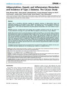
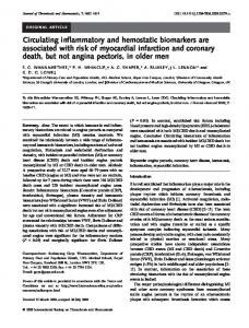
HMW adiponectin were associated with higher risk of HCC (IRR continuously per doubling of concentrations = 1.22; 95% CI = 1.02-1.46, P = 0.03; 1.90; 95% CI = 1.30-2.77, P = 0.001; 2.25; 95% CI = 1.43-3.54, P = 0.0005 and 2.09; 95% CI = 1.19-3.67, P = 0.01, respectively; Table 3). Higher levels of GLDH were also significantly associated with a higher risk of HCC (IRR = 1.62, 95% CI = 1.25-2.11, P = 0.0003; Table 3). There was no evidence for a nonlinear shape of these associations (Figure 2 Supplement). HMW-adiponectin, leptin and fetuin-a were not significantly associated with HCC risk in the multivariable-adjusted model. When additionally adjusted for GLDH, the associations remained unaltered, except for CRP, which was no longer statistically significant (Figure 1). Mutual adjustment of biomarkers also did not substantially affect the results, with the exception of non-HMW adiponectin, which was no longer significant after IL-6 was added to the multivariable model (IRR continuously per doubling of concentrations = 1.07; 95% CI: 0.30-3.82, P = 0.24). Higher CRP concentrations were associated with higher risk of GBTC (multivariable-adjusted IRR = 1.22; 95% CI = 1.05-1.42; P = 0.01; Table 4). This association remained statistically significant when the analyses were restricted to gallbladder cancer only (IRR = 1.55; 95% CI = 1.15-2.08; P = 0.003; Table 3 Supplement). Higher levels of GLDH were associated with a higher risk of IBD (IRR = 10.5; 95% CI = 2.2-50.9, P = 0.003; Table 5), but not with GBTC (IRR = 1.15; 95% CI = 0.95-1.40, P = 0.15; Table 4). The remaining biomarkers were not statistically significantly related to either GBTC or IBD cancers (Table 4; Table 5). Predictive capacity of biomarkers. Addition of CRP, IL-6, C-peptide, and non-HMW adiponectin to the multivariable model significantly increased the AUC for the prediction of HCC from 0.766 to 0.876 , whereas addition of the liver damage marker GLDH to the multivariable model raised the AUC from 0.769 to 0.813 (Figure 2) When inflammatory and metabolic biomarkers were added to the model, the IDI was 0.81, and the NRI was 0.63 (P < 0.0001), indicating strong
Hepatology
Hepatology
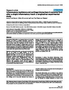
reclassification improvement; while when GLDH was added to the model, the IDI was 0.24 and the NRI was 0.46 (P = 0.07), indicating moderate improvement. Addition of CRP, IL-6, C-peptide, and non-HMW adiponectin to the multivariable model that additionally included α-fetoprotein (AFP) significantly increased the AUC for the prediction of HCC from 0.777 to 0.855, GLDH increased the AUC from 0.803 to 0.836 (Figure 3). When inflammatory and metabolic biomarkers were added to the model, the IDI was 0.43, and NRI(>0) was 0.44 (P = 0.0004), indicating moderate reclassification improvement; when GLDH was added to the model, the IDI was 0.10 and the NRI(>0) was 0.21 (P = 0.29), indicating weak improvement (Figure 3). Sensitivity Analyses. After exclusion of cases which occurred during the first two years of follow up, the associations of the biomarkers with HCC were not substantially changed, except for CRP and non-HMW adiponectin, which were no longer statistically significant (IRR, 1.10; 95% CI 0.88 – 1.37; P=0.12, and 1.63; 95% CI 0.86-3.05; P=0.12; Table 2 Supplement). The association with CRP was also attenuated and lost statistical significance after excluding cases with underlying HBsAg/anti-HCV infection (IRR, 1.17 (95% CI 0.95-1.45; P=0.12; Table 2 Supplement). We also did not observe any substantial differences in the results after excluding individuals with high alcohol consumption. Overall, no substantial differences in risk estimates were seen after exclusion of cases with prevalent diabetes, with the exception for the results for fetuin-a, which became statistically significant (IRR, 5.64; 95% CI 1.6019.89; Table 2 Supplement). Due to the small number of cases these analyses should be interpreted with caution. The associations were not altered when we restricted the analyses on HCC to histologically confirmed cases. Discussion In this prospective nested case-control study, higher circulating concentrations of IL-6, CRP, C-peptide, non-HMW adiponectin, and GLDH were significantly associated with higher Hepatology
Page 16 of 47
Page 17 of 47
Hepatology
risk of HCC, independent of established liver cancer risk factors and obesity parameters. Further, our data suggest these biomarkers to be able to improve the risk assessment of HCC, beyond established liver cancer risk factors, therefore suggesting their potential application for identification of individuals at high risk of cancer. In animal models it was shown that obesity may promote HCC development through elevated production of TNF and IL-6 (20). In clinical studies, higher levels of IL-6 and CRP have been found among patients with HCC when compared to controls (21, 22). Chronic inflammation is associated with persistent liver injury and consecutive regeneration, potentially leading to fibrosis and cirrhosis and consequently to the development of HCC (23). Chronic inflammation may also originate from hepatotropic viruses, toxins, or impaired autoimmunity (24). Mechanisms that link inflammation and liver cancer are not completely understood, but transcription factors of the NF-κB family and signal transducer and activator of transcription 3 (STAT3), cytokines such as IL-6, and ligands of the epidermal growth factor receptor family are pivotal players (24, 25). In line with our findings, a recent casecontrol study nested in a Japanese cohort with 188 HCC cases and 605 controls reported relative risks (95% CI) of 1.94 (0.72-5.51) for CRP, and 5.12 (1.54-20.1) for Il-6 for the highest tertile of biomarker distribution versus the lowest after multivariable adjustment (26). Interestingly, a recent study observed a lower risk of HCC among aspirin users, providing additional means for cancer prevention (27). Hyperinsulinemia is often present in patients with chronic hepatitis C, and is associated with more advanced HCV-related hepatic fibrosis (28). Clinical studies suggested that insulin resistance is significantly associated with HCC development in patients with chronic HCV infection (29, 30). Our data suggests that C-peptide as a marker of hyperinsulinemia is strongly positively associated with risk of HCC and IBD cancer, even after adjusting for HBC/HCV infection and inflammation, giving support to the hypothesis that hyperinsulinemia may increase risk of HCC and IBD cancer. High insulin levels may Hepatology
Hepatology
Page 18 of 47
directly promote cell proliferation and survival through the PI3K/Akt and Ras/MAPK pathways (31, 32). Insulin may also interact with leptin and adiponectin (see below). Adiponectin is involved in the regulation of energy homeostasis, vascular reactivity, inflammation, cell proliferation and tissue remodeling (33, 34). It primarly acts as an insulin sensitizing agent (35), but may also inhibit cancer cell growth (36), induce apoptosis (37), and thus be directly implicated in cancer (38). High adiponectin concentrations have been found to be associated with lower risks of prostate, breast, endometrial, colorectal (39) and pancreatic cancer (40). In contrast, in our study higher adiponectin levels were associated with higher risk of HCC. While this may be surprising given the beneficial aspects attributed to adiponectin, this is in line with previous studies that found adiponectin positively correlated with hepatic inflammation in patients with chronic liver disease (41) and with HCV-related HCC (42). We also observed that non-HMW adiponectin but not HMW adiponectin was significantly associated with risk of HCC. Furthermore, the association between non-HMW adiponectin and HCC risk was statistically largely accounted for by IL-6. Since lowmolecular forms of adiponectin are more closely associated with inflammation, we speculate whether IL-6 may act as a mediator in these associations. Leptin has angiogenic properties, promotes cell proliferation and migration, and interacts with growth factors, all of which could promote tumor growth (43). Evidence on the role of leptin in NAFLD and cancer risk is controversial with some studies showing positive associations and others showing null results (44),(45).
Our study does not support the
hypothesis that leptin levels are associated with liver cancer risk. On the basis of the mechanistic evidence obtained with cultured cells and tumor specimens, we speculate that local, rather than systemic, leptin concentrations may be important for tumor progression. In addition, leptin concentrations in plasma may be affected by the soluble leptin receptor (sOBR), a marker related to diabetes and cancer risk (46); however, future studies are warranted to examine whether sOB-R may be specifically related to liver cancer. Hepatology
Page 19 of 47
Hepatology
Fetuin-a is suggested to provide a link between fatty liver and insulin resistance (47, 48), thereby being potentially relevant for liver cancer. In our data, a significant association of fetuin-a with HCC risk was observed only after exclusion of participants with prevalent diabetes at baseline. Although these results may be due to a chance finding, we also speculate whether mechanisms other than insulin sensitivity may be more relevant here. High serum GLDH levels occur in liver diseases with hepatocyte necrosis as predominant event, such as toxic liver damage or hypoxic liver disease, and they have been useful in clinical practice in distinguishing between acute viral hepatitis and acute toxic liver necrosis or acute hypoxic liver disease (49). In our analysis, higher pre-diagnostic concentrations of GLDH were associated with higher risks of HCC and IBD. These data suggest that GLDH may be used as a marker of hepatic injury in liver cancer pathogenesis among ostensibly healthy subjects. Interestingly, in our analysis, the associations for IL-6, Cpeptide, and non-HMW adiponectin with HCC risk remained statistically significant after adjustment for GLDH, suggesting that prevalent undiagnosed liver injury may not account for these associations. Strengths of our study include the prospective design and the ability to control for established and putative liver cancer risk factors and for a variety of circulating metabolic markers. Anthropometric data were mostly measured rather than self-reported, which reduces the possibility of residual confounding by obesity. Limitations of our study include a relatively small number of incident cases, particularly for the analyses of the inflammatory biomarkers, which limited the possibility to perform detailed stratified and sensitivity analyses. The duration of follow-up was relatively short and concentrations of biomarkers may have been influenced by pre-existing undiagnosed disease. However, our risk estimates did not appreciably change after exclusion of patients who were diagnosed within the first two years of follow-up. Since most of our study participants were HBV/HCV negative our
Hepatology
Hepatology
findings are largely valid for HCC of non-viral etiology. Because histologically confirmed and probable HCC cases were included in the analyses, a potential misclassification of liver cancer cases may have occurred. However, when we performed analyses only with histologically confirmed HCC cases the results did not change. Additionally, since the distal part of the extrahepatic bile duct runs through the head of the pancreas, some of the cancers classified as GBTC may in fact be cancers of the pancreas and vice versa. Our results are based on single assessments of exposure variables within participants, and biomarkers may be susceptible to short-term variation, which would bias the results toward the null; however, most biomarkers have shown relatively high reliability over time (50). Due to the low prevalence of established risk factors (i.e. HBV/HCV infection, diabetes and alcohol consumption) in this study population, we were not able to evaluate whether biomarkers are specifically related to risk among persons with known risk factors, which may be a question of relevance to the clinical practice. We adjusted our analysis for a number of potential risk factors of liver cancer. Nevertheless, we cannot rule out the possibility of residual confounding. Further, given the observational nature, our study does necessarily prove causation. In conclusion, higher circulating concentrations of IL-6, CRP, C-peptide, non-HMW adiponectin, and GLDH were significantly associated with higher risk of HCC, independent of established liver cancer risk factors and obesity parameters. Further studies are warranted to investigate the role of these inflammatory and metabolic biomarkers as mediators of the relation between obesity and liver cancer, as well as to explore their potential applications for cancer prevention.
Hepatology
Page 20 of 47
Page 21 of 47
Hepatology
Figure Legends Figure 1. Association of metabolic biomarkers (continuously per doubling of concentrations) and risk of HCC in multivariable model before and after adjustment for GLDH as a marker of liver damage Abbreviations: Il-6, Interleuking-6, CRP, C-reactive protein, HMW, high molecular weight, GLDH, Glutamate dehydrogenase. a
Multivariable model taking into account matching factors: study center, gender, age (± 12
months), date (±2 months), fasting status (6hrs), and time of the day (±3 hours) at blood collection. Women were additionally matched according to menopausal status (pre-, peri-(unknown), postmenopausal) and exogenous hormone use (yes, no, missing) at blood donation. Further adjusted for education (no school degree or primary school, secondary school, high school, missing), smoking (never, former, current, missing), alcohol at baseline, drinking status at baseline (non-drinker, drinker), diabetes (no, yes, missing), coffee (g/day), HBsAg/anti-HCV (negative, positive, missing), BMI, and waist to height ratio (WHtR) adjusted for BMI. Note: The analyses we based on overall 293cases and 581controls for adiponectin, fetuin-a and leptin, 293 cases and 577 controls for CRP and GLDH, 277cases and 549 controls for Cpeptide, 214 cases and 419 controls for Il-6.
Hepatology
Hepatology
Figure 2. Predictive ability of inflammatory and metabolic biomarkersa and GLDH beyond the multivariable adjusted modelb Abbreviations: Il-6, Interleukin-6, CRP, C-reactive protein, HMW, high molecular weight, GLDH, Glutamate dehydrogenase. a
The biomarkers included in the model have been associated with HCC risk. These include
CRP, Il-6, C-peptide and non-HMW adiponectin. b
Multivariable model taking into account matching factors: study center, gender, age (± 12
months), date (±2 months), fasting status (6hrs), and time of the day (±3 hours) at blood collection. Women were additionally matched according to menopausal status (pre-, peri-(unknown), postmenopausal) and exogenous hormone use (yes, no, missing) at blood donation. Further adjusted for education (no school degree or primary school, secondary school, high school, missing), smoking (never, former, current, missing), alcohol at baseline, drinking status at baseline (non-drinker, drinker), diabetes (no, yes, missing), coffee (g/day), HBsAg/anti-HCV (negative, positive, missing), BMI, and waist to height ratio (WHtR) adjusted for BMI. Note: The analyses we based on overall 293 cases and 581controls for adiponectin, 293 cases and 577 controls for CRP and GLDH, 277cases and 549 controls for C-peptide. For this analysis, missing values for Il6 (33 cases, 72 controls) were substituted with sex- and casecontrol-specific median values.
Hepatology
Page 22 of 47
Page 23 of 47
Hepatology
Figure 3. Predictive ability of inflammatory and metabolic biomarkers and GLDH beyond the multivariable adjusted model and α-fetoprotein (AFP) levels Abbreviations: Il-6, Interleukin-6, CRP, C-reactive protein, HMW, high molecular weight, GLDH, Glutamate dehydrogenase. a
The biomarkers included in the model have been associated with HCC risk. These include
CRP, Il-6, C-peptide and non-HMW adiponectin. b
Multivariable model taking into account matching factors: study center, gender, age (± 12
months), date (±2 months), fasting status (6hrs), and time of the day (±3 hours) at blood collection. Women were additionally matched according to menopausal status (pre-, peri-(unknown), postmenopausal) and exogenous hormone use (yes, no, missing) at blood donation. Further adjusted for education (no school degree or primary school, secondary school, high school, missing), smoking (never, former, current, missing), alcohol at baseline, drinking status at baseline (non-drinker, drinker), diabetes (no, yes, missing), coffee (g/day), HBsAg/anti-HCV (negative, positive, missing), BMI, and waist to height ratio (WHtR) adjusted for BMI. Note: The analyses we based on overall 293 cases and 581controls for adiponectin, 293 cases and 577 controls for CRP and GLDH, 277cases and 549 controls for C-peptide. For this analysis, missing values for Il6 (33 cases, 72 controls) were substituted with sex- and casecontrol-specific median values.
Hepatology
Hepatology
REFERENCES 1.
Siegel R, Ward E, Brawley O, Jemal A. Cancer statistics, 2011: the impact of
eliminating socioeconomic and racial disparities on premature cancer deaths. CA Cancer J Clin 2011;61:212-236. 2.
Jemal A, Bray F, Center MM, Ferlay J, Ward E, Forman D. Global cancer statistics.
CA Cancer J Clin 2011;61:69-90. 3.
Caldwell SH, Crespo DM, Kang HS, Al-Osaimi AM. Obesity and hepatocellular
carcinoma. Gastroenterology 2004;127:S97-103. 4.
Baffy G, Brunt EM, Caldwell SH. Hepatocellular carcinoma in non-alcoholic fatty
liver disease: An emerging menace. J Hepatol 2012. 5.
Trichopoulos D, Bamia C, Lagiou P, Fedirko V, Trepo E, Jenab M, Pischon T, et al.
Hepatocellular carcinoma risk factors and disease burden in a European cohort: a nested casecontrol study. J Natl Cancer Inst 2011;103:1686-1695. 6.
Vineis P, Perera F. Molecular epidemiology and biomarkers in etiologic cancer
research: the new in light of the old. Cancer Epidemiol Biomarkers Prev 2007;16:1954-1965. 7.
Slimani N, Kaaks R, Ferrari P, Casagrande C, Clavel-Chapelon F, Lotze G, Kroke A,
et al. European Prospective Investigation into Cancer and Nutrition (EPIC) calibration study: rationale, design and population characteristics. Public Health Nutr 2002;5:1125-1145. 8.
International Statistical Classification of Diseases and Related Health Problems, Tenth
Revision – ICD-10. In. 2 ed. Geneva, Switzerland: World Health Organisation 2004 9.
International classification of diseases for oncology. In: Organisation WH, editor. 2
ed. Geneva, Switzerland; 1990. 10.
Riboli E, Hunt KJ, Slimani N, Ferrari P, Norat T, Fahey M, Charrondiere UR, et al.
European Prospective Investigation into Cancer and Nutrition (EPIC): study populations and data collection. Public Health Nutr 2002;5:1113-1124.
Hepatology
Page 24 of 47
Page 25 of 47
Hepatology
11.
Bowker AH. A test for symmetry in contingency tables. J Am Stat Assoc
1948;43:572-574. 12.
Prentice RL BN. Retrospective studies and failure time models. Biometrika
1978;65:153-158. 13.
O'Brien PJ, Slaughter MR, Polley SR, Kramer K. Advantages of glutamate
dehydrogenase as a blood biomarker of acute hepatic injury in rats. Lab Anim 2002;36:313321. 14.
Durrleman S, Simon R. Flexible regression models with cubic splines. Stat Med
1989;8:551-561. 15.
Pencina MJ, D'Agostino RB, Sr., Demler OV. Novel metrics for evaluating
improvement
in
discrimination:
net
reclassification
and
integrated
discrimination
improvement for normal variables and nested models. Stat Med 2012;31:101-113. 16.
Steyerberg EW, Pencina MJ, Lingsma HF, Kattan MW, Vickers AJ, Van Calster B.
Assessing the incremental value of diagnostic and prognostic markers: a review and illustration. Eur J Clin Invest 2012;42:216-228. 17.
DeLong ER, DeLong DM, Clarke-Pearson DL. Comparing the areas under two or
more correlated receiver operating characteristic curves: a nonparametric approach. Biometrics 1988;44:837-845. 18.
Mühlenbruch
K.
Ein
Makro
zur
Berechnung
von
Diskriminanz-
und
Reklassifizierungsstatistiken für die Verbesserung eines Prädiktionsmodells bei Anwendung der Cox-Regression. In: Hilbert A MR, eds. Proceedings der 16 Konferenz der SASAnwender in Forschung und Entwicklung (KSFE). Aachen: Shaker Verlag, 2012:249-62. 19.
Pencina MJ, D'Agostino RB, Pencina KM, Janssens AC, Greenland P. Interpreting
incremental value of markers added to risk prediction models. Am J Epidemiol 2012;176:473481.
Hepatology
Hepatology
20.
Park EJ, Lee JH, Yu GY, He G, Ali SR, Holzer RG, Osterreicher CH, et al. Dietary
and genetic obesity promote liver inflammation and tumorigenesis by enhancing IL-6 and TNF expression. Cell 2010;140:197-208. 21.
Budhu A, Wang XW. The role of cytokines in hepatocellular carcinoma. J Leukoc
Biol 2006;80:1197-1213. 22.
Porta C, De Amici M, Quaglini S, Paglino C, Tagliani F, Boncimino A, Moratti R, et
al. Circulating interleukin-6 as a tumor marker for hepatocellular carcinoma. Ann Oncol 2008;19:353-358. 23.
Sun B, Karin M. Obesity, inflammation, and liver cancer. J Hepatol 2012;56:704-713.
24.
Alison MR, Nicholson LJ, Lin WR. Chronic inflammation and hepatocellular
carcinoma. Recent Results Cancer Res 2011;185:135-148. 25.
Berasain C, Castillo J, Perugorria MJ, Latasa MU, Prieto J, Avila MA. Inflammation
and liver cancer: new molecular links. Ann N Y Acad Sci 2009;1155:206-221. 26.
Ohishi W, Cologne JB, Fujiwara S, Suzuki G, Hayashi T, Niwa Y, Akahoshi M, et al.
Serum Interleukin-6 associated with hepatocellular carcinoma risk: A nested case-control study. Int J Cancer 2013. 27.
Sahasrabuddhe VV, Gunja MZ, Graubard BI, Trabert B, Schwartz LM, Park Y,
Hollenbeck AR, et al. Nonsteroidal anti-inflammatory drug use, chronic liver disease, and hepatocellular carcinoma. J Natl Cancer Inst 2012;104:1808-1814. 28.
Souza AFMd, Pace FHdL, Chebli JMF, Ferreira LEVVdC. Insulin resistance in non-
diabetic patients with chronic hepatitis C: what does it mean? Arquivos Brasileiros de Endocrinologia & Metabologia 2011;55:412-418. 29.
Hung CH, Wang JH, Hu TH, Chen CH, Chang KC, Yen YH, Kuo YH, et al. Insulin
resistance is associated with hepatocellular carcinoma in chronic hepatitis C infection. World J Gastroenterol 2010;16:2265-2271.
Hepatology
Page 26 of 47
Page 27 of 47
Hepatology
30.
Salmon D, Bani-Sadr F, Loko MA, Stitou H, Gervais A, Durant J, Rosenthal E, et al.
Insulin resistance is associated with a higher risk of hepatocellular carcinoma in cirrhotic HIV/HCV-co-infected patients: Results from ANRS CO13 HEPAVIH. J Hepatol 2012;56:862-868. 31.
Donohoe CL, Doyle SL, Reynolds JV. Visceral adiposity, insulin resistance and
cancer risk. Diabetol Metab Syndr 2011;3:12. 32.
Doyle SL, Donohoe CL, Lysaght J, Reynolds JV. Visceral obesity, metabolic
syndrome, insulin resistance and cancer. Proc Nutr Soc 2012;71:181-189. 33.
Gunter MJ, Leitzmann MF. Obesity and colorectal cancer: epidemiology, mechanisms
and candidate genes. J Nutr Biochem 2006;17:145-156. 34.
Brochu-Gaudreau K, Rehfeldt C, Blouin R, Bordignon V, Murphy BD, Palin MF.
Adiponectin action from head to toe. Endocrine 2010;37:11-32. 35.
Yang WS, Chuang LM. Human genetics of adiponectin in the metabolic syndrome. J
Mol Med (Berl) 2006;84:112-121. 36.
Kim AY, Lee YS, Kim KH, Lee JH, Lee HK, Jang SH, Kim SE, et al. Adiponectin
represses colon cancer cell proliferation via AdipoR1- and -R2-mediated AMPK activation. Mol Endocrinol 2010;24:1441-1452. 37.
Byeon JS, Jeong JY, Kim MJ, Lee SM, Nam WH, Myung SJ, Kim JG, et al.
Adiponectin and adiponectin receptor in relation to colorectal cancer progression. Int J Cancer 2010. 38.
La Cava A. Adiponectin: a relevant player in obesity-related colorectal cancer? Gut
2013;62:483-484. 39.
Izadi V, Farabad E, Azadbakht L. Serum adiponectin level and different kinds of
cancer: a review of recent evidence. ISRN Oncol 2012;2012:982769.
Hepatology
Hepatology
40.
Bao Y, Giovannucci EL, Kraft P, Stampfer MJ, Ogino S, Ma J, Buring JE, et al. A
prospective study of plasma adiponectin and pancreatic cancer risk in five US cohorts. J Natl Cancer Inst 2013;105:95-103. 41.
Jonsson JR, Moschen AR, Hickman IJ, Richardson MM, Kaser S, Clouston AD,
Powell EE, et al. Adiponectin and its receptors in patients with chronic hepatitis C. J Hepatol 2005;43:929-936. 42.
Khattab MA, Eslam M, Mousa YI, Ela-adawy N, Fathy S, Shatat M, Abd-Aalhalim H,
et al. Association between metabolic abnormalities and hepatitis C-related hepatocellular carcinoma. Ann Hepatol 2012;11:487-494. 43.
Chan JL, Mantzoros CS. Role of leptin in energy-deprivation states: normal human
physiology and clinical implications for hypothalamic amenorrhoea and anorexia nervosa. Lancet 2005;366:74-85. 44.
Czaja MJ. Liver injury in the setting of steatosis: crosstalk between adipokine and
cytokine. Hepatology 2004;40:19-22. 45.
Garofalo C, Surmacz E. Leptin and cancer. J Cell Physiol 2006;207:12-22.
46.
Aleksandrova K, Boeing H, Jenab M, Bueno-de-Mesquita HB, Jansen E, van
Duijnhoven FJ, Rinaldi S, et al. Leptin and soluble leptin receptor in risk of colorectal cancer in the European Prospective Investigation into Cancer and Nutrition cohort. Cancer Res 2012;72:5328-5337. 47.
Stefan N, Hennige AM, Staiger H, Machann J, Schick F, Krober SM, Machicao F, et
al. Alpha2-Heremans-Schmid glycoprotein/fetuin-A is associated with insulin resistance and fat accumulation in the liver in humans. Diabetes Care 2006;29:853-857. 48.
Ix JH, Shlipak MG, Brandenburg VM, Ali S, Ketteler M, Whooley MA. Association
between human fetuin-A and the metabolic syndrome: data from the Heart and Soul Study. Circulation 2006;113:1760-1767.
Hepatology
Page 28 of 47
Page 29 of 47
Hepatology
49.
Schomaker S, Warner R, Bock J, Johnson K, Potter D, Van Winkle J, Aubrecht J.
Assessment of emerging biomarkers of liver injury in human subjects. Toxicol Sci 2013;132:276-283. 50.
Pischon T, Hotamisligil GS, Rimm EB. Adiponectin: stability in plasma over 36 hours
and within-person variation over 1 year. Clin Chem 2003;49:650-652.
Acknowledgment: We thank Ellen Kohlsdorf (EPIC-Potsdam, Germany) for her work on data management and technical assistance. We thank all participants in the EPIC study for their outstanding cooperation.
Conflicts of interest statement: None declared.
Hepatology
Hepatology
Page 30 of 47
Table 1: Selected baseline characteristics of incident cases of HCC, IBD and GBTC outside of the liver and their matched controls, the European Prospective Investigation into Cancer and Nutrition, 1992HCC Characteristic Number Female sex, % Age, years, mean (SD) LIVER CANCER RISK FACTORS Smoking status, n (%) Never smoker Former smoker Current smoker Education, n (%) No school degree or primary school Secondary school High school Body mass index†, kg/m2 , mean (SD) Waist circumference, cm, mean (SD) Waist-height-ratio, mean (SD) Chronic HBsAg/anti-HCV infection No, n (%) Yes, n (%) Missing, n (%) Diabetes No, n (%) Yes, n (%) Missing, n (%) Ethanol intake at baseline (g/day) ‡ None to low, n (%) Moderate, n (%) High, n (%) Coffee intake, g/d
BIOMARKERS CRP, mg/L, median (IQR)
Cases
Controls
125 32 60.1 (6.6)
250 31.6 60.1 (6.6)
34 (27.2) 41 (32.8) 48 (38.4)
105 (42.0) 97 (38.8) 47 (18.8)
52.2 29.2 16.0 28.1 (5.3) 97.1 (15.2) 0.57 (0.08)
47.8 30.0 19.6 26.9 (3.9) 92.6 (11.2) 0.54 (0.06)
82 (65.6) 40 (32) 3 (2.4)
231 (92.4) 13 (5.2) 6 (2.4)
105 (84) 16 (12.8) 4 (3.2)
225 (90) 16 (6.4) 9 (3.6)
71 (56.8) 27 (21.6) 27 (21.6)
GBTC P-paired
*
Cases
Controls
137 56.2 58.5 (7.5)
274 56.2 58.5 (7.5)
62 (45.2) 38 (27.7) 36 (26.3)
133 (48.5) 84 (30.7) 55 (20.1)
44.7 38.7 16.1 26.9 (4.7) 89.8 (14.3) 0.54 (0.08)
47.6 35.1 16.1 26.4 (3.9) 88.2 (12.6) 0.53 (0.07)
123 (89.8) 10 (7.3) 4 (2.9)
248 (90.5) 16 (5.8) 10 (3.7)
0.03
121 (88.3) 9 (6.6) 7 (5.1)
126 (50.4) 97 (38.8) 27 (10.8)