Jul 3, 2006 - Results. Ketoconazole inhibits rifampicin- and paclitaxel-mediated ..... For all experiments, bound protein complexes were washed, separated.
Oncogene (2007) 26, 258–268
& 2007 Nature Publishing Group All rights reserved 0950-9232/07 $30.00 www.nature.com/onc
ORIGINAL ARTICLE
Inhibition of drug metabolism by blocking the activation of nuclear receptors by ketoconazole H Huang1,2, H Wang1,2, M Sinz3, M Zoeckler3, J Staudinger4, MR Redinbo5, DG Teotico5, J Locker6, GV Kalpana1,7 and S Mani1,2,7 1 Albert Einstein Cancer Center, Albert Einstein College of Medicine, Bronx, NY, USA; 2Department of Medicine, Albert Einstein College of Medicine, Bronx, NY, USA; 3Bristol Myers Squibb Co., Wallingford, CT, USA; 4Department of Toxicology, University of Kansas, Lawrence, KS, USA; 5Department of Chemistry, University of North Carolina at Chapel Hill, Chapel Hill, NC, USA; 6 Department of Pathology, Albert Einstein College of Medicine, Bronx, NY, USA and 7Department of Molecular Genetics, Albert Einstein College of Medicine, Bronx, NY, USA
Individual variation in drug metabolism is a major cause of unpredictable side effects during therapy. Drug metabolism is controlled by a class of orphan nuclear receptors (NRs), which regulate expression of genes such as CYP (cytochrome)3A4 and MDR-1 (multi-drug resistance-1), that are involved in this process. We have found that xenobiotic-mediated induction of CYP3A4 and MDR-1 gene transcription was inhibited by ketoconazole, a commonly used antifungal drug. Ketoconazole mediated its effect by inhibiting the activation of NRs, human pregnenolone X receptor and constitutive androstene receptor, involved in regulation of CYP3A4 and MDR-1. The effect of ketoconazole was specific to the group of NRs that control xenobiotic metabolism. Ketoconazole disrupted the interaction of the xenobiotic receptor PXR with the co-activator steroid receptor co-activator-1. Ketoconazole treatment resulted in delayed metabolism of tribromoethanol anesthetic in mice, which was correlated to the inhibition of PXR activation and downmodulation of cyp3a11 and mdr-1 genes and proteins. These studies demonstrate for the first time that ketoconazole represses the coordinated activation of genes involved in drug metabolism, by blocking activation of a specific subset of NRs. Our results suggest that ketoconazole can be used as a pan-antagonist of NRs involved in xenobiotic metabolism in vivo, which may lead to novel strategies that improve drug effect and tolerance. Oncogene (2007) 26, 258–268. doi:10.1038/sj.onc.1209788; published online 3 July 2006 Keywords: drug metabolism; transcriptional regulation; orphan nuclear receptors
Correspondence: Dr S Mani, Department of Oncology, Albert Einstein Cancer Center, Albert Einstein College of Medicine, 1300 Morris Park Avenue, Chanin 302D-1, Bronx, NY 10461, USA. E-mail: smani@montefiore.org Received 4 May 2006; revised 30 May 2006; accepted 30 May 2006; published online 3 July 2006
Introduction Individual variation in drug metabolism is one of the major causes of treatment failures during cancer therapy (Undevia et al., 2005). Understanding the expression and activity of enzymes involved in metabolism and biotransformation may lead to the development of novel strategies to regulate this process. The expression of genes encoding the drug metabolizing enzymes is tightly and coordinately regulated at the level of transcription by the action of orphan nuclear receptors (NRs) (Kliewer et al., 2002; Sonoda et al., 2003; Eloranta et al., 2005). NRs (e.g., pregnenolone X receptor, PXR; constitutive androstene receptor, CAR) controlling drug metabolism belong to a superfamily of a number of closely related ligand-dependent and -independent transcription factors (Blumberg et al., 1998; Wei et al., 2000; Handschin and Meyer, 2005). These transcription factors are characterized by a C-terminal ligand binding domain and an N-terminal zinc-finger DNA binding domain. Their ligands are small endobiotic lipophilic compounds (e.g., bile acids, fatty acids) as well as larger xenobiotic compounds that infrequently act as inducers or activators (e.g., rifampicin and phenobarbital that activate hPXR and hCAR, respectively) and sometimes as repressors (e.g., highly chlorinated polychlorinated biphenyls that inhibit hPXR) of these NRs (Blumberg et al., 1998; Wei et al., 2000; Xie et al., 2003; Tabb et al., 2004; Handschin and Meyer, 2005). It is important to note that there is a cross-talk in receptor-mediated gene transcription by PXR, CAR and farnesol X receptor (FXR) (Xie et al., 2000; Wei et al., 2002; Gnerre et al., 2004). For example, all three NRs mediate CYP (cytochrome)3A4 transcription and in the absence of one or more receptors the other may maintain transcriptional homeostasis. We have previously demonstrated that microtubulebinding drugs serve as activating ligands of hPXR (22) and others have shown that a variety of xenobiotics activate CAR and FXR. All these agents combine to activate genes involved in drug metabolism (e.g., CYP3A4 and CYP2B6) and transport (e.g., MDR-1 (multi-drug resistance-1)) (Synold et al., 2001; Evans,
Ketoconazole, nuclear receptors and drug metabolism H Huang et al
259
2005; Handschin and Meyer, 2005; Mani et al., 2005). As patients are generally administered multiple drugs at any given time, it is difficult to predict the net transcriptional outcome and resultant effects on drug pharmacokinetics and pharmacodynamics. Therefore, finding pan-antagonists for PXR, CAR and FXR could provide a unique tool to control drug metabolism. As a prototype for pan-antagonist, we have analysed the effect of ketoconazole. It has been demonstrated previously that ketoconazole inhibits corticosteroidmediated hPXR activation and the interaction of silencing mediator of retinoid and thyroid hormone receptor (SMRT) (co-repressor) and steroid receptor coactivator-1 (SRC-1) (co-activator) with hPXR (Takeshita et al., 2002). Additionally, it has been established that ketoconazole inhibits the glucocorticoid receptor (Loose et al., 1983; Pascussi et al., 2001). However, the effect of ketoconazole on other NRs is largely unknown. Furthermore, even though ketoconazole blocks coregulator interactions, additional mechanisms may be involved at the receptor level. More importantly, it is unclear whether blocking NR activation by ketoconazole has any significant effect on drug metabolism in vivo.
In this report, we found that ketoconazole is a general inhibitor of activated PXR, CAR, liver activated receptor (LXR) and FXR. In addition, analysis of the molecular mechanism of inhibition of gene transcription demonstrated that ketoconazole specifically disrupts the interaction of NRs with co-regulators and does not affect DNA binding, ligand binding or receptor dimerization. Finally, we have demonstrated, for the first time, that ketoconazole can be used to control drug metabolism by utilizing in vivo mouse models. Results Ketoconazole inhibits rifampicin- and paclitaxel-mediated transcriptional activation of CYP3A4 and MDR-1 genes in cell culture To determine the effect of ketoconazole on xenobioticmediated transcription of CYP3A4 and MDR-1, we used HepG2 hepatocarcinoma cells, primary human hepatocytes and LS174T colon carcinoma cell lines. In HepG2 cells, rifampicin and paclitaxel induced 12.9and 10.1-fold increases in CYP3A4 mRNA expression,
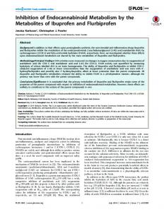
Figure 1 Inhibition of ligand-mediated activation of CYP3A4 and MDR-1 gene transcription by ketoconazole. (a–c) Ketoconazole inhibits the expression of CYP3A4 gene and protein. (a) Real-time RT–PCR analyses of total mRNA isolated from HepG2 cells. (b) Northern blot analysis of total mRNA isolated from human hepatocytes. (c) Immunoblot analysis of total proteins isolated from HepG2 cell microsome fractions. (d and e) Ketoconazole inhibits the expression of MDR-1 gene and protein in LS174T cells. (d) Northern blot analysis of total mRNA. (e) Immunoblot analysis of total membrane protein fractions of LS174T cells. The cells were treated for either 48 h (a–c) or 72 h. (d and e) with drug(s) as indicated and total mRNA and microsome- or membrane-associated proteins were isolated. b-Actin was used as a loading control for Northern analyses (b and d), Ponceau staining was used to ensure equal loading of microsome protein fractions (c) and Na þ /K þ ATPase was used as a loading control for membrane-associated protein fractions (e). Each RNA experiment was repeated at least three times. Immunoblot experiments were repeated three separate times. (c and e) Histograms reflect quantitative densitometric analysis of average fold change in CYP3A4 and MDR-1 bands normalized to the control (DMSO) from all three separate immunoblots, respectively. Columns, mean; bars, s.e.m. Oncogene
Ketoconazole, nuclear receptors and drug metabolism H Huang et al
260
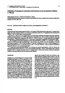
Figure 2 Inhibition of ligand-mediated activation of orphan NRs by ketoconazole. (a, b) Ketoconazole inhibits activation of PXR. Transient transcription assays were carried out in CV-1 cells co-transfected with plasmids expressing either pCMX-hPXR (a) or pCMX-mPXR (b) along with CYP3A4 luciferase reporter plasmid (�10 466 to þ 53). (c) Ketoconazole inhibits activation of CAR. Transient transcription analysis in HepG2 cells, co-transfected with pCI-mCAR expression plasmid and cyp2b10 reporter plasmid. Androstenol a and b were used as CAR inverse agonists (*Po0.001). (d) Ketoconazole inhibits activation of FXR. Transient transcription assays were carried out in CV-1 cells co-transfected with GAL4DB-FXR-LBD along with Tk-MH100 � 4-luc reporter plasmid. (e) Ketoconazole inhibits activation of LXR. Transient transcription assays were carried out in HepG2 cells co-transfected with GAL4DB-LXR-LBDa or b and Tk-MH100 � 4-luc (*, **Po0.03). T0901317 was used as an agonist ligand for LXR. (f, g) Ketoconazole does not inhibit the activation of PPARg or ERa. Transient transcription assays were carried out in CV-1 cells cotransfected with Gal4DB-PPARg-LBD and Tk-MH100 � 4-luc (f) or Gal4DB-ERa-LBD and Tk-MH100 � 4-luc (g). In all transient transcription assays indicated, plasmids and pSV-b-galactosidase control vector were co-transfected for 24 h. Subsequently, the cells were treated with drug(s) as indicated. All transfections were normalized for transfection efficiency using pSV-b-galactosidase in the presence or absence of drug(s) as indicated. The cells were harvested in equal aliquots at 24 h post-drug treatment for luciferase and b-galactosidase assays (for details, see Materials and methods). All experiments were carried out at least thrice in triplicate. Columns, mean; bars, s.e.m.
Oncogene
Ketoconazole, nuclear receptors and drug metabolism H Huang et al
261
respectively. This stimulation was largely eliminated by addition of ketoconazole, even though ketoconazole alone slightly increased basal level transcription (Figure 1a). Similar results were obtained in human hepatocytes (Figure 1b). We demonstrated that the effect of ketoconazole was reflected in steady-state protein levels. Immunoblot analysis using antibodies to CYP3A4 protein indicated that both paclitaxel and rifampicin significantly increased CYP3A4 protein levels (Figure 1c). As for mRNA, ketoconazole blocked protein induction to basal levels. Effect of ketoconazole was also assessed on the paclitaxel- and rifampicin-mediated induction of MDR-1 (P-glycoprotein) in LS174T cells, by both Northern and Western blot analysis (Figure 1d and e). As with CYP3A4, ketoconazole inhibited induction of MDR-1 to basal levels. These studies together indicated that ketoconazole inhibited paclitaxel- and rifampicin-induced transcriptional activation of CYP3A4 and MDR-1 genes in hepatocytes and LS174T colon cancer cell lines. Inhibition at the transcription level was associated with a decrease in protein levels by ketoconazole. Importantly, the inhibition by ketoconazole was close to the baseline (dimethylsulfoxide (DMSO) control), suggesting that ketoconazole may be an effective agent for simultaneously inhibiting biotransformation enzymes and transporters. Ketoconazole inhibits activation of PXR and related adopted orphan NR but not that of ERa and PPARg Previous studies have shown that ketoconazole inhibits ligand-mediated activation of hPXR (Takeshita et al., 2002; Mani et al., 2005). As rifampicin and paclitaxel are activating ligands of hPXR, and as hPXR coordinately regulates the transcription of both CYP3A4 and MDR-1, ketoconazole may act by inhibiting the activation of this receptor. To determine whether ketoconazole inhibited hPXR-mediated activation of CYP3A4 gene, transient transcription assays were carried out in CV-1 cells. Whereas rifampicin and paclitaxel activated hPXR, ketoconazole inhibited this activation to below basal levels (Figure 2a). Similarly, ketoconazole inhibited the pregnenolone 16 a-carbonitrile (PCN)-mediated activation of mouse PXR (Figure 2b). To determine if ketoconazole can inhibit the activation of other NRs, transient transfection assays were carried out in the same cell lines used for PXR experiments. First, the effect of ketoconazole was tested on mouse (m)CAR (Figure 2c). CAR is known to be activated in its basal (unliganded) state (Wei et al., 2000; Figure 2c). TCPOBOP, a known agonist of mCAR, further activated CAR (Figure 2c). Ketoconazole inhibited both basal and TCPOBOP-mediated activation of mCAR. The levels of inhibition by ketoconazole were comparable to that observed by a- or b-androstenol, which is a known inverse agonist of mCAR (Figure 2c). We found similar results for phenobarbitalactivated hCAR (data not shown).
The effect of ketoconazole was also tested on a related NR FXR. FXR can crosstalk with PXR and CAR and also serves to regulate both lipid and bile acid metabolism (Gnerre et al., 2004). Unlike CAR, FXR is basally repressed and it is activated by chenodeoxycholic
Figure 3 Inhibition by ketoconazole is mediated by disruption of xenobiotic receptor’s interaction with the co-activator SRC-1. Mammalian two-hybrid studies in (a) HepG2 and (b,c) CV-1 cells to study the interaction of NR with SRC-1 in the presence and absence of drug(s). (a) HepG2 cells were transfected with pairs of plasmids expressing the proteins as indicated in the graph and the luciferase activity was measured (*, **, Po0.0001; FL ¼ full length cDNA; RID ¼ receptor interacting domain; UASG x 4 is upstream activating sequence of gal4 x 4); CV-1 cells were transfected with pairs of plasmids expressing protein as indicated in (b) (*Po0.0001) and (c), and luciferase activity was measured. All transfections were normalized for transfection efficiency using pSVb-galactosidase in the presence or absence of drug(s) as indicated (refer to Materials and methods). All experiments were carried out at least thrice in triplicate. Columns, mean; bars, s.e.m. Oncogene
Ketoconazole, nuclear receptors and drug metabolism H Huang et al
262
acid (secondary bile acid) but not lithocholic acid (another bile acid by-product) (Figure 2d). We found that ketoconazole inhibited chenodeoxycholic-induced GAL4DB-FXR-mediated activation to the basal level (Figure 2d). We also tested the effect of ketoconazole on activation of both GAL4DB fusions of LXRa and b isoforms. T0901317, a known chemical agonist of LXRa and b, activated GAL4DB-LXR 14- and 6.7-fold, respectively. Ketoconazole inhibited T0901317-induced LXRa- and b-mediated activation (Figure 2e). The receptors tested above are involved in xenobiotic or bile acid metabolism. As examples of receptors not directly implicated in xenobiotic or bile acid metabolism, we tested the effect of ketoconazole on other receptors, peroxisome proliferator-activated receptor (PPAR)g and estrogen receptor (ER)a activation, involved in lipid homeostasis and estrogen signaling, respectively. Ketoconazole had no effect on either PPARg or ERa activation in CV-1 cells (Figure 2g). All these results indicated that although ketoconazole uniformly inhibited the ligand-mediated activation of adopted orphan NRs involved in xenobiotic or bile acid metabolism, it had no effect on other NRs such as PPARg or ERa. By contrast, ketoconazole alone exhibited variable effects (increase, decrease, no effect) on unliganded receptors. Ketoconazole disrupts ligand-mediated binding of NR to co-activator (SRC-1) in vivo It has been demonstrated that corticosteroid-mediated hPXR interaction with co-activator (SRC-1) is disrupted by ketoconazole (Takeshita et al., 2002). To determine whether ketoconazole can disrupt the rifampicin- and paclitaxel-induced binding of hPXR to SRC-1, we performed mammalian two-hybrid experiments (Figure 3). We found that both rifampicin and paclitaxel augmented the interaction of GAL4DB-SRC-1 with VP16-hPXR (full length) (Figure 3a). Ketoconazole significantly inhibited rifampicin- and paclitaxelmediated interaction of SRC-1 and full-length hPXR (Figure 3a). We also tested the interaction of GAL4DB-SRC-1RID with VP16–FXR in the presence and absence of
ketoconazole, using the mammalian two-hybrid system (Figure 3b). As reported before ((Fujino et al., 2003), in vitro kd ¼ 36.2 mM), we found that FXR does not interact strongly with SRC-1 in the absence of its ligand (Figure 3b). Although FXR did not show interaction with SRC-1, addition of chenodeoxycholic acid dramatically augmented SRC-1 and FXR interaction, which was inhibited by ketoconazole. SRC-1 is a known activator of ERa (Lee et al., 2002; Bourdoncle et al., 2005). Therefore, we tested the effect of ketoconazole on interaction of SRC-1 with ERa. Ketoconazole did not inhibit the estrogen-mediated interaction of ERa with SRC-1 (Figure 3c). These results are consistent with the lack of inhibition of ERamediated activation by ketoconazole. Together, the above results suggest that ketoconazole exhibits specificity in disrupting NR interaction with co-activator. Ketoconazole disrupts the interaction of SRC-1 with hPXR-LBD in vitro To confirm the results of the mammalian two-hybrid system, we carried out in vitro CARLA (co-activatordependent receptor ligand assay) to determine the effect of ketoconazole. We found that both rifampicin and paclitaxel (as compared with DMSO) significantly augmented the interaction of SRC-1 with hPXR in these assays (Figure 4a and b). However, ketoconazole inhibited these interaction to basal levels (Figure 4a and b, compare lane 6 to lanes 2 and 4). Ketoconazole alone did not significantly affect the basal level interaction of SRC-1 with hPXR (Figure 4a and b, compare lanes 1 and 6). Furthermore, ketoconazole had no affect on binding of SRC-1 with ERa in the presence of estradiol (Figure 4c, lane 2 vs 3). These results are consistent with and directly confirm the observations of our mammalian two-hybrid studies. Ketoconazole does not affect RXR:PXR heterodimer formation, binding to DNA or ligand binding To rule out the involvement of other mechanisms mediating ketoconazole action, we tested the effect of ketoconazole on binding of hPXR:RXR complex to specific DNA containing DR-3 or ER-6 sequences. It is well established that only the hPXR heterodimer pair
Figure 4 Ketoconazole specifically disrupts receptor—co-activator interaction in vitro but does not affect ligand binding, DNA binding and heterodimer formation. GST pull-down assays by using 35S-labeled human SRC-1-RID (a) or full-length hPXR (b) with purified GST-hPXR or GST-SRC-1, respectively. Histogram represents average quantitative densitometry of 35S-hPXR from two separate experiments and expressed as fold abundance of protein relative to lane 6 (DMSO). (c) GST pull-down assay using 35S-labeled human SRC-1-RID and GST-ERa. In vitro transcribed and translated 35S-labeled proteins were incubated with 5–7 mg of GST or GST fusion proteins in the presence or absence of drugs as indicated. For all experiments, bound protein complexes were washed, separated by SDS–PAGE and visualized by autoradiography. The input lane represents 25% of the protein in the binding assay. The data are a representative of two replicate experiments. (d) Elecromobility shift) assay to determine the effect of ketoconazole. Left panels: Effect of drug(s) on hPXR:RXR hetrodimer binding to CYP3A4(DR3) �7715 (DR-3/CYP3A4) (left) or CYP3A4 (ER6) �173 (ER-6/ CYP3A4) (middle). Five- or 50-fold excess competitor (cold) oligonucleotides were used to demonstrate specificity of binding (refer to Materials and methods for details). Right panel: Effect of drug(s) as indicated on different amounts (top panel used a 1:1 ratio of 1 � concentration of total protein (1:1 ratio of 0.17 � (lanes 1–4) and 0.3 � (lanes 5–8) concentration of total protein of hPXR:RXR protein mix). (e) Effect of ketoconazole on rifampicin binding to hPXR using scintilation proximity assay (SPA). Fifty percent inhibitory concentrations (IC50) for ketoconazole (dashed line) and rifampicin (solid line) are indicated for this assay. The estimated Kb for ketoconazole and rifampicin is 55.3 and 6.3 mM, respectively. Oncogene
Ketoconazole, nuclear receptors and drug metabolism H Huang et al
263
Oncogene
Ketoconazole, nuclear receptors and drug metabolism H Huang et al
264
binds to DNA (Lehmann et al., 1998). In the presence of ketoconazole, rifampicin or rifampicin combined with ketoconazole, the binding of hPXR:RXR to either DR-3 or ER-6 elements was unchanged (Figure 4d). The binding specificity of hPXR:RXR to DR-3 and ER-6 could be efficiently competed for by the use of cold DR-3 or ER-6 oligo in a concentration-dependent manner (Figure 4d). The DNA binding was unaffected even when the amount of hPXR:RXR was reduced (Figure 4d, right panel). These studies demonstrated that ketoconazole has no effect on either PXR heterodimerization or its DNA binding activity in vitro. We next tested the effect of ketoconazole on ligand binding by hPXR by using a scintillation proximity assay. In this assay, IC50 was measured for inhibition of binding of [3H]SR12813 to hPXR-LBD. For rifampicin, a known activating ligand of hPXR, the IC50 was 8.8 mM. In contrast, for ketoconazole, the IC50 was 74.4 mM. The Kb (estimated using the Cheng–Prusoff equation) for rifampicin and ketoconazole were 6.3 and 55.3 mM, respectively. These data indicated that at biologically effective concentrations ranging from 6 to 25 mM, it was unlikely that ketoconazole was able to compete with ligands (e.g., rifampicin) for binding to ligand binding pocket of hPXR. These results suggested that ketoconazole might act outside this pocket or in another domain or site on PXR impinging on its activation after ligand was bound to the LBD. Ketoconazole inhibits PXR-mediated loss of righting reflex in mice The in vitro assays demonstrate that ketoconazole inhibits NR activation. To determine if PXR-mediated activation can be inhibited in vivo, we performed loss of righting reflex (LORR) studies in age- and sex-matched C57BL/6 mice. In this assay, the consequences of activating PXR can be studied using mice challenged with 2,2,2-tribromoethanol (Avertin) anesthesia, where the drug-induced change in duration of LORR acts as a phenotypic measure of PXR target gene activity and xenobiotic metabolism (Xie et al., 2001; Dussault et al., 2003; Mani et al., 2005). We verified that the effects of ketoconazole were PXR-specific, by comparing wildtype ( þ / þ ) and PXR-null (�/�) mice. In PXR wild-type female mice, PCN treatment induced a short duration of LORR (Figure 5a). However, when ketoconazole was administered in combination with PCN, the LORR duration increased significantly (Figure 5a). Similar results were obtained when paclitaxel was used as a PXR ligand (Figure 5b). In PXR�/� mice, PCN did not alter the duration of LORR, indicating that PXR is the key mediator of this effect in wild-type mice. Ketoconazole, either in the presence or absence of PCN, also had no effect on the duration of LORR in PXR�/� mice. These results indicated that ketoconazole effects are specific to PXR in vivo (Figure 5c). In summary, the above studies indicated that ketoconazole can be used to inhibit PXRmediated drug metabolism in vivo. Oncogene
Ketoconazole blocks PXR-mediated induction of cyp3a and mdr-1 in mice Cyp3a11 and mdr-1 are targets of PXR. We tested to determine whether the effect of ketoconazole on LORR was associated with inhibition of these genes. Northern analysis of mRNA isolated from livers of PXR ( þ / þ ) mice indicated that both cyp3a11 and mdr-1 genes were induced by PCN (Figure 6a). Ketoconazole inhibited PCN-mediated induction of cyp3a11 and mdr-1 mRNA (Figure 6a, mouse #’s 10, 11, 12 vs 7, 8, 9). In PXR (�/�) animals, there was no significant induction or inhibition of cyp3a11 and mdr-1 mRNA levels with PCN or ketoconazole (Figure 6b). To further clarify the effect of ketoconazole on cyp3a and mdr-1 expression, equal amounts by weights of mouse livers (n ¼ 3) were pooled in each treatment category and subjected to Western blot analysis. PCN increased the expression of cyp3a (Figure 6c, lane 3 vs 1) and mdr-1 (Figure 6c, lane 3 vs 1) in PXR þ / þ mice but not in PXR�/� mice. When ketoconazole was administered with PCN, cyp3a (Figure 6c; lane 4 vs 3) and mdr-1 (Figure 6c; lane 4 vs 3) abundance decreased when compared to mice treated with PCN alone. These results clearly indicated that phenotypic effects of ketoconazole were associated with the protein expression of the cyp3a and mdr-1 genes. Overall, our studies are consistent with the hypothesis that ketoconazole is able to inhibit the ligand-mediated activation of PXR target genes and is associated with their physiological effects as demonstrated by the LORR assay.
Discussion Ketoconazole is an antifungal drug with therapeutic efficacy in prostate cancer and other benign and malignant neoplastic conditions (e.g., cutaneous papillomatosis) (Yamamoto et al., 2000; Hamaguchi et al., 2002; Tzanakakis et al., 2002; Kanda and Watanabe, 2002b; Berthold et al., 2005). Ketoconazole inhibits cytochrome P450-dependent enzyme 11-hydroxylase activity, which suppresses testosterone production leading to inhibition of testosterone-dependent prostate cancer cell growth (Loose et al, 1983). Its pleiotropic inhibitory activity is evident from the fact that several P450- and non-P450-dependent enzymes (e.g., CYP3A4, UGT, aryl-hydrocarbon hydroxylase and 7-ethoxyresorufin-deethylase) are inhibited, as are other enzymes such as adenylate cyclase, 5-lipoxygenase or calmodulindependent enzymes (Beetens et al., 1986; Stalla et al., 1988; Dresser et al., 2000; Venkatakrishnan et al., 2000; Kanda and Watanabe, 2002b; Yong et al., 2005). However, ketoconazole is less well established as a transcriptional regulator of genes involved in drug or cholesterol metabolism and cyclic adenosine 30 ,50 monophosphate signaling (Takagi et al., 1989; Kim, 1992; Ellsworth et al., 1994; Kanda and Watanabe, 2002a, b). For example, ketoconazole activated lowdensity lipoprotein receptor and microsomal epoxide hydrolase gene transcription by affecting its promoter
Ketoconazole, nuclear receptors and drug metabolism H Huang et al
265
Figure 5 Ketoconazole modulates anesthetic (tribromoethanolamine) metabolism in vivo. (a and b) LORR assays to determine the effect of ketoconazole in PXR wild-type mice. Six- to eight-weekold C57BL/6 female mice (n ¼ 8 per treatment group) that were randomized in separate cages received i.p. injection of drug(s) as indicated for 3 days (ketoconazole was administered for 4 days). LORR studies were performed at 24 and 48 h after xenobiotic ligand (either PCN or paclitaxel) injection. The data plotted demonstrate LORR duration (min) as assessed at 48 h (similar results were obtained with 24 h LORR assessment, data not shown). (c) LORR studies to determine the effect of ketoconazole in PXR�/� mice: 6- to 8-week-old PXR (�/�) C57BL/6 female mice (n ¼ 6 per treatment group) that were randomized in separate cages received i.p. injection of drug(s) as indicated. LORR studies were performed as in (a) and (b). The data plotted demonstrate LORR duration as assessed at 48 h. Columns, mean; bars, s.e.m.
activity (Kim, 1992). More recently, it was demonstrated that ketoconazole inhibited corticosterone-mediated PXR-driven promoter activity by affecting both corepressor and co-activator interactions with this adopted orphan NR (Takeshita et al., 2002). However, our study establishes, for the first time, that ketoconazole is a potent transcriptional inhibitor when present in the context of ligands or xenobiotics that activate multiple NRs. Our study demonstrates that ketoconazole inhibited the ligand-induced activation of PXR, FXR, LXR and MDR-1 but not that of ERa and PPARg.
Figure 6 Ketoconazole inhibits ligand-induced PXR-mediated activation of CYP450 and MDR-1 gene expression in vivo. (a) and (b) Northern blot analysis to determine the effect of Ketoconazole on cyp3a11 and mdr-1 mRNA expression in the livers of PXR ( þ / þ ) mice (a) and PXR (�/�) mice (b) treated with drugs. Three mice were randomly chosen from groups of 6–8 mice each used for LORR studies (as in Figure 5) and their livers pooled for protein isolation as published previously (Matheny et al, 2004). (c) Immunoblot analysis of the pooled samples from PXR þ / þ and PXR�/� animals treated with the drug. Each lane represents ‘pooled’ samples of equal amount by weight of livers used in Figure 7c. For example, the corn oil lane represents equal weight liver samples from mouse #’s 1–3 pooled, protein lysate extracted, then subjected to Western blot analysis.
Intriguingly, the receptors that are affected by ketoconazole are phylogenetically related and all are involved in xenobiotic metabolism indicating that perhaps there is subclass specificity with which ketoconazole mediates its activity. Furthermore, we have demonstrated that ketoconazole action does not involve disruption of Oncogene
Ketoconazole, nuclear receptors and drug metabolism H Huang et al
266
heterodimer formation or DNA binding. In addition, our study suggests that ketoconazole is unlikely to compete for ligand binding given that its in vitro estimated IC50 (74.4 mM) and Kb (55.3 mM) are several fold higher than the concentrations (6–25 mM) required to inhibit hPXR transcriptional activity and subsequent biologic effects. Thus, our studies establish that the inhibitory action of ketoconazole is completely correlated to its ability to disrupt receptor–co-activator interaction. Our studies for the first time demonstrate that ketoconazole inhibited PXR-dependent 2,2,2-tribromoethanol metabolism in mice, which is associated with reduced cyp3a and mdr-1 transcript and protein levels in the liver. Together, our studies have established the effect of ketoconazole as an inhibitor of PXR- and other NR-mediated transcription in vitro and in vivo. Although the impact of ketoconazole on liganded hPXR is consistently inhibitory in multiple assays, cell lines and systems tested, in some cases, we observed a slight activation or repression of basal (no ligand present) PXR transcription activity. Basal activation of hPXR by ketoconazole could be owing to its ability to disrupt co-repressor, SMRT, binding to hPXR (Takeshita et al., 2002; Johnson et al., 2006). Using a two-hybrid assay, we have generated similar data that suggest that ketoconazole also inhibits SMRT binding to hPXR (data not shown). Additionally, as multiple co-activator complexes have been described that bind to hPXR (e.g., PGC-1, HNF4), it is possible that ketoconazole may also disrupt their binding (Bhalla et al., 2004; Albers et al., 2005; Li and Chiang, 2005). Therefore, the net effect of ketoconazole may be such that it interferes with multiple hPXR–co-regulator complexes with the net dominant dynamic inhibition of co-activator binding (Sohn et al., 2003). Studies are ongoing in our laboratory to further refine the spectra of protein–protein interactions disrupted by ketoconazole in the context of an activating ligand to multiple orphan receptors. Our results illustrating the CYP3A4 and MDR-1 mRNA and protein expression levels in multiple cell lines tested uniformly demonstrate that ketoconazole inhibits its expression to near basal levels at concentrations of 25 mM when present with an agonist of PXR. This reduction to baseline level is highly significant in that these results indicate that ketoconazole may be able to prevent the aberrant and unnecessary activation of genes involved in metabolism by xenobiotics. In fact, our mouse studies are consistent with this hypothesis. Based on all our data, we propose a model for ketoconazole action. Following evidence lends support to our model. First, we find that ketoconazole inhibits ligand activated hPXR. Second, ketoconazole disrupts the interaction of liganded hPXR with co-activator. Third, biologically effective concentrations of ketoconazole are unlikely to disrupt ligand binding to PXR. Fourth, ketoconazole does not affect the DNA binding or dimerization of hPXR. Based on these observations, we have developed two working models to explain the molecular mechanism of inhibition by ketoconazole. In one model, ligand binding alters the conformation of the Oncogene
Figure 7 Possible models describing the effect of ketoconazole on NR-mediated gene transcription. (a) Ketoconazole is predicted to minimally affect unliganded (basally repressed) orphan or adopted NRs. However, upon binding of a ligand (or xenobiotic) that activates the receptor, ketoconazole acts to allosterically inhibit NR activation by binding to a surface on the receptor distinct from ligand binding, DNA binding or dimerization domains. (b) Ketoconazole binds to distinct sites on hPXR, which are facilitated by ligand (agonist) binding to the receptor, and it directly prevents binding of SRC-1 to the receptor.
NR, which then recruits or facilitates SRC-1 binding. Ketoconazole binds to hPXR at a site distinct from the ligand binding pocket and allosterically modifies hPXR structure such that SRC-1 is unable to bind to it. In the second model, ketoconazole binds to distinct site(s) on hPXR, which are induced upon binding of ligand (agonist) to the receptor, which in turn prevents the binding of SRC-1 to the receptor (Figure 7). Further studies are ongoing in our laboratory to distinguish between these models and to elucidate the mechanisms of action and binding of ketoconazole to activated hPXR and CAR.
Materials and methods Plasmids and reagents See Supplementary Information (under Materials and methods). Cell culture HepG2 cells (ATCC, Manassas, VA, USA) were maintained in RPMI 1640 supplemented with 10% fetal bovine serum (FBS); only passage 4 was used for transfection. CV-1 and LS174 (both from ATCC, Manassas, VA, USA) were maintained in minimum essential medium supplemented with 10% FBS. Where indicated, the media were supplemented with charcoal adsorbed FBS (Hyclone, Logan, UT, USA). Human hepatocyte(s) were purchased from In Vitro Technologies Inc. (Baltimore, MD, USA) and maintained in InVitroGrow HI medium.
Ketoconazole, nuclear receptors and drug metabolism H Huang et al
267 RNA preparation and Northern blot analysis Total RNA from LS174 cells and human hepatocytes or mouse livers were isolated using Trizol (Invitrogen, Carlsbad, CA, USA) and subjected to Northern blot analysis as described previously (Mani et al., 2005). The cDNA probes for human CYP3A4 or b-actin were generated by reverse transcriptase–polymerase chain reaction (RT—PCR) and described previously (Mani et al., 2005). Other probes were prepared from human hepatocytes or mouse liver total RNA using SuperScript one-step RT–PCR system (Invitrogen, Carlsbad, CA, USA) according to the manufacturer’s instructions. Blots were hybridized using ULTRAHyb solution (Ambion, Austin, TX, USA) with 32P-labeled cDNA probes (the same blot was striped off using Strip-EZ DNA Kit, Ambion Inc. and subsequently reprobed), using appropriate primers (see Supplementary Information, Materials and methods). Real-time RT–PCR Total RNA was isolated using the Qiagen RNeasy Mini kit (Qiagen Inc., Valencia, CA, USA). In order to eliminate amplification of contaminating chromosomal DNA in the realtime quantification, the isolated total RNA was treated with RNase-Free DNase (Promega, Madison, WI, USA) following the suppliers’ instructions. Reverse transcription was performed using Superscript first strand synthesis system (Invitrogen, Carlsbad, CA, USA). Real-time PCR for cDNA quantification was performed using TaqMan universal PCR master mix and TaqMan probes using VIC as the 50 reporter fluorochrome and tetramethylrhodamine (TAMRA) as the 30 quencher fluorochrome. Simultaneous quantification of the 18S RNA using a kit from ABI systems (Cat #: 4308329) allowed for normalization between samples. Details regarding the probe used and analysis are available in Supplementary Information (under Materials and methods). Immunoblotting The relative abundance of each specific protein in 25–50 mg of whole cell lysate or microsomal or membrane fraction was determined by Western blot analysis as described previously (Matheny et al., 2004; Mani et al., 2005). Details regarding cellular fractionation and antibodies used are available in (under Materials and methods). Human PXR ligand binding assay The assay was carried out in 96-well white plates in a total volume of 100 ml per well as described previously (Zhu et al., 2004). Curve fitting software, Grafit 5.0 (Erithacus Software Ltd, Surrey, UK) was used to generate IC50 values (see Information for details under Materials and methods). Co-activator-dependent receptor ligand assays Human SRC-1 or hPXR [35S]methionine-labeled proteins were prepared using in vitro transcribed and translated protein (TNT, Promega). The GST-SRC-1 or -hPXR or -ERa fusion protein was expressed in Escherichia coli BL21 cells and purified using glutathione-sepharose (Amersham Biosciences,
Piscataway, NJ, USA) as described previously (Lehmann et al., 1998; Ding and Staudinger, 2005). Details of this method are described in (under Materials and methods). Electromobility shift assays Human PXR and hRXR were synthesized using in vitro transcribed and translated protein (TNT, Promega) according to the manufacturer’s instructions and experiments performed as described previously (Makishima et al., 2002) (see Information for details under Materials and methods). Transient transcription and mammalian two-hybrid assays Cells were split onto 24-well plates at densities of 2– 8 � 104 cells/well the day before transfection. Transfections were performed using the Lipofectamine reagent (Invitrogen, Carlsbad, CA, USA) according to the manufacturer’s instructions and as described previously (Mani et al., 2005) (see Information for details under Materials and methods). Animal treatment and LORR in C57BL/6 mice (Selye, 1971; Miquel et al., 1999; Dussault et al., 2003; Mani et al., 2005) Adult female wild-type PXR ( þ / þ ) and PXR-null (�/�) mice were maintained on standard laboratory chow and were allowed food and water ad libitum (Staudinger et al., 2001). For details of these experiments, see Information (under Materials and methods).
Abbreviations CAR, constitutive androstene receptor; CYP, cytochrome; DMSO, dimethylsulfoxide; EcR, ecdysone receptor; EGFR, epidermal growth factor receptor; ELISA, enzyme-linked immunosorbent assay; FBS, fetal bovine serum; FXR, farnesol X receptor; GAPDH, glyceraldehyde-3-phosphate dehydrogenase; hPXR, human pregnenolone X receptor; LXR, liver activated receptor; MDR, multidrug resistance; MDR-1, multidrug resistance-1; PCN, pregnenolone 16 a-carbonitrile; PPAR, peroxisome proliferator-activated receptor; RAR, retinoic acid receptor; RXR, retinoid X receptor; SMRT, silencing mediator of retinoid and thyroid hormone receptor; SRC-1, steroid receptor co-activator-1; SXR, steroid and xenobiotic receptor; TR, thyroid receptor; UGT, uridine glucuronosyl transferase; VDR, vitamin D receptor. Acknowledgements We thank Drs Ronald Evans, Hilda Ye and I David Goldman for helpful and insightful discussions. Dr Kenny Ye from the Department of Epidemiology and Biostatistics of Albert Einstein College of Medicine provided statistical support and advised on statistical methods used in this manuscript. This study was supported by a grant from the Damon Runyon Cancer Research Foundation (CI: 15-02 to SM) and a grant from ACS (#CCG-104933) to GVK.
References Albers M, Kranz H, Kober I, Kaiser C, Klink M, Suckow J et al. (2005). Mol Cell Proteomics 4: 205–213. Beetens JR, Loots W, Somers Y, Coene MC, De Clerck F. (1986). Biochem Pharmacol 35: 883–891. Berthold DR, Sternberg CN, Tannock IF. (2005). J Clin Oncol 23: 8247–8252.
Bhalla S, Ozalp C, Fang S, Xiang L, Kemper JK. (2004). J Biol Chem 279: 45139–45147. Blumberg B, Sabbagh Jr W, Juguilon H, Bolado Jr J, Van Meter CM, Ong ES et al. (1998). Genes Dev 12: 3195–3205. Bourdoncle A, Labesse G, Margueron R, Castet A, Cavailles V, Royer CA. (2005). J Mol Biol 347: 921–934. Oncogene
Ketoconazole, nuclear receptors and drug metabolism H Huang et al
268 Ding X, Staudinger JL. (2005). J Pharmacol Exp Ther 312: 849–856. Dresser GK, Spence JD, Bailey DG. (2000). Clin Pharmacokinet 38: 41–57. Dussault I, Yoo HD, Lin M, Wang E, Fan M, Batta AK et al. (2003). Proc Natl Acad Sci USA 100: 833–838. Ellsworth JL, Carlstrom AJ, Deikman J. (1994). Biochim Biophys Acta 1210: 321–328. Eloranta JJ, Meier PJ, Kullak-Ublick GA. (2005). Methods Enzymol 400: 511–530. Evans RM. (2005). Mol Endocrinol 19: 1429–1438. Fujino T, Sato Y, Une M, Kanayasu-Toyoda T, Yamaguchi T, Shudo K et al. (2003). J Steroid Biochem Mol Biol 87: 247–252. Gnerre C, Blattler S, Kaufmann MR, Looser R, Meyer UA. (2004). Pharmacogenetics 14: 635–645. Hamaguchi T, Nagase M, Higuchi R, Takiuchi I. (2002). Nippon Ishinkin Gakkai Zasshi 43: 95–98. Handschin C, Meyer UA. (2005). Arch Biochem Biophys 433: 387–396. Johnson DR, Li CW, Chen LY, Ghosh JC, Chen JD. (2006). Mol Pharmacol 69: 99–108. Kanda N, Watanabe S. (2002a). J Invest Dermatol 119: 590–599. Kanda N, Watanabe S. (2002b). J Invest Dermatol 119: 174–181. Kim SG. (1992). Mol Pharmacol 42: 273–279. Kliewer SA, Goodwin B, Willson TM. (2002). Endocr Rev 23: 687–702. Lee HS, Miyauchi K, Nagata Y, Fukuda R, Sasagawa S, Endoh H et al. (2002). J Biochem (Tokyo) 131: 399–405. Lehmann JM, McKee DD, Watson MA, Willson TM, Moore JT, Kliewer SA. (1998). J Clin Invest 102: 1016–1023. Li T, Chiang JY. (2005). Am J Physiol Gastrointest Liver Physiol 288: G74–G84. Loose DS, Kan PB, Hirst MA, Marcus RA, Feldman D. (1983). J Clin Invest 71: 1495–1499. Makishima M, Lu TT, Xie W, Whitfield GK, Domoto H, Evans RM et al. (2002). Science 296: 1313–1316. Mani S, Huang H, Sundarababu S, Liu W, Kalpana G, Smith AB et al. (2005). Clin Cancer Res 11: 6359–6369. Matheny CJ, Ali RY, Yang X, Pollack GM. (2004). Drug Metab Dispos 32: 1008–1014. Miquel M, Correa M, Aragon CM. (1999). Pharmacol Biochem Behav 64: 89–93.
Pascussi JM, Drocourt L, Gerbal-Chaloin S, Fabre JM, Maurel P, Vilarem MJ. (2001). Eur J Biochem 268: 6346–6358. Selye H. (1971). J Pharm Sci 60: 1–28. Sohn YC, Kim SW, Lee S, Kong YY, Na DS, Lee SK et al. (2003). Mol Endocrinol 17: 366–372. Sonoda J, Rosenfeld JM, Xu L, Evans RM, Xie W. (2003). Curr Drug Metab 4: 59–72. Stalla GK, Stalla J, Huber M, Loeffler JP, Hollt V, Von Werder K et al. (1988). Endocrinology 122: 618–623. Staudinger JL, Goodwin B, Jones SA, Hawkins-Brown D, MacKenzie KI, LaTour A et al. (2001). Proc Natl Acad Sci USA 98: 3369–3374. Synold TW, Dussault I, Forman BM. (2001). Nat Med 7: 584–590. Tabb MM, Kholodovych V, Grun F, Zhou C, Welsh WJ, Blumberg B. (2004). Environ Health Perspect 112: 163–169. Takagi K, Alvarez JG, Favata MF, Trzaskos JM, Strauss III JF. (1989). J Biol Chem 264: 12352–12357. Takeshita A, Taguchi M, Koibuchi N, Ozawa Y. (2002). J Biol Chem 277: 32453–32458. Tzanakakis GN, Krambovitis E, Tsatsakis AM, Vezeridis MP. (2002). Int J Gastrointest Cancer 32: 23–30. Undevia SD, Gomez-Abuin G, Ratain MJ. (2005). Nat Rev Cancer 5: 447–458. Venkatakrishnan K, von Moltke LL, Greenblatt DJ. (2000). Clin Pharmacokinet 38: 111–180. Wei P, Zhang J, Dowhan DH, Han Y, Moore DD. (2002). Pharmacogenomics J 2: 117–126. Wei P, Zhang J, Egan-Hafley M, Liang S, Moore DD. (2000). Nature 407: 920–923. Xie W, Barwick JL, Simon CM, Pierce AM, Safe S, Blumberg B et al. (2000). Genes Dev 14: 3014–3023. Xie W, Radominska-Pandya A, Shi Y, Simon CM, Nelson MC, Ong ES et al. (2001). Proc Natl Acad Sci USA 98: 3375–3380. Xie W, Yeuh MF, Radominska-Pandya A, Saini SP, Negishi Y, Bottroff BS et al. (2003). Proc Natl Acad Sci USA 100: 4150–4155. Yamamoto A, Okubo Y, Oshima H, Oh-i T, Koga M. (2000). J Dermatol 27: 598–603. Yong WP, Ramirez J, Innocenti F, Ratain MJ. (2005). Clin Cancer Res 11: 6699–6704. Zhu Z, Kim S, Chen T, Lin JH, Bell A, Bryson J et al. (2004). J Biomol Screen 9: 533–540.
Supplementary Information accompanies the paper on the Oncogene website (http://www.nature.com/onc).
Oncogene