JBC Papers in Press. Published on April 25, 2018 as Manuscript RA117.000953 The latest version is at http://www.jbc.org/cgi/doi/10.1074/jbc.RA117.000953
Insights into the binding behavior of native and non-native cytochromes to Photosystem I from Thermosynechococcus elongatus
Adrian Kölsch1*, Mahdi Hejazi1, Kai R. Stieger2, Sven C. Feifel2, Jan F. Kern3, Frank Müh4, Fred Lisdat2, Heiko Lokstein5, Athina Zouni1* 1
*To whom correspondence should be addressed: A. Kölsch and Prof. A. Zouni, Humboldt-Universität zu Berlin, Institute for Biology, Biophysics of Photosynthesis, Philippstr. 13, 10115 Berlin, Germany. Telephone: +49 30209347930; FAX: +49 30209347934; E-mail:
[email protected] and
[email protected] **Abbrevations: bRC, bacterial reaction center; Chl, chlorophyll; cyt cHH, cytochrome c from horse heart; cyt c6, cytochrome c6; DDM, dodecyl-β-D-maltoside; DLS, dynamic light scattering; ITC, isothermal titration calorimetry; PS I, photosystem I; RC, reaction center; RH, hydrodynamic radius, W(A655) and W(B632), tryptophan 655 and 632 from PsaA and PsaB, respectively.
Running title: Binding of cytochromes to PS I Keywords: photosynthesis, photosystem I, cytochrome c, complex, docking, crystallography
Abstract The binding of photosystem I (PS I) from Thermosynechococcus elongatus to the native cytochrome (cyt) c6 and cyt c from horse heart (cyt cHH), is analyzed by oxygen consumption measurements, isothermal titration calorimetry (ITC), and rigid body docking combined with electrostatic computations of binding energies. While PS I has a higher affinity for cyt cHH than for cyt c6, the influence of ionic strength and pH on binding is different in the two cases. ITC and theoretical computations reveal the existence of unspecific binding sites for cyt cHH, besides one specific binding site close to P700. Binding to PS I
is found to be the same for reduced and oxidized cyt cHH. Based on this information, suitable conditions for a co-crystallization of cyt cHH with PS I have been found, resulting in crystals with a PS I:cyt cHH ratio of 1:1. A crystal structure at 3.4 Å resolution has been obtained, but cyt cHH cannot be identified in the electron density map because of unspecific binding sites and/or a high flexibility at the specific binding site. Modeling the binding of cyt c6 to PS I reveals a specific binding site where the distance and orientation of cyt c6 relative to P700 is comparable to cyt c2 from purple bacteria relative to P870. This work provides new insights into the binding modes of different cytochromes to PS I, thus facilitating 1
Downloaded from http://www.jbc.org/ at HUMBOLDT-UNIVERSITÄT ZU BERLIN on July 27, 2018
Humboldt-Universität zu Berlin, Institute for Biology, Biophysics of Photosynthesis, Philippstr. 13, 10115 Berlin, Germany 2 University of Applied Sciences Wildau, Institute for Applied Life Sciences, Biosystems Technology, Hochschulring 1, 15745 Wildau, Germany 3 Lawrence Berkeley National Laboratory, 1 Cyclotron Road, CA 94720 Berkeley, USA 4 Johannes Kepler University Linz, Institute for Theoretical Physics, Department of Theoretical Biophysics, Altenberger Str. 69, 4040 Linz, Austria 5 Charles University, Department of Chemical Physics and Optics, Ke Karlovu 3, CZ-121 16 Praha 2, Czech Republic
steps towards solving the PS I - cyt c co-structure and a more detailed understanding of natural electron transport processes. Introduction
Results Purity of isolated proteins Dynamic light scattering (DLS) reveals that the hydrodynamic radius (RH) of the purified PS I 2
Downloaded from http://www.jbc.org/ at HUMBOLDT-UNIVERSITÄT ZU BERLIN on July 27, 2018
Photosystem I (PS I) from the thermophilic cyanobacterium Thermosynechococcus (T.) elongatus is a membrane-bound, trimeric, 1 MDa multi-pigment-protein supercomplex. It converts light to electrochemical energy with a quantum efficiency of almost 100 %. Due to its high stability, it is a suitable system for biotechnological applications. Thus, the protein complex has been used in photobioelectrodes for the generation of photocurrents and the production of biofuels (1–4). The structure of PS I from T. elongatus was solved at 2.5 Å resolution in 2001 (5) and very recently for plants at 2.6 Å resolution (6). The cyanobacterial PS I consists of 9 transmembrane and 3 cytoplasmic subunits harboring 127 cofactors per monomer. The two core subunits PsaA and PsaB bind the majority of cofactors, including reaction center (RC) and antenna pigments. In the RC, light induced charge separation starts at the primary electron donor P700, a dimer of strongly interacting chlorophylls (Chl). The electron transport chain consists of two branches, with one of the branches being more active than the other (7). From either branch, the electrons are transferred to the iron-sulfur cluster FX. The electrons are finally accepted by the terminal iron-sulfur clusters FA and FB bound by the extrinsic subunit PsaC. In cyanobacteria and green algae, the soluble redox mediators cytochrome c6 (cyt c6) and plastocyanin (PC) donate electrons to oxidized P700 at the luminal side of the thylakoid membrane. The alternative expression of these homologous proteins is regulated by the availability of copper (8). In plants, however, solely PC occurs, whereas the cyanobacterium T. elongatus contains only cyt c6 (8, 9). Mutagenesis studies indicated that optimal binding of both electron mediators for electron transfer to P700+ occurs at a hydrophobic binding site, which is formed by two parallel tryptophan residues W655 from PsaA (W(A655)) and W632 from PsaB (W(B632)) (10). Besides this hydrophobic site, a second binding site exists in plant and algal PS I, which is based on electrostatic interactions due to positively charged side chains of PsaF. After binding to the charged site, PC re-orients itself to bind to the hydrophobic area and form the active complex (11, 12). This binding model is based on
kinetic data. Because of the strong, charged binding–site, plant and algal PS I form a stable complex with PC (13). In contrast, PsaF does not contribute to the binding of PC or cyt c6 in most cyanobacteria. For the latter organisms, kinetic data and NMR perturbation experiments (14) allowed elucidating the binding patch on cyt c6 for binding to PS I. However, no detailed structural information about the binding of cyt c6 to PS I is available. Such information is not only of fundamental scientific interest, it could also be helpful to improve biotechnological applications. PS I from different organisms has been used in this context for creating photobioelectrodes or light-switchable biosensors. In some of these systems, cytochromes have been utilized to achieve electron transport to PS I (3, 15). Recently, it has been found that the mitochondrial cyt c from horse heart (cyt cHH) can be used to couple PS I to electrodes in an efficient way (1, 2, 16). On account of the demonstrated functionality of these non-native hybrid systems, the question arises of how cyt cHH interacts with PS I, and whether this interaction is different from native cyt c6 under physiological conditions. Structural information is a prerequisite for answering these questions. In particular, X-ray crystallography requires cyt c-PS I co-crystals, in which cyt c is located in the specific binding site for electron transfer. For co-crystallization, conditions have to be found, under which a stable complex is formed. To this end, we focus in this study on an investigation of the binding properties of PS I from T. elongatus with cyt cHH and cyt c6 under a variety of buffer conditions for elucidating the binding site. In particular, we employed analysis of oxygen reduction measurements and isothermal titration calorimetry (ITC). Based on these binding studies, cyt cHH has been co-crystallized with PS I and a crystal structure analyzed, in which, however, cyt cHH is not visible. Hence, as an alternative, binding of cyt cHH and cyt c6 has been theoretically modelled by using rigid body docking combined with electrostatic calculations of binding energies. Docking complexes are found for both cytochromes, which are likely to resemble the actual cyt c binding site of cyanobacterial PS I.
Interaction of PS I with cyt cHH and cyt c6: Dependence on pH and ionic strength In order to evaluate the interaction of cyt cHH and cyt c6 with PS I, we analyzed their capability to act as an electron donor for the photocatalytic complex. Here, oxygen reduction has been used as detection tool. We investigated a pH range from 6 corresponding to physiological conditions (luminal pH) (20) to pH 8 for potential crystallization setups. This range also includes conditions under which photobioelectrodes are often used (1, 2). By analyzing the concentration dependent behavior of both proteins it is found that the Michaelis-Menten model is well suited to describe the kinetics. Here the enzyme is PS I, the substrate is cytochrome, the pre equilibrium is between PS I and cytochrome, and the catalytic reaction involves all electron transfer reactions. The turnover number (kcat) is represented by the rate of oxygen reduction. For cyt cHH, kcat and the Michaelis-Menten constant KM are highly dependent on pH (Table 1). In phosphate buffer at pH 6 to pH 8, kcat increases from 7 s-1 to 35 s-1 and KM from 12 µM to 31 µM. Besides pH, the type of buffer also affects the binding affinity. In
Tricine buffer, pH 8, KM is decreased by a factor of 6 compared to phosphate buffer. The turnover number is identical in both buffer types at pH 7.5 and pH 8. To assess which buffer type is suitable to achieve high kcat and/or low KM, the oxygen reduction rate of PS I with 16 µM cyt cHH was analyzed in different buffer types at pH 8 (Supplementary Fig. S3). Because kcat remains constant, the change in the reduction rate results from the change in the KM value. For each buffer used, the reduction rate decreases linearly with increasing buffer concentration in the range from 5 to 100 mM. The rate is highest in Tricine and Tris buffer, followed by HEPES, MOPS and lastly, phosphate buffer. This order seems to correlate with the ionic strength of the buffer solutions. Ions of different charge affect the binding properties between proteins differently. Therefore, the reduction rate of PS I with cyt cHH was analyzed in the presence of NaCl, KCl, NH4Cl, Na2SO4, CaCl2, MgCl2 and MgSO4. None of these ions induces a specific effect, but rather results in a decreased reduction rate. This appears to originate from the increasing ionic strength (Fig. 1). Consequently, divalent ions lead to a stronger decrease than monovalent ions at identical molar concentration. All these experiments demonstrate that increasing salt concentrations decrease the reduction rate by strongly altering KM, while kcat still remains constant. This clearly points to an electrostatic nature of the interaction between PS I and cyt cHH. An opposing trend is found for the interaction of PS I with its native electron donor cyt c6. In this case, the oxygen reduction rates of PS I in the presence of cyt c6 without additional salt-ions can be increased by decreasing the pH (Fig. 1). An increase of the ionic strength at pH 6 leads to a decrease of the reduction rate, whereas, at pH 8 an increase of the reduction rate was measured with a larger magnitude for divalent cations at 100 mM ionic strength then for monovalent ions or divalent anions. The addition of CaCl2 leads to a decreased reduction rate above 100 mM. Therefore, the highest reduction rate can be obtained at pH 8 at high ionic strength, except for CaCl2. The increase in reduction rate originates from an increasing kcat as well as a decreasing KM (Table 1). Co-crystal structure of PS I with cyt cHH PS I possesses a high affinity for cyt cHH at low ionic strength. and it can be crystallized by 3
Downloaded from http://www.jbc.org/ at HUMBOLDT-UNIVERSITÄT ZU BERLIN on July 27, 2018
ranges from 9 to 10 nm, with a polydispersity of less than 5 %, as expected for monodisperse, trimeric PS I (17, 18). The absence of PS I monomers and dimers is further verified by BNPAGE (Supplementary Fig. S1). The subunit composition of each PS I preparation was analyzed by mass spectrometry (Supplementary Table S1). 10 subunits of the PS I protein complex could be detected. Most of them are post-translationally modified (for more details see (19)). However, subunits A and B could only be detected by SDS-PAGE because of their high mass (data not shown). The cloning of the open reading frame tll1383 encoding cyt c6 resulted in a recombinant protein that carried a 6x His-tag at the C-terminus. The protein was extracted from the periplasm of E. coli, purified using a Ni-NTA column and, subsequently, an anionic exchange chromatography. 1 L of E. coli cells yields 5 mg protein. The purified protein was analyzed by SDS-PAGE (Supplementary Fig. S2) and mass spectrometry. The mass of the purified cyt c6 determined by MALDI-TOF shows the presence of a single peak at 11063 m/z, which is in good agreement with the calculated mass of 11061 g/mol (cyt c6 with a 6x His-tag and a heme group).
sites can be obtained (Table 2). These data suggest that the majority of the produced heat originates from the specific binding site. For the second type of binding sites, n2 > 1 is obtained, suggesting a rather complex binding behavior. The cyt cHH binding seems to be independent of the redox state with equal numbers of binding sites and dissociation constants of 19 and 25 µM for the oxidized and reduced proteins, respectively. In contrast to cyt cHH, the heat of cyt c6 binding is exothermic indicating a different binding mechanism. Also, the binding properties of cyt c6 to PS I are dependent on the oxidation state: while a binding is found for reduced cyt c6, the thermogram of oxidized cyt c6 equals the heat of dilution (Fig. 3). The integrated heat signals of reduced proteins saturate at a lower cyt c6 : PS I ratio compared to cyt cHH, indicating a higher affinity. The values calculated from a fit assuming one binding site are found in Table 2, but due to the low heat of binding compared to the high heat of dilution, absolute values should be taken with care. An increased PS I concentration at elevated ionic strength (200 mM MgSO4) did not improve the signal (Supplementary Fig. S9).
Different binding modes of cyt cHH and cyt c6
Analysis of unspecific binding sites of cyt cHH and cyt c6
We used ITC to analyze the binding behavior of cyt c to PS I. The proteins need to be soluble throughout the measurement and in high concentration. 25 mM NaCl at pH 8.0 was found suitable for ITC measurements (Supplementary Table S3). To test the influence of the redox state of cyt cHH on the binding, the measurements were performed either in the presence (reduced cyt cHH and PS I) or absence (oxidized cyt cHH and PS I) of sodium ascorbate. Due to the low binding affinity and protein concentration, the number of binding sites (n) cannot be derived with certainty. For both redox conditions, a fit to the binding curve with n = 1 or n = 2 binding sites results in a large error (Fig. 2). This means that at least a second type of binding sites is necessary to describe the experimental data (Table 2). The heterogeneity of the binding can also be visualized by depicting the data in a logarithmic binding curve (Supplemental Fig. S8). Assuming a dissociation constant (KD) of the specific binding site, where the electron transfer occurs, to be equal to the KM value from the MichaelisMenten kinetic analysis, a reasonable set of parameters for a model of two types of binding
In order to investigate this complex binding behavior, potential binding sites (further referred to as docking sites) were calculated by FTDock and pyDock3 (23, 24). Figure 4 and Supplementary Figure S10 give an overview of the positions of docking sites with negative binding energy. The binding energy ranges from -14.4 to +123.4 kcal/mol and from -28.3 to +55.8 kcal/mol for docking sites of cyt cHH located at the cytoplasmic and luminal side, respectively. This result suggests that binding of cyt cHH to PS I occurs preferentially at the luminal side. Accordingly, the binding sites identified by ITC, including the specific and unspecific ones, can be expected to be located at the luminal side. Although, cyt cHH is a non-native electron donor to PS I, an accumulation of docking sites (henceforth denoted as cluster) at the luminal side of PS I close to P700 is found (Fig. 4, left). Similarly, docking sites of cyt c6 are found on both, the luminal side (-31.6 to +51.0 kcal/mol) and cytoplasmic side (-20.7 to +65.9 kcal/mol). The docking sites of cyt c6 at the luminal side with strongly negative binding energies are less dispersed compared to the ones for cyt cHH with 4
Downloaded from http://www.jbc.org/ at HUMBOLDT-UNIVERSITÄT ZU BERLIN on July 27, 2018
“salting in” at low pH (21, 22). We have combined this knowledge and crystallized PS I in the presence of cyt cHH with MES-NaOH, pH 6.0 and low MgSO4 concentrations. Green crystals grow within a week. Each crystal contains both PS I and cyt cHH as analyzed by MALDI-TOF (Supplementary Fig. S4). The cyt cHH content of the crystals was analyzed for crystal batches grown at different cyt cHH : PS I-ratios (Supplementary Fig. S5). Crystals containing a 1:1 ratio of both proteins are achieved by growing at a 5 fold excess of cyt cHH. Crystals do not grow at higher cyt cHH concentration. At 10 fold excess of cyt cHH, no nucleation occurs even at 0 mM MgSO4. The crystals diffract to 3.4 Å resolution with 97 % completeness. Unit cell parameters are identical to the ones from PS I-crystals grown without cyt cHH (Supplementary Table S2).We cannot yet assign an electron density for cyt cHH at 3.4 Å resolution (Supplementary Fig. S6). Nevertheless, we are able to detect the subunit cyt cHH after the X-ray measurements of PS Icyt cHH crystals by subsequent MALDI-TOF analysis of these crystals (Supplementary Fig. S7). In contrast to cyt cHH, no PS I-cyt c6 cocrystals with high cyt c6 saturation were achieved.
the majority of these sites organized in a cluster close to P700 (Fig. 4, right). As expected for cyanobacterial PS I, none of the docking sites are in close vicinity to PsaF. Elucidating the specific cyt c binding site of PS I
Discussion Activity and affinity of PS I for cyt c The PS I oxygen reduction rate with both cytochromes is highly dependent on the pH and the ionic strength. These effects are in agreement with the P700+ re-reduction rates from time resolved spectroscopy with cyt c6 (25, 26). The binding affinity of PS I for cyt c6 is increased by increasing ionic strength at pH 8, but not at pH 6, which is close to the physiological pH (20). The isoelectric point (pI) of 6x His-taged cyt c6 can be estimated to 6.5 based on the amino acid sequence and assuming a reduced heme group using the compute pI tool from ExPASy (27). Without the His-tag, the pI is estimated to be 5.5. Thus, in both cases, cyt c6 is close to zero net charge at pH 6, while it is negatively charged at pH 8. If we assume that the luminal side of PS I is negatively charged at both pH values (given, that it is negatively charged at pH 7 (16)), it follows that there is a repulsive interaction between PS I and 5
Downloaded from http://www.jbc.org/ at HUMBOLDT-UNIVERSITÄT ZU BERLIN on July 27, 2018
The most interesting binding site is the one where the electron transfer from cyt c to P700 occurs (specific binding site). At this site, the heme group of cyt c and P700 have to be in close proximity. In the case of cyt c6, the 100 docking sites with the strongest interaction display binding energies in the range of -31 to -15 kcal/mol. For 25 out of these 100 sites, the smallest distance between carbon atoms of the heme group of cyt c6 and tryptophan residues W(A655) and W(B631) of PS I is below 10 Å. An NMR analysis of cyt c6 - PS I interaction in Nostoc sp. PCC 7119 revealed certain amino acid residues of cyt c6, which are likely part of the binding interface (14). Nostoc sp. cyt c6 shares a high sequence identity with cyt c6 of T. elongatus. 13 of the 25 docking sites identified above are in agreement with the NMR results, with hemetryptophan distances of 2.5 to 8.9 Å. Binding of cyt c6 and PS I is dependent on ionic strength and pH, as shown above. Therefore, the electrostatic binding energy for these 13 docking sites was calculated for three different values of ionic strengths at pH 6 and 8 using the Poisson-Boltzmann equation (Supplementary Fig. S11). Re-calculating the electrostatic binding energy reveals that the binding energy of most of the docking sites is decreased to less positive values by increasing the ionic strength at pH 8 but not at pH 6 (Supplementary Fig. S11). The closest of these docking sites has a heme-tryptophan distance of 2.5 Å and a binding energy of -15.5 kcal/mol (Fig. 5, bottom). The distance between the iron from the heme group and the magnesium of the two P700 chlorophylls is 21.4 Å and 21.3 Å, respectively. In this specific docking site, cyt c6 is in close proximity to a luminal loop of PsaA. The negatively charged amino acid residue D628 from this loop is at 7.4 Å distance from the negatively charged residue E34 from cyt c6 leading to a repulsive interaction at low ionic strength (Fig. 5). The amino acid residues which form the interface between T. elongatus cyt c6 and PS I are shown in Supplementary Table S4. It has to be mentioned that the absolute distances shown in Supplementary Table S4 have to be taken with caution, because the expected perturbation of amino acid residues upon binding is not described
by rigid body docking. Out of the 19 amino acid residues shown in this table, only 3 are not perturbed in cyt c6 from Nostoc sp. upon binding to PS I (14). Since cyt cHH is a non-native binding partner, it does not necessarily have to bind in the native binding site. The 300 cyt cHH docking sites with strongest binding have binding energies in the range of -28 to -15 kcal/mol. 36 of these 300 docking sites have heme-tryptophan distances of less than 10 Å between carbon atoms. After recalculating the electrostatic binding energy by using the Poisson-Boltzmann equation, 7 docking sites remain, which show pH and ionic strength dependence in good agreement with the analysis of kinetic parameters (see above). The electrostatic binding energy is strongly negative in the absence of salt-ions and increases to about 0 kcal/mol at an ionic strength corresponding to 100 mM MgSO4 (Supplementary Fig. S11). Out of these 7 cyt cHH docking sites, the one with the most negative binding energy (-25.8 kcal/mol) is the one in closest proximity to P700. Here, the distance between the heme group and the parallel tryptophan residues W(A655)/W(B631) is 4.5 Å (Fig. 5, top). The distances of the iron from the heme group and the magnesium ions from P700 is 24.3 and 24.9 Å, respectively. The distances between the closest side chains of PS I and cyt cHH are shown in Supplementary Table S5. There is no salt bridge between residues, suggesting that the electrostatic interactions are mainly non-specific.
Binding affinity of oxidized and reduced cyt c to PS I To elucidate why cyt cHH is not identified in the crystal structure, we have analyzed the binding behavior of cyt cHH by ITC. Cyt cHH binds to PS I at more than one site. The positive enthalpy (Table 2) reveals that the binding of cyt cHH to PS I is endothermic. Positive enthalpies for the electrostatic binding of cyt cHH were reported and are likely to originate from the displacement of bound water molecules (35). Another observation by ITC is that the binding is independent of its redox state in contrast to cyt c6. This behavior renders cyt cHH a suitable redox mediator in biotechnological applications. Co-crystal structure of PS I with cyt cHH A co-crystal-structure of cyt cHH with PS I was solved, but no electron density was found for cyt cHH. This may have the following reasons. In the crystal, cyt cHH is highly disordered or flexible. In Supplementary Figure S12, a part of the PS I crystal lattice is shown. Here, the PS I trimers form layers with the membrane planes
oriented parallel to each other. The crystal contacts are formed by the cytoplasmic subunit PsaE and luminal helices of the subunit PsaF. A volume is present between the trimers, in which no electron density is visible. Part of this volume is occupied with detergent belts (17). The remaining volume contains an aqueous phase, including an area close to the luminal surface of PS I, highlighted in blue in Supplementary Figure S12. The cyt cHH can be expected to be located in this volume. As illustrated by the randomly chosen docking state shown in Supplementary Figure S12, cyt cHH cannot form protein contacts with other PS I trimers. In such flexible environments, a high resolution crystal structure is usually necessary to visualize the cocrystallized protein (36). If there is more than one binding site for cyt cHH, the occupancy of the specific binding site at P700 will not be 100 % even in a 1 : 1 co-crystal. In this respect, variation of the protein ratio could have an influence as shown for cyt cHH:peroxidase co-crystals (37, 38). By using isothermal titration calorimetry and rigid body docking, we have revealed that there is more than one cyt cHH binding site at PS I under low ionic strength. These binding sites are likely to spread over the whole luminal side of PS I (Fig. 4) and mostly would not interfere with the crystal contacts. Increasing the cyt cHH : PS I ratio might be necessary to achieve a full occupancy for the binding site at P700. However, cyt cHH disturbs the crystal formation. Therefore, saturating the binding site at P700 is not possible under the crystallization conditions used in this study. Even if cyt cHH is bound to the site close to P700 to 100 %, it could occur at different conformers or orientations rendering it invisible in the crystal structure. This possibility is supported by the theoretical binding studies (Supplementary Fig. S10). The specific cyt c binding site at PS I The specific binding sites of cyt c6 and cyt cHH are the ones with closest proximity of the heme group to W(A655) as analyzed by rigid body docking. Calculating the binding energy of the closest docking sites at different pH values and ionic strengths results in changes which are in good agreement with the measured oxygen reduction rates for both cytochromes. Both cytochromes bind more strongly to W(A655) then to W(B631) (Fig. 5) as was also shown for cyt c6 from Chlamydomonas reinhardtii (39). It is found that the PS I - cyt cHHcomplex has a more negative binding energy than 6
Downloaded from http://www.jbc.org/ at HUMBOLDT-UNIVERSITÄT ZU BERLIN on July 27, 2018
cyt c6 at pH 8 which is almost absent at pH 6. This can explain the ionic strength dependence found for the two different pH values. At both, pH 8 and pH 6, increasing the ionic strength decreases the binding affinity of cyt cHH to PS I. As the pI of cyt cHH is 10.5 (28), the protein is positively charged at the investigated pH values. Therefore, decreasing the ionic strength favors binding of cyt cHH. In this study, KM values of T. elongatus PS I of up to 33 µM (pH 8, high ionic strength) and 5 µM (pH 8, low ionic strength) could be achieved for the native and non-native cytochrome, respectively (Table 1). Both values are comparable to the affinity of plastocyanins and cytochromes in plants, algae and other cyanobacteria (7 to 125 µM, (14, 29– 32)). The binding affinity of the homologous cytochrome c2 (cyt c2) to photosynthetic, bacterial reaction centers (bRC) is 1 µM (33). In this case, co-crystallization of cyt c2 with bRC was successful (34). The latter results indicate how strong the affinity has to be for a successful cocrystallization. The present data confirm that cyt cHH can form a stable complex with PS I at low ionic strength (2). This motivated us to attempt a co-crystallization of cyt cHH with PS I. The low ionic strength necessary for complex formation matches the known crystallization conditions of PS I (5).
Conclusions and outlook We have analyzed the binding behavior of a native and a non-native cytochrome to PS I from the cyanobacterium T. elongatus. While the highest turnover number is found for the cyt c6 PS I complex, the highest affinity is detected for cyt cHH. Both proteins show a very different dependence of the interaction with PS I on the ionic strength. For cyt cHH, this points to a mainly electrostatically determined binding mode to the photo-active protein complex. This information is not only of fundamental interest, but can also be used to improve biotechnological applications. Because selfassembled photobioelectrodes often need low ionic strength, cyt cHH is well suited as a mediator for the assembly of PS I. Other arguments for the use of cyt cHH are the high turnover number and the similar binding behavior of oxidized and reduced protein. Theoretical modelling of cyt c – PS I interactions reveals docking sites for cyt c6 that highly resembles the native binding site of cyt c2 with bRC. In addition, the modelling provides a rationale for the inability to detect cyt cHH in cocrystals as it suggests a variety of binding sites. To improve the modeling with regard to pH dependence and accuracy of computed binding energies, future work will also consider the protonation states of titratable groups in the proteins that may be different from the ones assumed in the present work. Improved co-crystal structures will ultimately serve to understand the electron transfer reaction. The present data suggest that PS I should be cocrystallized with cyt cHH at higher cyt cHH concentration with low ionic strength at pH 6, which might be achieved by using an alternative precipitation agent such as polyethylene glycol. First crystals diffracting at medium resolution have been obtained. Although cyt c6 binds to PS I at a conserved binding site while no unspecific binding occurs, the binding affinity of cyt c6 to PS I is weaker and further investigations are needed to find suitable conditions for the cocrystallization. The present results serve as a guideline in this respect. Experimental Procedures Chemicals and Enzymes All chemicals were of analytical grade or higher and purchased from Sigma Aldrich (Germany). Cytochrome c from horse heart was purchased from Sigma Aldrich (Germany) with 95 % purity 7
Downloaded from http://www.jbc.org/ at HUMBOLDT-UNIVERSITÄT ZU BERLIN on July 27, 2018
the PS I - cyt c6 complex, which is in good agreement with the higher affinity of PS I for cyt cHH. The distance between the heme group and P700 is smaller for the PS I - cyt c6 complex, than for the PS I - cyt cHH-complex. As the positioning of the heme group is slightly different for both complexes, different turnover numbers can be expected. Indeed, PS I has a higher turnover number using cyt c6 as electron donor (Table 1). At low ionic strength and pH 8, the binding energy of PS I and cyt c6 is repulsive. This repulsive interaction partially arises from negatively charged side chains on the luminal loop of PsaA and cyt c6, as was shown for the interaction of PS I with PC (40) and as revealed by rigid body docking (Fig. 5). The screening effect is stronger for divalent cations then for monovalent ions of the same ionic strength (Fig. 1), suggesting that divalent cations can form a bridge between these side chains. Previously, a co-structure of PS I with cyt c6 was achieved by rigid body docking for the diatom Phaeodactylum (P.) tricornutum (31), here the docking sites with the most negative energy result from interaction of cyt c6 with PsaF. However, the closest docking site is different and has less negative binding energy. In contrast to this diatom, cyt c6 from T. elongatus does not show the complex kinetics that can be explained with an additional docking site at PsaF (25, 26, 41). Indeed, the docking sites described in the present work that show short heme - P700 distances are found within the top 100 ranks with the most negative binding energies, and no binding site close to PsaF with a high binding energy can be identified. This is in agreement with the binding properties in most cyanobacteria (12, 42). In contrast to cyanobacterial and algal PS I, the co-crystal structure of the bRC with cyt c2 from Rhodobacter sphaeroides is known (34). bRCs are structurally homologous to cyanobacterial photosystems (43). Fig. 6 shows a comparison between the modeled PS I – cyt c6 complex and the co-crystal structure of the bRC – cyt c2 complex. Both complexes differ only in a small rotation of the cytochrome, while having identical heme - P700/P870 distances. This suggests that the specific binding site, where the electron transfer occurs, diverged only slightly during evolution. The positioning of the heme group relative to the active center remains conserved, while the sequence identity of the amino acid residues on the protein surface is low.
for the majority of experiments and with 99 % purity for crystal structure analysis. The detergent n-dodecyl-β-D-maltoside (DDM) was purchased from Glycon (Germany). The plasmid pEC86, harboring the genes for heme maturation, was kindly provided by L. Thöny-Meyer (44). Isolation of Proteins
Cloning and expression of cytochrome c6 in E. coli The coding gene for cytochrome c6 from T. elongatus (tll1283) was amplified by PCR using the primers 5’-CTCGAGGCCTGCCCAACCCTT-3’ and 5’-CATATGGCTGACCTAGCCCATGGT-3’ containing restriction sites for NdeI and XhoI (underlined), respectively. Chromosomal DNA
Polyacrylamide Gel Electrophoresis Purity of the isolated cyt c6 was verified by SDSPAGE with 15 % polyacrylamide using 0.5 to 10 µg protein according to (48). PS I was analyzed by BN-PAGE with a polyacrylamide gradient from 3 % to 9 % according to (49). PS I crystals corresponding to 5 µg Chl were dissolved in solubilization buffer containing 0.2 % DDM and 100 mM NaCl. The gel was destained by 10 % acetic acid. Dynamic light scattering (DLS) Homogeneity of purified trimeric PS I samples was verified by DLS. PS I crystals were dissolved 8
Downloaded from http://www.jbc.org/ at HUMBOLDT-UNIVERSITÄT ZU BERLIN on July 27, 2018
Cultivation of T. elongatus and membrane protein extraction were performed as reported previously (45). For the purification of PS I, the protein extract was applied to two steps of anion exchange chromatography. In the first step, PS I was separated from PS II by a column packed with Toyo Pearl DEAE 650 S (GE Healthcare, Germany) equilibrated with buffer A (20 mM MES-NaOH, pH 6.0, 5 % glycerol (v/v), 20 mM CaCl2 and 0.02 % DDM (w/v)). After washing with buffer A containing 5 mM MgSO4, proteins were eluted with a linear gradient from 5 to 100 mM MgSO4 in buffer A. PSII was eluted at 30 mM MgSO4, whereas PS I was eluted at 55 mM MgSO4. The PS I fractions were pooled and diluted with buffer B (5 mM MES-NaOH, pH 6.0, 0.02 % DDM) to a conductivity of 6 mS/cm and applied to a Q-SepharoseTM (GE Healthcare, Germany) column, pre-equilibrated with buffer B containing 60 mM MgSO4. The column was washed with 2 column volumes of buffer B containing 60 mM MgSO4 and the proteins were eluted with a linear gradient of MgSO4 in buffer B. Trimeric PS I eluted at 150 mM MgSO4. The PS I trimer fractions were concentrated in an Amicon stirred filtration cell using a Biomax 100 membrane (Millipore, Germany) and were crystallized by slowly diluting against buffer B (protocol modified from (21)). The crystals were washed in buffer B, re-dissolved by addition of 30 mM MgSO4 and again crystallized as before. The concentration of the RC (equivalent to the concentration of P700) was determined by ε680 = 5.5 µM-1cm-1 and the concentration of PS Ibound Chl a by ε680 = 57.1 mM-1cm-1 in 25 mM Tris-HCl pH 8.0, 100 mM NaCl and 0.02 % DDM (46).
isolated from T. elongatus served as a template. The resulting PCR product was subcloned in a pJET1.2 vector (Thermofisher, Germany) and verified by DNA sequencing (SMB, Germany). Subsequently, cloning was performed into a pET22b expression vector (Novagen, Germany) and transformed into E. coli BL21-Star strain. For the maturation of cytochrome c6 in E. coli, the pEC86 vector was also introduced. For heterologous expression, cells were grown in 1 l of LB media containing 100 µg ml-1 ampicillin and 10 µg ml-1 chloramphenicol at 37 °C for 16 h. The addition of IPTG was not necessary. Harvested cells were incubated in 20 % sucrose (w/v), 1 mM EDTA, 25 mM Tris-HCl, pH 8.0, for 30 min on ice. Subsequently the cells were centrifuged at 12,000 g for 10 min at 4 °C. The cell pellet was re-solubilized in cold 10 mM TrisHCl, pH 8.0 containing 5 mM MgSO4 to isolate the periplasmatic proteins. After centrifugation (10000 g, 10 min, 4 °C), the supernatant was adjusted to buffer C (500 mM NaCl, 20 mM imidazole and 20 mM phosphate buffer, final pH = 7.5) and applied to a Ni-NTA column (Rotigarose-His/Ni, Carl-Roth, Germany). The column was washed with 10 volumes of buffer C and the protein was eluted with a linear gradient at 140 mM imidazole. The cyt c6 containing fractions were pooled and dialyzed against 1 mM Na-ascorbate in 25 mM Tricine-NaOH, pH 7.2. For further purification, cyt c6 was applied to a Toyo Pearl DEAE 650 S (GE Healthcare, Germany), washed with 5 column volumes of 25 mM Tricine-NaOH, pH 7.2 and 10 mM NaCl. Cyt c6 was eluted with a linear gradient of 10-30 mM NaCl in 25 mM Tricine-NaOH, pH 7.2. Cyt c concentration was spectrophotometrically determined in the presence of 5 mM Na-ascorbate (ε550 = 29.5 mM-1cm-1 for cyt cHH and ε553 = 25 mM-1cm-1 for cyt c6 (42, 47)).
in 25 mM Tricine-NaOH, pH 8.0, 100 mM NaCl, 0.02 % DDM to a protein concentration between 5 and 10 µM P700 and were filtered through a 0.45 µm membrane. Measurements were performed on a DynaPro NanoStar (Wyatt, USA) with a 787 nm laser at 20 °C in a disposable 4 µl cuvette. MS-Analysis
PS I activity measurements The oxygen reduction rate of PS I was measured with a Clark type electrode (Oxygraph+, Hansatech, Germany). Except where stated, the standard reaction mixture contained 25 mM Tricine-NaOH, pH 8.0, 0.02 % DDM, 2 mM Naascorbate and 300 µM methyl viologen with either 16 µM cyt cHH or 2 µM cyt c6, at 20 °C. The reaction mixture was stirred under illumination of > 500 µmol photons*m-2*s-1 for 30 s. The reaction was started by addition of PS I (5 µg/ml chlorophyll) and the initial velocity of the reaction was determined. For the determination of KM and kcat, the data were analysed in terms of Michaelis-Menten kinetics, using cyt cHH or cyt c6 as substrate. All measurements were done with three different PS I preparations. Isothermal titration calorimetry (ITC) For the ITC measurements, PS I crystals were washed twice with H2O containing 0.02 % DDM and subsequently dissolved to buffer D (25 mM Tricine-NaOH, pH 8.0, 0.02 % DDM, 25 mM NaCl). To remove remaining PS I crystals in the solution, the sample was centrifuged for 5 min and filtered through a 0.45 µm membrane. Cyt cHH powder was dissolved in buffer D. Cyt c6 was dialized against buffer D in a centrifugal concentrator (5000 MWCO), except for the 0.02 % DDM which was added after the final concentrating step. Experiments under reducing conditions were carried out by addition of 5 mM Na-ascorbate (final concentration) in the dark. Experiments were performed at 20 °C with VPITC (MicroCal, Northampton, USA) in a 1.45 ml cell. Baseline subtraction was done by NITPIC
Co-crystallization of the PS I - cyt cHH supercomplex and X-ray diffraction analysis For co-crystallization, 7.5 µM P700 were mixed with 37.5 µM cyt cHH in buffer B containing 40 mM MgSO4. Samples were dialyzed successively against buffer B (5 mM MES-NaOH, pH 6.0 with 0.02 % DDM) containing 5, 3 and 2 mM MgSO4 in dialysis buttons (Hampton Research, USA) with a 2000 MWCO membrane (CarlRoth, Germany) at 20 °C. 200-500 µm long, but thin and often hollow, needle shaped crystals grew within 3 - 5 days and were used for microseeding: The crystals were crushed in a seed tool kit (Hampton Research, USA) by vortexing for 5 min. The seed stock was centrifuged for 5 min at 16,000 g and the supernatant was diluted hundredfold with buffer B. 1 µl of the diluted supernatant was added to 40 µl of 7.5 µM P700 with 37.5 µM cyt cHH in buffer G containing 3 mM MgSO4. Crystals grown over night were needle-shaped with dimensions from 200 x 30 µm to 800 x 100 µm and diffracted up to 3.4 Å resolution. For cryo-protection the buffer was exchanged against buffer B containing 0.25 to 1.75 M sucrose in 0.25 M steps with 5 – 10 min incubation time for each step and 2 h for the last step. Crystals were frozen in liquid nitrogen. Diffraction data were collected from single crystals at beamline 14.1 at BESSY II electron storage ring, operated by Helmholtz-Zentrum Berlin (Berlin-Adlershof, Germany) and at beamline 8.2.1 at the Advanced Light Source (ALS, Berkeley, USA) (52). Data were integrated by XDS and XDSAPP (53, 54). The model was based on the 2.5 Å resolution structure from PS I (PDB-ID: 1JB0) (5) and refined by iterative cycles of REFMAC5 (55) followed by hand building in COOT (56). Analysis of cyt cHH content in PS I - cyt cHH cocrystals The presence of cyt cHH in the co-crystals was qualitatively analyzed by MALDI-MS. Single PS I - cyt cHH co-crystals with sizes from 200 x 30 to 800 x 100 µm from different batches were selected and transferred to buffer B. Each crystal was washed 6 times by exchanging the supernatant against buffer B (dilution factor > 10, each step) and subsequently dissolved in buffer B containing 20 mM MgSO4. The supernatant from these crystallization batches was treated in the same manner as control samples. MS analysis 9
Downloaded from http://www.jbc.org/ at HUMBOLDT-UNIVERSITÄT ZU BERLIN on July 27, 2018
The subunit composition of PS I samples was analyzed by MALDI-TOF. 0.5 µl of 2 µM purified PS I was mixed with 0.5 µl of sinapinic acid in 40 % (w/v) acetonitrile and 0.1 % (v/v) TFA on the target. MALDI-TOF mass spectra were recorded on a Microflex spectrometer (Bruker, Germany) in linear, positive-ion mode.
1.1.5 (50, 51), and data analysis with Origin 7 software.
Protein-Protein Docking Simulation The cyt c binding site of PS I was analyzed by rigid body docking using FTDock and rescoring by pyDock3 (23, 24).
The crystal structure of cyt cHH (PDB-ID: 1hrc), the solution structure of cyt c6 (PDB-ID: 1c6s) and the crystal structure of PS I (PDB-ID: 1jb0) were prepared as topology files using tLEaP in AmberTools16 with the ff99SB force field including force field information for the heme group. After FTDock sampling a POPGmembrane was simulated around PS I using CHARMM membrane builder (57). Cytochromes which have a center to membrane distance of less than their radius of gyration are considered to clash with the membrane and were therefore neglected. The electrostatic binding energies of selected docking sites were recalculated using the Adaptive Poisson-Boltzman Solver for different MgSO4 concentrations and pH (APBS 1.4 (58)). The temperature was set to 293.15 K, the dielectric constant of the particles to 10 and that of water to 80.1. The pdb files were prepared by pdb2pqr 2.1 (59). The electrostatic binding energy was calculated as 𝐸𝐸 𝑏𝑏𝑏𝑏𝑏𝑏𝑏𝑏𝑏𝑏𝑏𝑏𝑏𝑏 = 𝐸𝐸𝑐𝑐𝑐𝑐𝑐𝑐𝑐𝑐𝑐𝑐𝑐𝑐𝑐𝑐 − 𝐸𝐸𝑃𝑃𝑃𝑃 𝐼𝐼 − 𝐸𝐸𝑐𝑐𝑐𝑐𝑐𝑐 .
Acknowledgement We gratefully acknowledge financial support by the Bundesministerium für Bildung und Forschung, BMBF, Germany (Biotechnologie 2010+, project: 031A154B) and the Deutsche Forschungsgemeinschaft through the cluster of excellence ‘Unifying Concepts in Catalysis’ coordinated by the Technische Universität Berlin (Project E2/E3). H.L. also acknowledges support by the Czech Science Foundation, GACR (#P501/12/G055). We thank Prof. Holger Dobbek (Humboldt-Universität zu Berlin, Germany) for kindly providing access to ITC and Dr. Joerg Fettke (University of Potsdam, Germany) for kind access and support to the Bruker Microflex spectrometer. We thank Dr. Brian Jimenez-Garcia and Dr. Juan Fernandez-Recio for support with the docking calculations. We thank the beamline staff of the BESSY II electron storage ring (Berlin-Adlershof, Germany) and Martin Bommer (Humboldt-Universität zu Berlin, Germany) for their support. The Berkeley Center for Structural Biology is supported in part by the National Institutes of Health, National Institute of General Medical Sciences, and the Howard Hughes Medical Institute. The Advanced Light Source is a Department of Energy Office of Science User Facility under Contract No. DE-AC02-05CH11231. Conflict of Interest The authors declare that they have no conflicts of interest with the contents of this article. Author contributions M.H. conceived and performed the cloning of the heterolog cyt c6 in E. coli. A.K. and M.H. isolated the proteins. A.K. carried out the analysis solely or in collaboration with M.H. (MALDI-TOF, oxygen reduction rates), J.F.K. (crystal structure), F.M. and K.R.S. (docking). The experiments were designed 10
Downloaded from http://www.jbc.org/ at HUMBOLDT-UNIVERSITÄT ZU BERLIN on July 27, 2018
was done as described above. Additionally, for one crystal an X-ray diffraction dataset at 3.5 Å resolution was measured and the sucrose concentration in the crystal was successively reduced afterwards by washing with 1.5, 1.0, 0.5, 0 and 0 M sucrose in buffer B for analysis by MALDI-TOF. The supernatant of the single crystals was exchanged for 10 µl buffer B containing 10 mM MgSO4 for solubilization. 0.5 µl of each sample was measured as described in MS-analysis. Quantitative analysis of the cyt cHH content was carried out by washing a batch of crystals 6 times in buffer B, subsequently dissolving the batch in buffer B containing 100 mM MgSO4 and separating the cyt cHH from PS I using a centrifugal concentrator with a 100,000 MWCO membrane (Sartorius Stedim, Germany). The PS I and cyt cHH concentrations were analyzed by their chlorophyll and heme absorption bands at 680 and 550 nm, respectively.
by A.K., M.H., H.L., S.C.F., F.L. and A.Z. All authors reviewed the results and approved of the final version. References 1. 2. 3. 4. 5.
7. 8. 9.
10. 11. 12. 13. 14. 15. 16. 17. 18. 19. 20. 21. 22. 23. 24. 25.
Downloaded from http://www.jbc.org/ at HUMBOLDT-UNIVERSITÄT ZU BERLIN on July 27, 2018
6.
Stieger, K. R., Feifel, S. C., Lokstein, H., Hejazi, M., Zouni, A., and Lisdat, F. (2016) Biohybrid architectures for efficient light-to-current conversion based on photosystem I within scalable 3D mesoporous electrodes. J. Mater. Chem. A. 4, 17009–17017 Stieger, K. R., Ciornii, D., Kölsch, A., Hejazi, M., Lokstein, H., Feifel, S. C., Zouni, A., and Lisdat, F. (2016) Engineering of supramolecular photoactive protein architectures: the defined co-assembly of photosystem I and cytochrome c using a nanoscaled DNA-matrix. Nanoscale. 8, 10695–10705 Lubner, C. E., Applegate, A. M., Knörzer, P., Ganago, A., Bryant, D. A., Happe, T., and Golbeck, J. H. (2011) Solar hydrogen-producing bionanodevice outperforms natural photosynthesis. Proc. Natl. Acad. Sci. 108, 20988–20991 Nowaczyk, M. M., and Plumeré, N. (2016) Short circuit at the chlorophyll. Nat. Chem. Biol. 12, 990 Jordan, P., Fromme, P., Witt, H. T., Klukas, O., Saenger, W., and Krauß, N. (2001) Three-dimensional structure of cyanobacterial photosystem I at 2.5 Å resolution. Nature. 411, 909–917 Mazor, Y., Borovikova, A., Caspy, I., and Nelson, N. (2017) Structure of the plant photosystem I supercomplex at 2.6 Å resolution. Nat. Plants. 3, 17014 Guergova-Kuras, M., Boudreaux, B., Joliot, A., Joliot, P., and Redding, K. (2001) Evidence for two active branches for electron transfer in photosystem I. Proc. Natl. Acad. Sci. 98, 4437–4442 Wood, P. M. (1978) Interchangeable Copper and Iron Proteins in Algal Photosynthesis. Eur. J. Biochem. 87, 9–19 Nakamura, Y., Kaneko, T., Sato, S., Ikeuchi, M., Katoh, H., Sasamoto, S., Watanabe, A., Iriguchi, M., Kawashima, K., Kimura, T., Kishida, Y., Kiyokawa, C., Kohara, M., Matsumoto, M., Matsuno, A., Nakazaki, N., Shimpo, S., Sugimoto, M., Takeuchi, C., Yamada, M., and Tabata, S. (2002) Complete Genome Structure of the Thermophilic Cyanobacterium Thermosynechococcus elongatus BP-1. DNA Res. 9, 123–130 Sommer, F., Drepper, F., and Hippler, M. (2002) The Luminal Helix l of PsaB Is Essential for Recognition of Plastocyanin or Cytochrome c6 and Fast Electron Transfer to Photosystem I in Chlamydomonas reinhardtii. J. Biol. Chem. 277, 6573–6581 Hervás, M., De La Rosa, M., and Tollin, G. (1992) A comparative laser-flash absorption spectroscopy study of algal plastocyanin and cytochrome c552 photooxidation by photosystem I particles from spinach. Eur. J. Biochem. 203, 115–120 Hervás, M., Navarro, J. A., Diaz, A., Bottin, H., and De la Rosa, M. A. (1995) Laser-Flash Kinetic Analysis of the Fast Electron Transfer from Plastocyanin and Cytochrome c6 to Photosystem I. Experimental Evidence on the Evolution of the Reaction Mechanism. Biochemistry (Mosc.). 34, 11321–11326 Haehnel, W., Ratajczak, R., and Robenek, H. (1989) Lateral distribution and diffusion of plastocyanin in chloroplast thylakoids. J. Cell Biol. 108, 1397–1405 Díaz-Moreno, I., Díaz-Quintana, A., Molina-Heredia, F. P., Nieto, P. M., Hansson, Ö., De la Rosa, M. A., and Karlsson, B. G. (2005) NMR Analysis of the Transient Complex between Membrane Photosystem I and Soluble Cytochrome c6. J. Biol. Chem. 280, 7925–7931 Utschig, L. M., Silver, S. C., Mulfort, K. L., and Tiede, D. M. (2011) Nature-Driven Photochemistry for Catalytic Solar Hydrogen Production: A Photosystem I–Transition Metal Catalyst Hybrid. J. Am. Chem. Soc. 133, 16334–16337 Stieger, K. R., Feifel, S. C., Lokstein, H., and Lisdat, F. (2014) Advanced unidirectional photocurrent generation via cytochrome c as reaction partner for directed assembly of photosystem I. Phys. Chem. Chem. Phys. 16, 15667–15674 Golub, M., Hejazi, M., Kölsch, A., Lokstein, H., Wieland, D. C. F., Zouni, A., and Pieper, J. (2017) Solution structure of monomeric and trimeric photosystem I of Thermosynechococcus elongatus investigated by small-angle X-ray scattering. Photosynth. Res. 133, 163–173 Mukherjee, D., May, M., and Khomami, B. (2011) Detergent–protein interactions in aqueous buffer suspensions of Photosystem I (PS I). J. Colloid Interface Sci. 358, 477–484 El-Mohsnawy, E., Kopczak, M. J., Schlodder, E., Nowaczyk, M., Meyer, H. E., Warscheid, B., Karapetyan, N. V., and Rögner, M. (2010) Structure and Function of Intact Photosystem 1 Monomers from the Cyanobacterium Thermosynechococcus elongatus. Biochemistry (Mosc.). 49, 4740–4751 Kramer, D. M., Sacksteder, C. A., and Cruz, J. A. (1999) How acidic is the lumen? Photosynth. Res. 60, 151–163 Krauß, N., Schubert, W.-D., Klukas, O., Fromme, P., Witt, H. T., and Saenger, W. (1996) Photosystem I at 4 A resolution represents the first structural model of a joint photosynthetic reaction centre and core antenna system. Nat Struct Mol Biol. 3, 965–973 Fromme, P., and Witt, H. T. (1998) Improved isolation and crystallization of photosystem I for structural analysis. 10th Eur. Bioenerg. Conf. 1365, 175–184 Gabb, H. A., Jackson, R. M., and Sternberg, M. J. E. (1997) Modelling protein docking using shape complementarity, electrostatics and biochemical information1. J. Mol. Biol. 272, 106–120 Cheng, T. M.-K., Blundell, T. L., and Fernandez-Recio, J. (2007) pyDock: Electrostatics and desolvation for effective scoring of rigid-body protein–protein docking. Proteins Struct. Funct. Bioinforma. 68, 503–515 Hatanaka, H., Sonoike, K., Hirano, M., and Katoh, S. (1993) Small subunits of Photosystem I reaction center complexes from Synechococcus elongatus. I. Is the psaF gene product required for oxidation of cytochrome c-553? Biochim. Biophys. Acta BBA - Bioenerg. 1141, 45–51 11
26. 27. 28. 29. 30. 31. 32.
34. 35. 36. 37. 38. 39. 40. 41. 42. 43. 44. 45. 46. 47. 48. 49. 50. 51.
12
Downloaded from http://www.jbc.org/ at HUMBOLDT-UNIVERSITÄT ZU BERLIN on July 27, 2018
33.
Nguyen, K., Vaughn, M., Frymier, P., and Bruce, B. D. (2017) In vitro kinetics of P700+ reduction of Thermosynechococcus elongatus trimeric Photosystem I complexes by recombinant cytochrome c6 using a Joliot-type LED spectrophotometer. Photosynth. Res. 131, 79–91 Bjellqvist, B., Hughes, G. J., Pasquali, C., Paquet, N., Ravier, F., Sanchez, J.-C., Frutiger, S., and Hochstrasser, D. (1993) The focusing positions of polypeptides in immobilized pH gradients can be predicted from their amino acid sequences. ELECTROPHORESIS. 14, 1023–1031 Malmgren, L., Olsson, Y., Olsson, T., and Kristensson, K. (1978) Uptake and retrograde axonal transport of various exogenous macromolecules in normal and crushed hypoglossal nerves. Brain Res. 153, 477–493 Drepper, F., Hippler, M., Nitschke, W., and Haehnel, W. (1996) Binding Dynamics and Electron Transfer between Plastocyanin and Photosystem I. Biochemistry (Mosc.). 35, 1282–1295 Zygadlo, A., Jensen, P. E., Leister, D., and Scheller, H. V. (2005) Photosystem I lacking the PSI-G subunit has a higher affinity for plastocyanin and is sensitive to photodamage. Biochim. Biophys. Acta BBA - Bioenerg. 1708, 154–163 Bernal-Bayard, P., Pallara, C., Carmen Castell, M., Molina-Heredia, F. P., Fernández-Recio, J., Hervás, M., and Navarro, J. A. (2015) Interaction of photosystem I from Phaeodactylum tricornutum with plastocyanins as compared with its native cytochrome c6: Reunion with a lost donor. Biochim. Biophys. Acta BBA - Bioenerg. 1847, 1549–1559 van Thor, J. J., Geerlings, T. H., Matthijs, H. C. P., and Hellingwerf, K. J. (1999) Kinetic Evidence for the PsaEDependent Transient Ternary Complex Photosystem I/Ferredoxin/Ferredoxin:NADP+ Reductase in a Cyanobacterium. Biochemistry (Mosc.). 38, 12735–12746 Moser, C. C., and Dutton, P. L. (1988) Cytochrome c and c2 binding dynamics and electron transfer with photosynthetic reaction center protein and other integral membrane redox proteins. Biochemistry (Mosc.). 27, 2450–2461 Axelrod, H. L., Abresch, E. C., Okamura, M. Y., Yeh, A. P., Rees, D. C., and Feher, G. (2002) X-ray Structure Determination of the Cytochrome c2: Reaction Center Electron Transfer Complex from Rhodobacter sphaeroides. J. Mol. Biol. 319, 501–515 Pettigrew, G. W., Goodhew, C. F., Cooper, A., Nutley, M., Jumel, K., and Harding, S. E. (2003) The Electron Transfer Complexes of Cytochrome c Peroxidase from Paracoccus denitrificans. Biochemistry (Mosc.). 42, 2046–2055 Shimada, S., Shinzawa‐Itoh, K., Baba, J., Aoe, S., Shimada, A., Yamashita, E., Kang, J., Tateno, M., Yoshikawa, S., and Tsukihara, T. (2016) Complex structure of cytochrome c–cytochrome c oxidase reveals a novel protein–protein interaction mode. EMBO J. 10.15252/embj.201695021 Poulos, T. L., Sheriff, S., and Howard, A. J. (1987) Cocrystals of yeast cytochrome c peroxidase and horse heart cytochrome c. J. Biol. Chem. 262, 13881–13884 Pelletier, H., and Kraut, J. (1992) Crystal structure of a complex between electron transfer partners, cytochrome c peroxidase and cytochrome c. Science. 258, 1748–1755 Sommer, F., Drepper, F., Haehnel, W., and Hippler, M. (2004) The Hydrophobic Recognition Site Formed by Residues PsaA-Trp651 and PsaB-Trp627 of Photosystem I in Chlamydomonas reinhardtii Confers Distinct Selectivity for Binding of Plastocyanin and Cytochrome c6. J. Biol. Chem. 279, 20009–20017 Navarro, J. A., Hervás, M., Sun, J., De la Cerda, B., Chitnis, P. R., and De la Rosa, M. A. (2000) Negatively charged residues in the H loop of PsaB subunit in Photosystem I from Synechocystis sp. PCC 6803 appear to be responsible for electrostatic repulsions with plastocyanin. Photosynth. Res. 65, 63–68 Proux-Delrouyre, V., Demaille, C., Leibl, W., Sétif, P., Bottin, H., and Bourdillon, C. (2003) Electrocatalytic Investigation of Light-Induced Electron Transfer between Cytochrome c6 and Photosystem I. J. Am. Chem. Soc. 125, 13686–13692 Hervás, M., Ortega, J. M., Navarro, J. A., De la Rosa, M. A., and Bottin, H. (1994) Laser flash kinetic analysis of Synechocystis PCC 6803 cytochrome c6 and plastocyanin oxidation by Photosystem I. Biochim. Biophys. Acta BBA Bioenerg. 1184, 235–241 Fischer, W. W., Hemp, J., and Johnson, J. E. (2016) Evolution of Oxygenic Photosynthesis. Annu. Rev. Earth Planet. Sci. 44, 647–683 Arslan, E., Schulz, H., Zufferey, R., Künzler, P., and Thöny-Meyer, L. (1998) Overproduction of the Bradyrhizobium japonicum c-Type Cytochrome Subunits of the cbb3 Oxidase in Escherichia coli. Biochem. Biophys. Res. Commun. 251, 744–747 Kern, J., Loll, B., Lüneberg, C., DiFiore, D., Biesiadka, J., Irrgang, K.-D., and Zouni, A. (2005) Purification, characterisation and crystallisation of photosystem II from Thermosynechococcus elongatus cultivated in a new type of photobioreactor. Biochim. Biophys. Acta BBA - Bioenerg. 1706, 147–157 Müh, F., and Zouni, A. (2005) Extinction coefficients and critical solubilisation concentrations of photosystems I and II from Thermosynechococcus elongatus. Biochim. Biophys. Acta BBA - Bioenerg. 1708, 219–228 Van Gelder, B. F., and Slater, E. C. (1962) The extinction coefficient of cytochrome c. Biochim. Biophys. Acta. 58, 593– 595 Laemmli, U. K. (1970) Cleavage of Structural Proteins during the Assembly of the Head of Bacteriophage T4. Nature. 227, 680–685 Wittig, I., Braun, H.-P., and Schagger, H. (2006) Blue native PAGE. Nat Protoc. 1, 418–428 Keller, S., Vargas, C., Zhao, H., Piszczek, G., Brautigam, C. A., and Schuck, P. (2012) High-Precision Isothermal Titration Calorimetry with Automated Peak-Shape Analysis. Anal. Chem. 84, 5066–5073 Scheuermann, T. H., and Brautigam, C. A. (2015) High-precision, automated integration of multiple isothermal titration calorimetric thermograms: New features of NITPIC. Methods. 76, 87–98
52. 53. 54. 55. 56. 57. 58. 59.
Table 1 Oxygen consumption measurements of PS I with cyt cHH and cyt c6 as electron donor in either 5 mM phosphate buffer or 25 mM Tricine buffer at specified pH and addition of ions. Kinetic constants have been determined by applying the Michaelis-Menten model. Standard deviations result from three independent measurements. buffer cyt cHH phosphate buffer phosphate buffer phosphate buffer phosphate buffer Tricine phosphate buffer Tricine Tricine + 25 mM NaCl Tricine + 10 mM MgSO4 cyt c6 Tricine Tricine + 10 mM MgSO4 Tricine + 200 mM MgSO4 *Value taken from (2).
pH 6.0 6.5 7.0* 7.5 7.5 8.0 8.0 8.0 8.0 8.0 8.0 8.0
KM [µM] 11.5 ± 2.8 14.2 ± 2.7 22.8 ± 2.8 23.5 ± 2.3 4.9 ± 0.5 30.5 ± 3.0 5.0 ± 0.7 10.8 ± 1.7 44.7 ± 3.6 290 ± 45 65 ± 9 33.3 ± 1.3
kcat [s-1] 7.1 ± 1.3 8.5 ± 0.7 20.3 ± 1.0 28.9 ± 1.1 27.7 ± 1.5 34.9 ± 3.1 34.3 ± 1.9 34.8 ± 1.4 31.2 ± 0.9 55 ± 9 143 ± 11 159 ± 2
Table 2 Binding parameters derived from ITC measurements. Datasets for oxidized and reduced proteins are analyzed with either 1 or 2 sets of binding sites. Standard deviations are determined from three independent measurements (n: number of binding sites, KD: dissociation constant, ∆H: binding enthalpy). 13
Downloaded from http://www.jbc.org/ at HUMBOLDT-UNIVERSITÄT ZU BERLIN on July 27, 2018
Mueller, U., Darowski, N., Fuchs, M. R., Forster, R., Hellmig, M., Paithankar, K. S., Puhringer, S., Steffien, M., Zocher, G., and Weiss, M. S. (2012) Facilities for macromolecular crystallography at the Helmholtz-Zentrum Berlin. J. Synchrotron Radiat. 19, 442–449 Kabsch, W. (2010) XDS. Acta Crystallogr. Sect. D. 66, 125–132 Krug, M., Weiss, M. S., Heinemann, U., and Mueller, U. (2012) XDSAPP: a graphical user interface for the convenient processing of diffraction data using XDS. J. Appl. Crystallogr. 45, 568–572 Murshudov, G. N., Skubak, P., Lebedev, A. A., Pannu, N. S., Steiner, R. A., Nicholls, R. A., Winn, M. D., Long, F., and Vagin, A. A. (2011) REFMAC5 for the refinement of macromolecular crystal structures. Acta Crystallogr. Sect. D. 67, 355–367 Emsley, P., Lohkamp, B., Scott, W. G., and Cowtan, K. (2010) Features and development of Coot. Acta Crystallogr. Sect. D. 66, 486–501 Jo, S., Kim, T., Iyer, V. G., and Im, W. (2008) CHARMM-GUI: A web-based graphical user interface for CHARMM. J. Comput. Chem. 29, 1859–1865 Baker, N. A., Sept, D., Joseph, S., Holst, M. J., and McCammon, J. A. (2001) Electrostatics of nanosystems: Application to microtubules and the ribosome. Proc. Natl. Acad. Sci. 98, 10037–10041 Dolinsky, T. J., Nielsen, J. E., McCammon, J. A., and Baker, N. A. (2004) PDB2PQR: an automated pipeline for the setup of Poisson–Boltzmann electrostatics calculations. Nucleic Acids Res. 32, W665–W667
cyt cHH oxidized cyt cHH reduced cyt c6 reduced sets of sites+ 1 2 1 2 1 n1 1.5 ± 0.1 1* 1.5 ± 0.2 1* 1* KD1 [µM] 18.8 ± 0.2 11 * 24.8 ± 1.2 11 * 21 ± 3 ΔH1 [cal/mol] 5320 ± 70 8500 ± 280 4530 ± 250 7290 ± 360 -1370 ± 40 n2 2.0 ± 0.2 6.4 ± 1.2 KD2 [µM] 28.4 ± 1.0 39.4 ± 5.4 ΔH2 [cal/mol] -480 ± 120 -220 ± 100 + Number of types of binding sites, presumed in the fit. *n1 and KD1 were set to 1 and 11 µM, respectively, as derived from Michaelis-Menten kinetics.
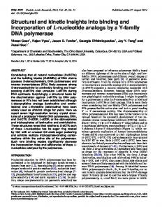
Figure 1. Oxygen reduction rates of PS I with 16 µM cyt cHH (top) and cyt c6 (bottom) at pH 8 (left) and pH 6 (right) as a function of ionic strength. Monovalent (NaCl, black) and divalent (MgCl2, red) cations are depicted as circles and squares, respectively. For cyt c6, pH 8 (bottom, left) differences between the applied salts become prominent. Therefore, a further differentiation of the salts is shown: NaCl (black), Na2SO4 (yellow), NH4Cl (blue), MgCl2 (red), CaCl2 (grey), MgSO4 (cyan). NaCl, MgCl2 and CaCl2 are connected by a line in their corresponding color. All measurements were performed in either 25 mM Tricine-NaOH (pH 8) or 5 mM MES-NaOH (pH 6) with 2 mM ascorbic acid and 300 µM methyl viologen at 20 °C. The concentration of buffer ions and counter ions, which contribute to the ionic strength, was calculated by using the Henderson-Hasselbalch equation with a pKA of 8.2 and 6.2 for Tricine and MES buffers, respectively. Standard deviations result from three to nine independent measurements.
Figure 2. Isothermal titration calorimetry of PS I with cyt cHH. A: Thermogram for exemplary background measurements (top), oxidized (middle) and reduced (bottom) proteins. B: Integrated heats of titrations after background subtraction in the presence (red) or absence (black) of 5 mM ascorbate. High cyt cHH : P700 ratios are omitted for a better overview. Fits (top) and residuals (bottom) are shown for 1 set of binding sites with n = 1.0 (dashed line) and for one set of binding sites with n = 1.5 (solid line). Parameters obtained from the models are shown in Table 2. Measurements were performed at 20 °C in 25 mM Tricine buffer, pH 8.0 with 25 mM NaCl and 0.02 % DDM. Each titration step consisted of 5 µl injected volume from 1 mM cyt cHH.
Figure 3. Isothermal titration calorimetry of PS I with cyt c6. A: Thermogram for exemplary background measurements (top, red), oxidized (middle) and reduced (bottom) proteins. B: Integrated heats of titrations after background subtraction in the presence (red, reduced) or absence (black, oxidized) of 5 mM ascorbate. The fit of the reduced data is shown for 1 set of binding sites with n = 1.0. Parameters obtained from the model are shown in Table 2. After substraction of the heat of dilution, the oxidized data converge to negative values at high cyt c6 : PS I ratio (-0.1 kcal/mol, not shown) and are thus not analyzed by a model. Measurements were performed at 20 °C in 25 mM Tricine-NaOH, pH 8.0 with 25 mM NaCl and 0.02 % DDM. Each titration step consisted of 5 µl injected volume from 1 mM cyt c6. 14
Downloaded from http://www.jbc.org/ at HUMBOLDT-UNIVERSITÄT ZU BERLIN on July 27, 2018
Figure Legends
Figure 4. Molecular docking simulation of monomeric PS I with cyt cHH (left) and cyt c6 (right). Each sphere represents the position of a docked cyt c. The binding energy, calculated by pyDock, is highlighted by a color code. Docking states with less than -20 kcal/mol are highlighted by an increased sphere size.
Figure 6. Superposition of the potential cyt c6 binding site to the known cyt c2 binding site of the bRC from Rhodobacter sphaeroides (PDB-ID: 1l9b (34)). The superposition was achieved by aligning the heme groups. Right view rotated by 90° with respect to the left view. The distance of the heme-iron from cyt c6 to the Mg2+ ions of P700 are 21.4 and 21.3 Å, respectively. This distances are identical to the distances between the heme-iron of cyt c2 and the Mg2+ ions of P870 (pink) from bRC (21.3 and 21.2 Å, respectively).
Figure 1
15
Downloaded from http://www.jbc.org/ at HUMBOLDT-UNIVERSITÄT ZU BERLIN on July 27, 2018
Figure 5. Potential cyt cHH (top) and cyt c6 (bottom) binding site of PS I. Shown are the docking sites which most likely resemble the specific cyt c binding site of PS I. The heme group (red) of cyt cHH and cyt c6 point towards the luminal tryptophan residues W(A655) and W(B631) (blue) and P700 (green) of PS I. The distances between the heme groups and the closest tryptophan are highlighted by a black, dotted line. Cyt c6 does not interact with PsaF (purple), but is close to the luminal loop of PsaA (yellow). The carboxyl group of E34 from cyt c6 is at a distance of 7.4 Å from the carboxyl group of D628 from PsaA (grey, dotted line).
Downloaded from http://www.jbc.org/ at HUMBOLDT-UNIVERSITÄT ZU BERLIN on July 27, 2018
Figure 2
16
Downloaded from http://www.jbc.org/ at HUMBOLDT-UNIVERSITÄT ZU BERLIN on July 27, 2018
Figure 3
17
Downloaded from http://www.jbc.org/ at HUMBOLDT-UNIVERSITÄT ZU BERLIN on July 27, 2018
Figure 4
18
Downloaded from http://www.jbc.org/ at HUMBOLDT-UNIVERSITÄT ZU BERLIN on July 27, 2018
Figure 5
Figure 6
19
Insights into the binding behavior of native and non-native cytochromes to Photosystem I from Thermosynechococcus elongatus Adrian Kölsch, Mahdi Hejazi, Kai R. Stieger, Sven C. Feifel, Jan F. Kern, Frank Müh, Fred Lisdat, Heiko Lokstein and Athina Zouni J. Biol. Chem. published online April 25, 2018
Access the most updated version of this article at doi: 10.1074/jbc.RA117.000953
Click here to choose from all of JBC's e-mail alerts
Downloaded from http://www.jbc.org/ at HUMBOLDT-UNIVERSITÄT ZU BERLIN on July 27, 2018
Alerts: • When this article is cited • When a correction for this article is posted
Supplementary Information
Insights into the binding behavior of native and non-native cytochromes to Photosystem I from Thermosynechococcus elongatus
Adrian Kölsch1*, Mahdi Hejazi1, Kai R. Stieger2, Sven C. Feifel2, Jan F. Kern3, Frank Müh4, Fred Lisdat2, Heiko Lokstein5, Athina Zouni1* 1
Humboldt-Universität zu Berlin, Institute for Biology, Biophysics of Photosynthesis, Philippstr. 13, 10115 Berlin, Germany 2 University of Applied Sciences Wildau, Institute for Applied Life Sciences, Biosystems Technology, Hochschulring 1, 15745 Wildau, Germany 3 Lawrence Berkeley National Laboratory, 1 Cyclotron Road, CA 94720 Berkeley, USA 4 Johannes Kepler University Linz, Institute for Theoretical Physics, Department of Theoretical Biophysics, Altenberger Str. 69, 4040 Linz, Austria 5 Charles University, Department of Chemical Physics and Optics, Ke Karlovu 3, CZ-121 16 Praha 2, Czech Republic
*To whom correspondence should be addressed: A. Kölsch and Prof. A. Zouni, Humboldt-Universität zu Berlin, Institute for Biology, Biophysics of Photosynthesis, Philippstr. 13, 10115 Berlin, Germany. Telephone: +49 30209347930; FAX: +49 30209347934; E-mail:
[email protected] and
[email protected]
S-1
Table S1 MALDI-MS analysis of purified PS I. Following purification and crystallization, PS I crystals were dissolved in 5 mM MES-NaOH, pH 6.0 and 30 mM MgSO4. MS spectra were recorded in linear mode. The standard deviation is given for 12 independent PS I preparations. PsaM
PsaX
PsaI
PsaJ
PsaE
PsaK
PsaC
3424
4101
4166
4767
8389
8480
8800
15113 15370 16251
3424 3970 4195 4796 ±2 ±2 ±2 ±2 *Post translational modifications are described in (1).
8261 ±3
8392 ±3
8672 ±3
15116 15232 16125 ±7 ±12 ±9
Calculated mass* (Da) Determined mass (Da)
PsaF
PsaD
PsaL
Figure S1: Hydrodynamic radius of trimeric PS I measured by dynamic light scattering (DLS, left) and blue native (BN) PAGE of trimeric PS I (T, 1000 kDa) in comparison to monomeric PS I (M, 340 kDa) and dimeric photosystem II (PSII, 750 kDa) (right). For DLS, PS I crystals were dissolved in 100 mM NaCl, 0.02 % DDM and 25 mM Tricine-NaOH, pH 8 to 5 µM P700 and filtered through a 0.2 µm filter. For BN-PAGE, PS I crystals corresponding to 5 µg of chlorophyll were dissolved in BN-solubilisation buffer containing 0.2 % DDM and applied to a gradient gel containing 3 to 9 % polyacrylamide. S-2
Figure S2: SDS-PAGE of purified cyt c6 and cyt cHH at ≥ 95 % and ≥ 99 % purity, obtained from Sigma Aldrich (Germany). Homogeneity of the proteins was analyzed according to the protocol of Laemmli (2). Samples were denatured in sample buffer at 95 °C for 5 min and applied to a 15 % poly-acryl-amide gel.
S-3
Figure S3: PS I - cyt cHH oxygen reduction activity as a function of the buffer concentration. The activity was highest in Tricine (red circles) and Tris (black squares) buffer, while it was lower in HEPES (red diamond), MOPS (black cross) and phosphate buffer (black triangles) at pH 8 with 16 µM cyt cHH. Standard deviations result from three independent measurements.
S-4
Figure S4. Section of MALDI-TOF analysis of single PS I - cyt cHH co-crystals grown in 5 independent batches (grey spectra). For comparison, spectra of cyt cHH (95 % purity) and PS I are shown in red and green, respectively. Crystals were washed six times prior to analysis to avoid contamination by free cyt cHH. Neither cyt cHH nor PS I are present in the supernatant from the last washing step (black spectrum).
S-5
Figure S5. Ratio of cyt cHH to P700 in PS I-cyt cHH co-crystals. Batches of crystals were washed, dissolved and cyt cHH was separated from PS I for photometric quantification. Standard deviations result from three to twelve independent measurements.
Table S2 X-ray data collection and refinement statistics. Values in parenthesis are for the high resolution shell. Statistics Wavelength (Å) Space Group Cell Dimensions a, b, c (Å) α, β, γ (°) Resolution (Å) Multiplicity I/σI Rmeas Completeness (%) No. of reflections Rwork/Rfree Rmsd Bond lengths (Å) Rmsd Bond angles (°)
Values 0.999 P63 281.4, 281.4, 165.6 90, 90, 120 47.36 – 3.42 (3.63 - 3.42) 15.88 8.79 (1.75) 0.42 (1.60) 99.2 (97.0) 99609 0.2611/0.3169 0.0162 5.8874 S-6
Figure S6: Electron density map (grey) of a PS I – cyt cHH co-crystal at 3.4 Å resolution with the respective PS I model (green) at 2 σ contour level. Crystal contacts are formed between PsaE on the cytoplasmic side and PsaF on the luminal side of PS I. All electron density can be assigned to PS I, and no density is found for cyt cHH.
S-7
Figure S7: MALDI-TOF analysis of a single PS I cyt cHH co-crystal after structure analysis at BESSY II at 3.5 Å resolution. The cyt cHH peak is clearly visible, while not all subunits were found by MALDI-TOF, although they were present in the electron density map (e.g. PsaF).
S-8
Table S3: Solubility of PS I at high protein concentration and low salt concentration. PS I crystals were dissolved in Tricine pH 8.0 buffer containing 0.02 % DDM and 100 mM NaCl. The solution was slowly diluted to 30 µM P700 and its final NaCl concentration. The solution was filtered into a disposable cuvette and measured by dynamic light scattering. A high polydispersity (Pd) results from protein aggregation, while a low Pd indicates monodisperse proteins. Standard deviations result from three independent measurements. NaCl [mM] 10 15 20 25 30 35
Pd at 30 µM P700 [%] 18 ± 3 10 ± 1 5±1 5±1 5±1
S-9
Fig. S8: ITC data from Fig. 4 in semilogarithmic representation. Shown is the summed energy of the titrations against the cyt cHH : P700 ratio for measurements in oxidized (black squares) and reduced (red circles) conditions. The black vertical bars represent a ratio of 1 and 2, while the red bar represents the ratio of 1.5, which was calculated to be the number of binding sites by a model for one set of binding sites (Table 2).
S-10
Figure S9: Isothermal titration calorimetry of PS I with cyt c6. Thermogram for exemplary measurements with oxidized (black) and reduced (red) proteins. Measurements were performed at 20 °C in 25 mM Tricine buffer, pH 8.0 with 200 mM MgSO4 and 0.02 % DDM. Each titration step consisted of 5 µl injected volume from 1 mM cyt c6 in 50 µM P700. Measurements under reducing conditions were performed in the presence of 5 mM ascorbic acid and measurements under oxidizing conditions in the absence or presence of up to 15 mM K3(CN)6Fe(III). A strong baseline shift appeared in all measurements under these conditions, indicating the presence of a reaction that cannot be described by a simple binding enthalpy model.
S-11
Figure S10: Cyt cHH (top) and cyt c6 (bottom) can bind at the P700-docking site in different orientations. The depicted docking states have less than 10 Å C-C distance between the heme group and the luminal tryptophan residues W(A655) and W(B632) and more than -15 kcal/mol binding energy. The angle was calculated between two lines, formed by the geometrical center of the cytochrome (g) with the iron from the heme group and by g with the Mg-ions of P700. The docking states shown in Figure 6 are highlighted in red.
S-12
Figure S11: Re-calculation of electrostatic energy of the docking sites shown in Figure 6. Cyt cHH (filled symbols) and cyt c6 (open symbols) were calculated at pH 6 (circles) and pH 8 (squares) in the presence of 0, 10 and 100 mM MgSO4 by using the PoissonBoltzmann equation. The protein dielectric constant was set to 10, and the temperature to 20 °C. For cyt c6 a 6x His-tag with random conformation was added to the proteinstructure. Calculations performed by the adaptive Poisson-Boltzmann solver (APBS 1.4) with amber force field.
S-13
Table S4 Distance between cyt c6 and PS I residues derived from the docking site shown in Figure 6. Residue cyt c6 Residue PS I Distance [Å] expected perturbation * ARG64 PsaB GLN636 1.5 significant MET26 PsaA ILE634 2.2 not homolog MET19 PsaA ASN638 2.2 significant ALA16 PsaA ARG651 2.3 not homolog ALA57 PsaA SER659 2.4 significant VAL25 PsaA ARG750 2.4 strong GLY12 PsaB ASN639 2.5 significant HEM88 PsaA TRP655 2.5 SER11 PsaB ASN642 2.6 strong ASN13 PsaB GLN636 2.6 strong GLY20 PsaB PHE644 2.6 not significant GLY63 PsaB LYS738 2.7 not significant LEU65 PsaB GLN636 2.8 significant VAL24 PsaA ASP652 2.9 strong ALA27 PsaA ILE634 3.0 strong ALA15 PsaB PHE644 3.0 significant CYS17 PsaA TRP655 3.0 significant PRO59 PsaA TRP655 3.2 not significant PHE61 PsaB GLN636 3.3 strong ASN23 PsaA THR635 3.4 significant *NMR signals of cyt c6 amino acids from Nostoc sp. PCC 7119 which were perturbed after addition of PS I (3).
S-14
Table S5 Distance between cyt cHH and PS I residues derived from the docking site shown in Figure 6. Residue cyt cHH THR28 LYS25 ILE81 GLN16 HEM105 GLN12 PHE82 LYS72 GLY77 LYS79 ALA83 PRO76 LYS27 VAL11 CYS17 HIS26 THR49 ASP50 THR78 ALA51
Residue PS I PsaB TRP631 PsaA ILE634 PsaB GLN636 PsaA ARG651 PsaB SER635 PsaB PHE644 PsaB GLN636 PsaB GLN636 PsaB GLU609 PsaB LEU632 PsaB GLN636 PsaB GLU609 PsaA GLN660 PsaB PHE644 PsaA TRP655 PsaA GLN660 PsaB ASN611 PsaF LYS16 PsaB GLU609 PsaB ASN611
Distance [Å] 2.6 2.7 2.7 3.0 3.3 3.3 3.5 3.5 3.7 3.8 4.0 4.1 4.6 4.8 4.9 5.3 5.4 5.6 5.7 5.7
S-15
Figure S12: Photosystem I crystal lattice (green). The dotted lines mark the area where the DDM detergent belt may be localized. The remaining area where cyt cHH could be positioned is highlighted by the blue box. A random docking state with cyt cHH being close to P700 is inserted into the crystal lattice (red). Since there are no clashes of the docked cyt cHH with neighboring PS I monomers, the lattice of a co-crystal does not need to be altered as compared to the PS I crystal lattice. The lattice was visualized by using the symexp command from PyMOL, with the crystal structure PDB-ID: 1jb0 (4).
Supplemental References 1. El-Mohsnawy, E., Kopczak, M. J., Schlodder, E., Nowaczyk, M., Meyer, H. E., Warscheid, B., Karapetyan, N. V., and Rögner, M. (2010) Structure and Function of Intact Photosystem 1 Monomers from the Cyanobacterium Thermosynechococcus elongatus. Biochemistry. 49, 4740–4751 2. Laemmli, U. K. (1970) Cleavage of Structural Proteins during the Assembly of the Head of Bacteriophage T4. Nature. 227, 680–685 3. Díaz-Moreno, I., Díaz-Quintana, A., Molina-Heredia, F. P., Nieto, P. M., Hansson, Ö., De la Rosa, M. A., and Karlsson, B. G. (2005) NMR Analysis of the Transient Complex between Membrane Photosystem I and Soluble Cytochrome c6. J. Biol. Chem. 280, 7925–7931 4. Jordan, P., Fromme, P., Witt, H. T., Klukas, O., Saenger, W., and Krauß, N. (2001) Three-dimensional structure of cyanobacterial photosystem I at 2.5 Å resolution. Nature. 411, 909–917 S-16