contributing to the binding of heparin hexasaccharide. ... Surface Plasmon Resonance imaging (SPRi) binding assays. ... precipitated with 4 volumes of acetone at -20 °C for 1 h and centrifuged 10 min at 16 ..... Two-hybrid, far Western blotting.
Insights into the structure and dynamics of lysyl oxidase propeptide, a flexible protein with numerous partners Sylvain D. Vallet, Adriana E. Miele, Urszula Uciechowska-Kaczmarzyk, Adam Liwo, Bertrand Duclos, Sergey A. Samsonov, Sylvie Ricard-Blum SUPPLEMENTARY FIGURES Supplementary Figure 1. Deglycosylation of the propeptide of human lysyl oxidase by PNGase F. Supplementary Figure 2. Analysis of the propeptide of human lysyl oxidase by circular dichroism with and without heparin. Supplementary Figure 3. Analysis of the propeptide of human lysyl oxidase by SEC-SAXS. Supplementary Figure 4. Models the propeptide of human lysyl oxidase and SAXS data Supplementary Figure 5. Topology of the models of the propeptide of human lysyl oxidase generated with PROMOTIF software. Supplementary Figure 6. Arginine residues of the propeptide of human lysyl oxidase contributing to the binding of heparin hexasaccharide. Supplementary Figure 7. Surface Plasmon Resonance imaging (SPRi) binding assays. Supplementary Figure 8. Surface Plasmon Resonance (SPR) and Bio-Layer Interferometry (BLI) binding assays. a) Sensorgrams collected by SPR. b) Kinetic analysis performed by SPR for plasminogen (62.5-1000 nM) and tropoelastin (2.5-40 µM) and by BLI for anastellin. Experimental (black) and fitted (red) data. SUPPLEMENTARY TABLES Supplementary Table 1. Changes in the radius of gyration (Rg) of LOX-PP during MD simulation in the presence of a heparin hexasaccharide. Supplementary Table 2. Changes in the radius of gyration (Rg) of LOX-PP during MD simulation in the absence of a heparin hexasaccharide. Supplementary Table 3. Secondary structure of LOX-PP/heparin hexasaccharide complexes in the MD trajectories after 10 ns and 20 ns of simulation based on DSSP analysis obtained from the cpptraj module of the AMBER package. Supplementary Table 4. Free energy contribution (kcal/mol) of arginine and negatively charged residues for the five models of LOX-PP complexed with heparin hexasaccharide. Supplementary Table 5. List of LOX-PP partners reported in the literature with the detection methods and the PMIDs reporting the interactions. Supplementary Table 6. List of biomolecules tested in binding assays performed by Surface Plasmon Resonance (SPR) imaging, SPR and Bio-Layer Interferometry (BLI). Supplementary Table 7. List of LOX-PP partners identified in this study by SPRi, SPR, and BLI binding assays. Negative interactions are also indicated.
SUPPLEMENTARY METHOD: COARSE-GRAINED SIMULATIONS
1
38 kDa
2
3
kDa 130 100 70 55 40 35
25
17 kDa 15
10
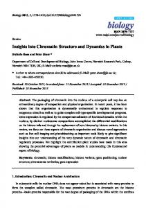
Supplementary Figure 1. Deglycosylation of the propeptide of human lysyl oxidase by PNGase F. LOX-PP (8.2 µg) incubated without (lane 1) or with PNGase F (lane 2) as described below. Lane 3: molecular weight markers. 18 µl of LOX-PP solution at 0.92 mg/ml in HBS (16.6 µg) was incubated at 55°C for 10 min with 2 µl 400 mM DTT, and then at 37°C for 1 h with 2 µl PNGase F according to the manufacturer’s instructions (New England’s Biolab, P0706). The reaction medium was then precipitated with 4 volumes of acetone at -20 °C for 1 h and centrifuged 10 min at 16 000 × g. Finally, the pellet was resuspended in 20 µl of reducing loading buffer and analyzed by SDSPAGE on a 15 % polyacrylamide gel.
2
Supplementary Figure 2. Analysis of the propeptide of human lysyl oxidase by circular dichroism with and without heparin. Averaged circular dichroism spectra (n=5) of LOX-PP at 2 µM (60 µg/ml) in 10 mM potassium phosphate pH 7.4. LOX-PP was analyzed in absence and presence of 2 µM of heparin hexasaccharide (dp6) or full-length heparin immediately after mixing or after one-hour incubation at room temperature.
3
Supplementary Figure 3. Analysis of the propeptide of human lysyl oxidase by SECSAXS. SEC-SAXS experiments were performed on the SWING beamline (French National Synchrotron Facility SOLEIL, Saint-Aubin, France, 20170906). 50 µl of LOX-PP (219 µM) were injected in HBS on a Superdex 200 Increase 5/150 GL column at a flow rate of 0.2 ml/min. The parameters used for SAXS analysis were the following: acquisition 1 frame/s, detector distance: 2.087 m, wavelength: 1.033 Å, detector: Eiger 4M (beam center 1020 × 646 pixel). Foxtrot was used for data reduction and frame selection. Buffer spectra collected before the void volume were subtracted from LOX-PP spectra using CHROMIXS (Panjkovich and Svergun, 2018). Useful data range was defined by SHANUM (Konarev and Svergun, 2015). PRIMUS (Konarev et al., 2003) was used to calculate a) the radius of gyration of LOX-PP using the Guinier plot with a sRg limit < 1.02 and b) Dmax using the distance distribution function. Konarev, P. V. & Svergun, D. I. A posteriori determination of the useful data range for smallangle scattering experiments on dilute monodisperse systems. IUCrJ 2, 352-360 (2015). Konarev, P. V., Volkov, V. V., Sokolova, A. V., Koch, M. H. J. & Svergun, D. I. PRIMUS: a Windows PC-based system for small-angle scattering data analysis. J. Appl. Cryst. 36, 12771282 (2003). Panjkovich, A. & Svergun, D. I. CHROMIXS: automatic and interactive analysis of chromatography-coupled small-angle X-ray scattering data. Bioinformatics 34, 1944-1946 (2018).
4
Supplementary Figure 4. Models the propeptide of human lysyl oxidase and SAXS data. Gaussian-smoothed Cα-distance distribution from SAXS data for LOX-PP and for the five models predicted using the UNRES force field.
5
Supplementary Figure 5. Topology of the models of the propeptide of human lysyl oxidase generated with PROMOTIF software.
6
Supplementary Figure 6. Arginine residues of the propeptide of human lysyl oxidase contributing to the binding of heparin hexasaccharide. Visualization of the ten arginine residues (sticks) predicted to contribute the most to the heparin hexasaccharide binding according to molecular mechanics energies combined with the generalized Born and surface area continuum (MM-GBSA) solvation (free energy per residue).
7
Supplementary Figure 7. Surface Plasmon Resonance imaging (SPRi) binding assays. The partners of the propeptide of human lysyl oxidase (ligands) were spotted in triplicate over the arrays and the propeptide of human lysyl oxidase was used as the analyte. Sensorgrams were smoothed on 6 neighboring data points and adjusted to the baseline. One representative sensorgram of each triplicate is shown. The recirculation of the propeptide of lysyl oxidase over the arrays is visualized by dotted lines.
8
Supplementary Figure 8. Surface Plasmon Resonance (SPR) and Bio-Layer Interferometry (BLI) binding assays. a) Sensorgrams collected by SPR. b) Kinetic analysis performed by SPR for plasminogen (62.5-1000 nM) and tropoelastin (2.5-40 µM) and by BLI for anastellin. Experimental (black) and fitted (red) data.
9
Supplementary Table 1. Changes in the radius of gyration (Rg) of LOX-PP during MD simulation in the presence of a heparin hexasaccharide. Model
Cluster
1
1
2
2
3
1
1
2
4
1
2
3
4
5
1
Rg (nm) at 10 ns
Rg (nm) at 20 ns
1.92±0.04
1.78±0.03
2.62±0.05
2.05±0.04
2.05±0.03
1.96±0.01
2.44±0.04
2.68±0.06
2.87±0.07
2.27±0.03
3.21±0.06
3.11±0.05
2.25±0.05
2.50±0.08
2.34±0.05
2.16±0.02
2.11±0.04
2.31±0.04
2.22±0.08
2.02±0.04
2.28±0.03
1.92±0.02
1.91±0.03
1.77±0.03
2.47±0.05
2.39±0.03
2.09±0.09
2.15±0.05
2.20±0.03
2.25±0.04
3.19±0.03
3.26±0.02
2.37±0.04
1.90±0.03
2.24±0.06
2.07±0.03
2.96±0.03
2.94±0.04
2.03±0.06
2.24±0.07
3.05±0.05
3.30±1.00
2.31±0.03
2.28±0.08
2.16±0.05
2.17±0.04
2.45±0.05
2.79±0.02
3.21±0.05
3.38±0.08
3.28±0.03
3.30±0.05
3.28±0.03
2.84±0.03
2.13±0.04
2.06±0.05
1.99±0.06
1.83±0.02
2.25±0.04
2.12±0.06
10
Supplementary Table 2. Changes in the radius of gyration (Rg) of LOX-PP during MD simulation in the absence of a heparin hexasaccharide.
LOX-PP model
Rg (nm) Initial
10 ns
20 ns
1
2.93
2.63 ± 0.05
2.16 ± 0.04
2
2.74
2.84 ± 0.07
3.0 ± 0.10
3
2.58
1.86 ± 0.03
1.81 ± 0.01
4
2.73
2.05 ± 0.05
2.24 ± 0.04
5
2.56
1.96 ± 0.04
1.89 ± 0.02
11
Supplementary Table 3. Secondary structure of LOX-PP/HP complexes in the MD trajectories after 10 ns and 20 ns of simulation based on DSSP analysis obtained from the cpptraj module of AMBER package, which considers all types of helices as helices. (DSSP: Dictionary of Secondary Structure of Proteins) Model
Cluster
1
1
2
2
3
1
1
2
4
1
2
3
4
5
1
DSSP 10 ns
DSSP 20 ns
-helix (%) -sheet (%) -helix (%)
-sheet (%)
11.5
5.4
11.5
8.2
15.6
4.7
8.8
6.2
6.8
8.2
15
4.7
15
3.4
10.2
9.5
10.8
10.8
15
9.5
5.4
12.2
6.2
10.2
10.8
4.7
12.3
5.4
12.3
5.4
12.3
8.8
7.5
8.2
6.2
11.5
11.5
17.6
10.8
14.5
6.8
12.3
8.8
12.3
12.2
11.5
11.5
14.3
11.5
17.6
13.2
15.6
6.8
12.3
6.8
11.5
12.3
11.5
10.2
15.6
8.2
8.8
11.5
8.2
10.8
15.6
12.3
13.6
12.3
8.2
14.3
6.2
11.5
10.2
10.8
6.8
12.3
10.8
12.3
12.3
14.3
4.7
12.3
6.2
10.8
8.8
10.2
9.5
7.5
13.6
8.2
12.3
6.2
14.3
7.5
13.2
11.5
13.6
10.8
10.8
4.7
7.5
6.8
15
13.6
7.5
10.8
4.2
13.2
7.5
8.8
5.4
8.2
8.2
10.2
8.2
17.2
5.4
11.5
4.7
12
Supplementary Table 4. Free energy contribution (kcal/mol) of arginine, histidine and negatively charged residues for the five models of LOX-PP complexed with HP hexasaccharide. Mean values are obtained from all MD simulations. Residue
Model 1
Model 2
Model 3
Model 4
Model 5
R33
-1.56
-1.33
-8.23
-0.40
-0.69
R43
-4.90
-4.45
-5.07
-0.37
-0.73
R68
-6.01
-14.96
-4.66
-0.47
-0.93
R69
-1.38
-2.88
-0.97
-0.46
-1.10
R70
-6.05
-7.22
-1.01
-0.49
-1.03
R88
-7.58
-15.01
-0.74
-0.71
-0.95
R95
-0.95
-0.89
-0.97
-0.80
-1.06
R98
-2.30
-1.00
-6.38
-0.99
-1.18
R103
-7.66
-3.10
-6.42
-5.34
-14.50
R105
-9.17
-2.11
-9.90
-2.09
-1.19
R116
-3.90
-6.60
-0.78
-15.24
-0.69
R118
-3.02
-2.14
-0.94
-12.05
-0.74
R122
-4.02
-0.84
-0.91
-1.34
-0.90
R133
-2.17
-0.73
-3.34
-3.79
-0.79
R135
-0.88
-0.66
-1.21
-6.10
-0.85
R141
-0.92
-0.63
-2.71
-2.79
-0.77
R158
-0.58
-0.59
-0.68
-0.90
-5.04
R162
-0.56
-0.80
-0.68
-1.01
-2.98
H123
-0.16
-0.05
0.04
0.05
0.05
D71
1.42
1.50
0.93
0.52
1.26
D96
1.12
0.98
1.27
0.91
1.11
D164
0.48
0.81
0.60
1.21
1.55
E34
1.07
2.56
4.18
0.37
0.65
E49
1.32
0.99
1.98
0.42
0.80
E136
0.98
0.64
1.02
2.09
0.83
E143
0.73
0.70
0.93
1.17
0.80
E150
0.62
0.64
0.69
0.85
0.88
13
Supplementary Table 5. List of LOX-PP partners reported in the literature with the detection methods and the PMIDs reporting the interactions. LOX-PP partners reported in the literature
Interaction detection methods
PMIDs
Apoptotic protease-activating factor 1 Collagen I alpha1 chain Collagen I alpha2 chain Collagen III alpha1 chain Double-strand break repair protein MRE11 Fibronectin Fibulin-4 Heparin (6 and 16 kDa)
Pull down Two-hybrid Two-hybrid Two-hybrid Pull down, co-immunoprecipitation Two-hybrid Co-immunoprecipitation Bio-layer interferometry
21536655 21690299 21690299 21690299 24882580 21690299 19855011 DOI: 10.1039/9781788010283-00398
Heat shock 70 kDa protein 1A
Pull down, co-immunoprecipitation
21536655
Heat shock 70 kDa protein 1B Heat shock cognate 71 kDa protein MMP-2 MMP-10 Protein UXT RAF proto-oncogene serine/threonine-protein kinase
Pull down, co-immunoprecipitation Pull down Enzymatic assay iTRAQ-TAILS** Two-hybrid Pull down
21536655 21536655 8636146 24281761 28106301 21536655
Receptor-type tyrosine-protein phosphatase kappa
Two hybrid, co-immunoprecipitation
21690299
SH3 domain-containing kinase-binding protein 1 Tropoelastin Tubulin alpha-3C/D chain Tubulin beta chain
Co-immunoprecipitation Two-hybrid, far Western blotting Pull down Pull down
24167568 16251195 21536655 21536655
** iTRAQ-TAILS: Isobaric tags for relative and absolute quantitation -Terminal amine isotopic labeling of substrates.
Supplementary Table 6. List of biomolecules tested in binding assays performed by Surface Plasmon Resonance (SPR) imaging, SPR and BioLayer Interferometry (BLI). ChEBI: Chemical Entities of Biological Interest, CPX: Complex Portal identifier, PRO_features are from UniProtKB.
Biomolecules tested in binding assays -synuclein Adiponectin Aggrecan Agrin Angiopoietin-like protein 4 (AngPTL-4), mutant Lys163Ala, Arg164Ala
Biomolecule ChEBI, UniprotKB, or Complex Portal identifiers (1st-last amino acid residues of the proteins and protein fragments used in binding assays P37840 Q15848 P13608 O00468 (1260-2045)
References (commercial sources or PMID/doi) Boston Biochem, SP-480 Sigma-Aldrich, SRP4901 Sigma-Aldrich, A1960 R&D Systems, 6624-AG
Q9BY76 (26-406)
R&D Systems, 4487-AN
Anthrax toxin receptor 1 (Tumor endothelial marker 8, TEM-8)
Q9H6X2-2 (1-368)
Abnova, H00084168-P01
-2 microglobulin Biglycan Brevican Calreticulin Catalase Chondroitin sulfate Coagulation factor X Collagen I Collagen II Collagen III Collagen IV Collagen V
P61769 P21809 Q96GW7 (23-911) P27797 P00432 CHEBI:37397 P00742 CPX-1650 CPX-3105 CPX-1714 CPX-1723 CPX-1727
Sigma-Aldrich, M4890 Sigma-Aldrich, B8041 R&D Systems, 4009-BC Abcam, ab91577 Sigma-Aldrich, C9322 Sigma-Aldrich, C8529 Sigma-Aldrich, 233282 Sigma-Aldrich, C7774 Sigma-Aldrich, C1188 Sigma-Aldrich, C4407 Sigma-Aldrich, C7521 Sigma-Aldrich, C3657
Collagen VI
CPX-1736
GeneTex, GTX27538
Collagen VI (vWF1 domain)
P12109 (20-256)
Expressed in the laboratory (PMID 24117177)
Collagen VI (vWF2-3 domain)
P12109 (593-1028)
Expressed in the laboratory (PMID 24117177, 28106549)
Collagen XIII ectodomain
CPX-1754 (62-717)
Expressed in the laboratory (PMID: 28106549)
Collagen XVII ectodomain
CPX-1758 (490-1497)
Collagen XVIII (NC1 domain)
CPX-1759 (1443-1754)
Collagen XXIII ectodomain Collagen XXV ectodomain Connective tissue growth factor (CTGF) Discoidin domain-containing receptor 1 ectodomain (DDR1) Discoidin domain-containing receptor 2 ectodomain (DDR2) Decorin Dermatan sulfate Dermatopontin Extracellular matrix protein 1 (ECM1) Endostatin (expressed in Pichia pastoris)
CPX-1764 (111-540) CPX-1766 (113-654) P29279
Expressed in the laboratory (PMID: 20861347, 24117177, 28106549) Expressed in the laboratory (PMID: 10449407, 14585835, 19542224, 19502598, 24478075, 24117177, 28106549) Expressed in the laboratory Expressed in the laboratory RayBiotech, 228-10290-2
Q5ST11 (21-416)
R&D Systems, 2396-DR
Q16832 (24-399)
R&D Systems, 2538-DR
P07585 (17-359) CHEBI:18376 Q07507 Q16610 (20-540) P39060 (PRO_0000005794)
R&D Systems, 143-DE Sigma-Aldrich, C3788 Abcam, ab158310 R&D Systems, 3937-EC Sigma-Aldrich, E8154
Endostatin (expressed in HEK293 cells)
P39060 (PRO_0000005794)
Endostatin, mutant Asp1675Asn
P39060 (PRO_0000005794)
Endostatin, mutant Arg1598Ala, Arg1710Ala
P39060 (PRO_0000005794)
Enolase Epidermal growth factor (EGF) Epidermal growth factor receptor (EGFR) Epigen Fibroblast growth factor-2 Fibromodulin Fibronectin (cellular) Fibronectin (plasma)
P01133 (PRO_0000007541) P00533 (25-647) Q6UW88 P09038 (PRO_0000008933) P13605 P02751 P02751
Expressed in the laboratory (PMID: 14585835, 19542224, 19502598, 24117177, 24478075, 28106549, DOI: 10.1039/9781788010283-00398) Expressed in the laboratory (PMID: 19502598) Expressed in the laboratory (PMID: 19502598) Sigma-Aldrich, E6126 Sigma-Aldrich, E9644 RayBiotech, 228-10367-2 Sigma-Aldrich, SRP4969 PromoKine, C60240 Sigma-Aldrich, F6921 Sigma-Aldrich, F2518 Sigma-Aldrich, F2006
Fibronectin fragment III1-C (anastellin)
P02751 (PRO_0000390479, 631-702)
Sigma-Aldrich, F3542
Fibulin 4 Glypican 1 Glypican 2 Glypican 3 Glypican 5 Glypican 6 Heparan sulfate Heparin (high molecular weight) Heparin (low molecular weight, 3 kDa) Hyaluronan
O95967 (26-443) P35052 (24-530) Q8N158 (18-553) P51654 P78333 (25-554) Q9Y625 (24-355) CHEBI:28815 CHEBI:28304 CHEBI:28304 CHEBI:16336
USCN, RPF421HU01 R&D Systems, 4519-GP R&D Systems, 2304-GP R&D Systems, 2119-GP R&D Systems, 2607-G5 R&D Systems, 2845-GP Celsus Lab, HO-3105 Sigma-Aldrich, H3393 Sigma-Aldrich, H3400 Acros Organics, 25177
Hyaluronan (25-75 kDa) Hyaluronidase-1 Integrin 41 ectodomain Integrin 51 ectodomain Integrin v3 ectodomain Integrin v5 ectodomain Laminin-111 Latent transforming growth factor beta-1 (TGF1)
CHEBI:16336 Q12794 CPX-1802 CPX-1794 CPX-1795 CPX-1796 CPX-3008 P01137
Latent transforming growth factor binding protein 1 (4-7 EGF-like domains)
Q14766 (873-1037)
Latent transforming growth factor binding protein 1 (9-14 EGF-like domains)
Q14766 (1079-1328)
Leukocyte-associated immunoglobulin-like receptor-1 (LAIR1)
Q6GTX8 (22-163)
Lysyl oxidase propeptide (LOX-PP) (Pro24Leu)
P28300 (PRO_0000018520)
Lysyl oxidase homolog 2 (LOXL2) Lysyl oxidase homolog 3 (LOXL3) Lumican Macrophage receptor MARCO Matrix metalloproteinase-2 (MMP-2) Neurexin-1 Neurocan Neuroglycan C
Q9Y4K0 P58215 P51884 (19-338) Q9UEW3 (79-520) P08253 (30-660) P58400 (51-363) P55066 (23-637) O95196 (31-420)
Sigma-Aldrich, S0326 R&D Systems, 7358-GH R&D Systems, 5668-A4 R&D Systems, 3230-A5 R&D Systems, 3050-AV R&D Systems, 2528-AV Sigma-Aldrich, L2020 Sino Biological, 10804-H08H Generous gift from Dr. L. Perrin-Cocon and Dr. V. Lotteau (International Center for Infectiology Research, Lyon, France) (PMID: 24117177) Generous gift from Dr. L. Perrin-Cocon and Dr. V. Lotteau (International Center for Infectiology Research, Lyon, France) (PMID: 24117177) R&D Systems, 2664-LR Expressed in the laboratory (DOI: 10.1039/9781788010283-00398) R&D Systems, 2639-AO R&D Systems, 6069-AO R&D Systems, 2846-LU R&D Systems, 7586-MA R&D Systems, 902-MPN R&D Systems, 5268-NX R&D Systems, 5800-NC R&D Systems, 5685-NG
Neuropilin-1 (NRP1) ectodomain
O14786, 23-815
Osteonectin (SPARC) Osteopontin (with bovine serum albumin) Osteoprotegerin
P09486 P10451 O00300 (22-401)
Procollagen C-proteinase enhancer 1 (PCPE-1)
Q15113
Platelet-derived growth factor receptor alpha (PDGFR) Platelet-derived growth factor receptor beta (PDGFR), mutant Glu241Asp Periostin Perlecan Plasminogen Reelin Superoxide dismutase Syndecan-1 ectodomain Syndecan-2 ectodomain Syndecan-3 ectodomain Syndecan-4 ectodomain Thrombospondin-1 (TSP-1) Transglutaminase-2 (TG-2) (guinea pig) Transglutaminase-2 (TG-2) (human) Transglutaminase-2 (TG-2) (human)
Expressed in the laboratory (PMID, 28106549) Immundiagnostik AG, A4225AG.1 Sigma-Aldrich, O4264 R&D Systems, 185-OS Generous gift from Dr E. Kessler (Goldschleger Eye Research Institute, Tel Aviv University Faculty of Medicine Sheba Medical Center, Tel-Hashomer Israel) (PMID: 15834133, 24117177, 28106549)
P16234 (24-524)
R&D Systems, 322-PR
P09619 (33-530)
R&D Systems, 385-PR
Q15063 (22-836) Q05793 P00747 Q60841 (1221-2661) Q4Q597 P18827 (18-251) P34741 (19-144) O75056 (48-383) P31431 (19-145) CPX-1785 P08587 P21980 P21980
R&D Systems, 3548-F2 Sigma-Aldrich, H4777 R&D Systems, 1939-SE R&D Systems, 3820-MR Expressed in the laboratory R&D Systems, 2780-SD R&D Systems, 2965-SD R&D Systems, 3539-SD R&D Systems, 2918-SD Immundiagnostik AG, AW1011AG.1 Sigma-Aldrich, T5398 Immundiagnostik AG, AK3010AG.1 Zedira, T022
Transglutaminase-2 (TG-2), mutant Cys277Ser Tropoelastin Tropomyosin Tumor necrosis factor (TNF) Vascular endothelial growth factor (VEGF)
P21980 P15502 P01375 (77-233) CPX-1977
Zedira, T018 Sigma-Aldrich, T0706 Sigma-Aldrich, T2400 R&D Systems, 210-TA Sigma-Aldrich, V7259
Vascular endothelial growth factor receptor-2 (VEGFR-2)
P35968 (20-764)
R&D Systems, 357-KD
Vitronectin
P04004
R&D Systems, 2349-VN
Supplementary Table 7. List of LOX-PP partners identified in this study by SPRi, SPR, and BLI binding assays. Negative interactions are also indicated (nt: not tested)
Biomolecules tested in binding assays (no: no binding, yes: binding, nt: not tested) -synuclein Adiponectin Aggrecan Agrin Angiopoietin-like protein 4 (AngPTL-4), mutant Lys163Ala, Arg164Ala
Interaction detection method SPRi (Surface Plasmon SPR (Surface BLI (Bio-Layer Resonance imaging) Plasmon Resonance) Interferometry) no nt nt no nt nt no nt nt no nt nt no
nt
nt
Anthrax toxin receptor 1 (Tumor endothelial marker 8, TEM-8)
yes
nt
nt
-2 microglobulin Biglycan Brevican Calreticulin Catalase Chondroitin sulfate Coagulation factor X Collagen I Collagen II Collagen III Collagen IV
no no no no no yes no yes no no no
nt nt nt nt nt nt nt yes nt nt nt
nt nt nt nt nt nt nt nt nt nt nt
Collagen V Collagen VI
no no
nt nt
nt nt
Collagen VI (vWF1 domain)
yes
nt
nt
Collagen VI (vWF2-3 domain)
no
nt
nt
Collagen XIII ectodomain
no
nt
nt
Collagen XVII ectodomain
no
nt
nt
Collagen XVIII (NC1 domain)
no
nt
nt
Collagen XXIII ectodomain Collagen XXV ectodomain Connective tissue growth factor (CTGF) Discoidin domain-containing receptor 1 ectodomain (DDR1) Discoidin domain-containing receptor 2 ectodomain (DDR2) Decorin Dermatan sulfate Dermatopontin Extracellular matrix protein 1 (ECM1) Endostatin (expressed in Pichia pastoris)
no no no
nt nt nt
nt nt nt
no
nt
nt
no
nt
nt
no yes yes no no
nt nt yes nt nt
nt nt nt nt nt
Endostatin (expressed in HEK293 cells)
no
nt
nt
Endostatin, mutant Asp1675Asn Endostatin, mutant Arg1598Ala, Arg1710Ala Enolase Epidermal growth factor (EGF) Epidermal growth factor receptor (EGFR) Epigen Fibroblast growth factor-2 Fibromodulin Fibronectin (cellular) Fibronectin (plasma)
no no no yes no no no yes yes no
nt nt nt yes nt nt nt nt yes nt
nt nt nt nt nt nt nt nt nt nt
Fibronectin fragment III1-C (anastellin)
yes
yes
yes
Fibulin 4 Glypican 1 Glypican 2 Glypican 3 Glypican 5 Glypican 6 Heparan sulfate Heparin (high molecular weight) Heparin (low molecular weight, 3 kDa) Hyaluronan Hyaluronan (25-75 kDa) Hyaluronidase-1 Integrin 41 ectodomain Integrin 51 ectodomain
no no no no no no yes yes yes yes yes no no no
nt nt nt nt nt nt nt yes nt nt nt nt nt nt
nt nt nt nt nt nt nt nt nt nt nt nt nt nt
Integrin v3 ectodomain Integrin v5 ectodomain Laminin-111 Latent transforming growth factor beta-1 (TGF1)
no no no no
nt nt nt nt
nt nt nt nt
Latent transforming growth factor binding protein 1 (4-7 EGF-like domains)
yes
nt
nt
Latent transforming growth factor binding protein 1 (9-14 EGF-like domains)
yes
nt
nt
Leukocyte-associated immunoglobulin-like receptor-1 (LAIR1)
no
nt
nt
Lysyl oxidase propeptide (LOX-PP) (Pro24Leu)
no
nt
nt
Lysyl oxidase homolog 2 (LOXL2) Lysyl oxidase homolog 3 (LOXL3) Lumican Macrophage receptor MARCO Matrix metalloproteinase-2 (MMP-2) Neurexin-1 Neurocan Neuroglycan C Neuropilin-1 (NRP1) ectodomain Osteonectin (SPARC) Osteopontin (with bovine serum albumin) Osteoprotegerin Procollagen C-proteinase enhancer 1 (PCPE-1) Platelet-derived growth factor receptor alpha (PDGFR)
yes no no no yes no no no no no no no no
nt nt nt nt nt nt nt nt nt nt nt nt nt
nt nt nt nt nt nt nt nt nt nt nt nt nt
no
nt
nt
Platelet-derived growth factor receptor beta (PDGFR), mutant Glu241Asp Periostin Perlecan Plasminogen Reelin Superoxide dismutase Syndecan-1 ectodomain Syndecan-2 ectodomain Syndecan-3 ectodomain Syndecan-4 ectodomain Thrombospondine-1 (TSP-1) Transglutaminase-2 (TG-2) (guinea pig) Transglutaminase-2 (TG-2) (human) Transglutaminase-2 (TG-2) (human) Transglutaminase-2 (TG-2), mutant Cys277Ser Tropoelastin Tropomyosin Tumor necrosis factor (TNF) Vascular endothelial growth factor (VEGF)
no
nt
nt
no no yes no yes no no no no no no yes no yes no no no no
nt nt yes nt nt nt nt nt nt nt nt yes nt nt yes nt nt nt
nt nt nt nt nt nt nt nt nt nt nt nt nt nt nt nt nt nt
Vascular endothelial growth factor receptor-2 (VEGFR-2)
no
nt
nt
Vitronectin
no
nt
nt
SUPPLEMENTARY METHOD: COARSE-GRAINED SIMULATIONS We performed multiplexed replica exchange molecular dynamics simulations1 using the coarse grained UNRES force field2–4 with restraints derived from the present SAXS experiments, which were enabled in our recent work5. The coarse-grained UNRES model2,3 is a reduced model of polypeptide chains, in which a polypeptide chain is represented by a sequence of α-carbon (Cα) atoms linked by virtual bonds with attached united side chains and united peptide group. United peptide groups and united side chains serve as interaction sites. The UNRES force field has been derived as a Restricted Free Energy (RFE) function of an allatom polypeptide chain and the surrounding solvent, where the all-atom energy function is averaged over the degrees of freedom that are reduced when passing from the all-atom to the simplified coarse-grained system6,7. The RFE is further decomposed into factors derived from interactions within and between united interaction sites. Expansion of the factors into generalized Kubo’s cumulants enable to derive approximate analytical expressions for the respective terms, including the multibody or correlation terms, which are derived in other force fields from structural databases or on a heuristic basis8. In this work, we used the latest variant of UNRES calibrated with a set of 7 proteins4 and the maximum-likelihood method developed in our laboratory9. The pseudo-energy function U used for this approach is given by equation 1 where the UUNRES is UUNRES energy function, Vtempl the template-restraint penalty term, VSAXS SAXS-restrained term, wtempl and wSAXS are the weights of the respective restraint terms. In our work the wtempl and wSAXS were set to 1 and 100 respectively5. U = UUNRES + wtempl Vtempl + wSAXS VSAXS (1) In order to sample conformational space more efficiently molecular dynamics method (MREMD) was applied1,10. In this approach several replicas of the system were simulated at different temperatures (T0, T1, …, TM) and after a defined timestep an exchange of temperatures between neighboring replicas (j=i+1) was attempted11. In the MREMD calculations, we run trajectories at 20 replicas, four trajectories per each temperature. The temperatures ranged from 210 to 500 K, spaced 10 K until 350 K from 350 K to 500 spaced 20 K. This range of temperatures covered the region of the folding-unfolding transition. Each trajectory consisted of 3 107 MD steps, and the replicas were exchanged every 2 104 MD steps. The temperature was controlled by the Berendsen thermostat with the coupling constant 48.9 fs12. The last 100 snapshots were taken into further analysis. The obtained conformations were analyzed with weighted histogram analysis method WHAM13. The heat-capacity profile was calculated to obtain the temperature at which the ensemble is analyzed, Ta, taken as 20 K below the temperature of the major heat-capacity peak14,15. The probabilities of the conformations were calculated at Ta based on the WHAM analysis and minimum variance clustering14,15. Then cluster analysis was used to dissect the conformational ensembles and to identify the most populated regions of the conformational space. The ensembles were grouped into five clusters ranked from the largest and containing the most probable structure (rank 1) to the cluster with the least probable structures 15 (rank 5). The probabilities of the clusters were calculated by summing the probabilities of the conformations constituting a cluster which were, in turn, calculated at Ta based on the results of WHAM14,15. For each cluster, the conformation closest to the centroid was determined, and
considered representative of the entire cluster14,15. Finally, the five coarse-grained models were converted to all-atom structures by using the PULCHRA16 and SCWRL17 knowledge-based algorithms for all-atom backbone and all-atom side-chain reconstruction, respectively. The conversion algorithms use Cα and side-chain pseudoatom from the coarse-grained models and reconstruct the full backbone and side-chain structures based on the experimental data on backbone and side-chain low energy conformations available in the Protein Data Bank (https://www.rcsb.org/). The final refinement for all-atom models was carried out by performing energy minimization and short MD runs with AMBER 1618. References 1. Rhee, Y. M. & Pande, V. S. Multiplexed-replica exchange molecular dynamics method for protein folding simulation. Biophys. J. 84, 775–786 (2003). 2. Liwo, A., Czaplewski, C., Ołdziej, S. & Scheraga, H. A. Computational techniques for efficient conformational sampling of proteins. Curr. Opin. Struct. Biol. 18, 134–139 (2008). 3. Liwo, A. et al. A unified coarse-grained model of biological macromolecules based on meanfield multipole-multipole interactions. J. Mol. Model. 20, 2306 (2014). 4. Krupa, P. et al. Maximum Likelihood Calibration of the UNRES Force Field for Simulation of Protein Structure and Dynamics. J. Chem. Inf. Model. 57, 2364–2377 (2017). 5. Karczyńska, A. S. et al. Prediction of protein structure with the coarse-grained UNRES force field assisted by small X-ray scattering data and knowledge-based information. Proteins 86, 228–239 (2018). 6. Liwo, A., Czaplewski, C., Pillardy, J. & Scheraga, H. A. Cumulant-based expressions for the multibody terms for the correlation between local and electrostatic interactions in the unitedresidue force field. J. Chem. Phys. 115, 2323–2347 (2001). 7. Sieradzan, A. K., Makowski, M., Augustynowicz, A. & Liwo, A. A general method for the derivation of the functional forms of the effective energy terms in coarse-grained energy functions of polymers. I. Backbone potentials of coarse-grained polypeptide chains. J. Chem. Phys. 146, 124106 (2017). 8. Kubo, R. Generalized Cumulant Expansion Method. J. Phys. Soc. Jpn. 17, 1100–1120 (1962). 9. Zaborowski, B. et al. A Maximum-Likelihood Approach to Force-Field Calibration. J. Chem. Inf. Model. 55, 2050–2070 (2015). 10. Hansmann, U. H. E. & Okamoto, Y. Comparative study of multicanonical and simulated annealing algorithms in the protein folding problem. Physica A 212, 415–437 (1994). 11. Czaplewski, C., Kalinowski, S., Liwo, A. & Scheraga, H. A. Application of Multiplexed Replica Exchange Molecular Dynamics to the UNRES Force Field: Tests with alpha and alpha+beta Proteins. J. Chem. Theory Comput. 5, 627–640 (2009). 12. Swope, W. C., Andersen, H. C., Berens, P. H. & Wilson, K. R. A computer simulation method for the calculation of equilibrium constants for the formation of physical clusters of molecules: Application to small water clusters. J. Chem. Phys. 76, 637–649 (1982). 13. Kumar, S., Bouzida, D., Swendsen, R. H., Kollman, P. A. & Rosenberg, J. M. THE weighted histogram analysis method for free‐energy calculations on biomolecules. I. The method. J. Comput. Chem. 13, 1011–1021 (1992).
27
14. Krupa, P. et al. Prediction of Protein Structure by Template-Based Modeling Combined with the UNRES Force Field. J. Chem. Inf. Model. 55, 1271–1281 (2015). 15. Liwo, A. et al. Modification and optimization of the united-residue (UNRES) potential energy function for canonical simulations. I. Temperature dependence of the effective energy function and tests of the optimization method with single training proteins. J. Phys. Chem. B. 111, 260–285 (2007). 16. Rotkiewicz, P. & Skolnick, J. Fast procedure for reconstruction of full-atom protein models from reduced representations. J. Comput. Chem. 29, 1460–1465 (2008). 17. Wang, Q., Canutescu, A. A. & Dunbrack, R. L. SCWRL and MolIDE: computer programs for side-chain conformation prediction and homology modeling. Nat. Protoc. 3, 1832–1847 (2008). 18. Case, D. A. et al. AMBER 2017. University of California, San Francisco (2017).
28