RESEARCH ARTICLE
Integration of AI-2 Based Cell-Cell Signaling with Metabolic Cues in Escherichia coli Arindam Mitra¤a, Christopher D. Herren¤b, Isha R. Patel¤c, Adam Coleman¤d, Suman Mukhopadhyay¤e* Virginia-Maryland Regional College of Veterinary Medicine, University of Maryland, College Park, Maryland, United States of America
a11111
OPEN ACCESS Citation: Mitra A, Herren CD, Patel IR, Coleman A, Mukhopadhyay S (2016) Integration of AI-2 Based Cell-Cell Signaling with Metabolic Cues in Escherichia coli. PLoS ONE 11(6): e0157532. doi:10.1371/journal.pone.0157532 Editor: Utpal Pal, University of Maryland, College Park, UNITED STATES Received: February 10, 2016 Accepted: June 1, 2016 Published: June 30, 2016 Copyright: This is an open access article, free of all copyright, and may be freely reproduced, distributed, transmitted, modified, built upon, or otherwise used by anyone for any lawful purpose. The work is made available under the Creative Commons CC0 public domain dedication. Data Availability Statement: All relevant data are within the paper and its Supporting Information files. Funding: This work was in part supported by USDANRI-CSREES Competitive Grant 2004-35204-14749, USDA-Animal Health 2002-1106-0195318 and Maryland Agriculture Experimental Station grant from the University of Maryland. The funders had no role in study design, data collection and analysis, decision to publish, or preparation of the manuscript.
¤a Current address: Department of Microbiology, Adamas University, Kolkata, West Bengal, India ¤b Current address: Division of Biology, Kansas State University, Manhattan, KS, United States of America ¤c Current address: Division of Molecular Biology, Office of Applied Research and Safety Assessment (HFS025), Center for Food Safety and Applied Nutrition, Food and Drug Administration, Laurel, MD, United States of America ¤d Current address: Division of Newborn and Childhood Screening, Maryland Department of Health and Mental Hygiene, Baltimore, MD, United States of America ¤e Current address: Division of Microbiology and Infectious Diseases, NIAID/NIH, Bethesda, MD, United States of America *
[email protected]
Abstract The quorum sensing molecule Autoinducer-2 (AI-2) is generated as a byproduct of activated methyl cycle by the action of LuxS in Escherichia coli. AI-2 is synthesized, released and later internalized in a cell-density dependent manner. Here, by mutational analysis of the genes, uvrY and csrA, we describe a regulatory circuit of accumulation and uptake of AI2. We constructed a single-copy chromosomal luxS-lacZ fusion in a luxS + merodiploid strain and evaluated its relative expression in uvrY and csrA mutants. At the entry of stationary phase, the expression of the fusion and AI-2 accumulation was positively regulated by uvrY and negatively regulated by csrA respectively. A deletion of csrA altered message stability of the luxS transcript and CsrA protein exhibited weak binding to 5’ luxS regulatory region. DNA protein interaction and chromatin immunoprecipitation analysis confirmed direct interaction of UvrY with the luxS promoter. Additionally, reduced expression of the fusion in hfq deletion mutant suggested involvement of small RNA interactions in luxS regulation. In contrast, the expression of lsrA operon involved in AI-2 uptake, is negatively regulated by uvrY and positively by csrA in a cell-density dependent manner. The dual role of csrA in AI-2 synthesis and uptake suggested a regulatory crosstalk of cell signaling with carbon regulation in Escherichia coli. We found that the cAMP-CRP mediated catabolite repression of luxS expression was uvrY dependent. This study suggests that luxS expression is complex and regulated at the level of transcription and translation. The multifactorial regulation supports the notion that cell-cell communication requires interaction and integration of multiple metabolic signals.
Competing Interests: The authors have declared that no competing interests exist.
PLOS ONE | DOI:10.1371/journal.pone.0157532 June 30, 2016
1 / 19
Regulation of luxS
Introduction Quorum sensing is a process of cell-to-cell communication in bacteria via freely diffusible molecules called autoinducers, which modulates gene expression in a population density-dependent manner [1, 2]. Many physiological processes and group behaviors such as motility, swarming, exopolysaccharide synthesis, stress survival, biofilm formation and virulence in bacteria are mediated by quorum sensing (QS) [2–8]. Interference with quorum sensing by quorum sensing inhibitors (QSI) can block infection processes and consequently have the potential to tackle infectious disease caused by antibiotic resistant pathogens [9]. E. coli is known to synthesize at least three types of autoinducers of which autoinducer-2 (AI-2) is generated as a byproduct of the activated methyl cycle and requires the action of enzyme, LuxS in a key step of the process[10, 11]. The gene luxS encodes S- ribosyl homocysteinase that interconverts S-ribosyl homocysteine to homocysteine generating a furnanone borate ester, the active autoinducer, AI-2. AI-2 is thought to be a metabolic cue as it is generated from a central metabolic pathway. Homologs of luxI, the AI-1 synthase, are not found in E. coli and consequently Acyl-homoserine lactone (AI-1) is not detected in cell-free supernatant from cultures of E. coli, however a AI-1 receptor is present [11]. The luxS gene is conserved across many gram-positive and gram-negative bacterial pathogens and it is thought to be acquired by horizontal transfer million years ago [12–14]. Consequently synthesis of AI-2 is thought to be universal across both gram-positive and gram-negative bacterial species and thus AI-2 is considered as universal signaling molecule. Accumulation of AI-2 is controlled by a homolog of ribose uptake transport system, Lsr, (luxS regulated), which imports AI-2 from the external environment. Induction of the Lsr system at high cell density minimizes the levels of AI-2 from the extracellular milieu [15, 16]. Several quorum sensing circuits are well established in many clinically important pathogens, many of which integrate with two-component regulatory systems [17–21]. Two-component regulatory systems (TCS) are unique bacterial signaling systems that facilitate adaptation in a rapidly changing environment [22–25]. TCS consists of a sensor kinase that senses and transmits external signals to its cognate response regulator by phosphorelay; the response regulator upon phosphorylation regulates gene expression usually by transcription activation [26]. TCS are attractive choice as drug targets for pathogenic bacteria as several of them are strongly are associated with virulence [27]. E. coli harbors more than thirty two-component regulatory systems, many of which are linked with pathogenesis [28]. The fimbrial gene regulations are an important contributing factor in virulence and establishing an infection. Previously, we have elucidated regulation of biofilm formation, adhesion, motility and virulence genes by an important two-component regulatory system, the BarA/UvrY/CsrA pathway in extra-intestinal pathogenic Escherichia coli. Particularly, we found that in avian pathogenic Escherichia coli (APEC) and uropathogenic Escherichia coli (UPEC), uvrY stimulates transcription of fimbrial and virulence genes [29–31]. Because the BarA/UvrY/CsrA pathway is strongly associated with virulence, we hypothesized that QS might be one of the mechanism by which virulence and other pleiotropic roles could be mediated through the pathway in E. coli. Several other observations led us to hypothesis about the association of this TCS and AI-2 based quorum sensing. Similar to the association of virulence with the BarA/UvrY/CsrA TCS pathway, the luxS gene is also linked to pathogenesis of Enteropathogenic and Enterohemarrhagic E. coli [32, 33]. Our previous studies have also demonstrated that luxS contributes to pathogenicity in APEC [34]. Furthermore, UvrY is a LuxR type transcriptional regulator which is commonly associated with quorum sensing [35]. Expression of small RNA CsrB and CsrC small RNAs are under the positive control of the BarA-UvrY-CsrA pathway in a cell density mediated manner. Phenotypes such as biofilm formation, swarming motility and virulence associated with the BarA-UvrY-CsrA pathway are dependent on community associations of
PLOS ONE | DOI:10.1371/journal.pone.0157532 June 30, 2016
2 / 19
Regulation of luxS
microbes [31]. Most importantly, both the BarA/UvrY/CsrA pathway and AI-2 based quorum sensing contributes to biofilm formation in E. coli [36]. These observations led us to study the regulatory effect of the pathway on luxS based quorum sensing. In this study, we investigated the role of the uvrY and csrA genes in regulation of synthesis and uptake of AI-2 and luxS gene expression. Based on our results, we propose a regulatory circuit that controls quorum sensing at the transcriptional level via uvrY and post transcriptional level via CsrA. These findings suggest a principal role of BarA-UvrY-CsrA system in establishing early infection in pathogenic proteobacteria via quorum sensing and mediating a switch from a planktonic state to biofilm mode of persistence.
Materials and Methods Bacterial strains, plasmids, media and growth conditions Bacterial strains, plasmids, and bacteriophages used in this study are listed in Table 1. E. coli was grown in Luria-Bertani (LB) medium (10 gl-1 Tryptone, 5 gl-1 Yeast Extract, 10 gl-1 Sodium Chloride, pH 7) and strains harboring λ fusions were grown in Tryptone Broth (TB) (10gl-1 Tryptone, 5gl-1 Sodium Chloride, pH 7). Selection of phage λ lysates and plating’s were done on R medium (10 gl-1 tryptone, 1 gl-1 yeast extract, 5 gl-1 NaCl, 1 mM CaCl2 and 0.1% glucose). M9 minimal medium with or without 0.1% Casamino acids was used for glucose induction assay. V. harveyi strains were grown in AB medium (17.5 gl-1 NaCl, 12.3 gl-1 MgSO4, 2 gl-1 Casamino acids, pH 7.5) supplemented with 10 mM potassium phosphate (pH 7.0), 1 mM L-arginine and 1% glycerol. The antibiotics were added in the given concentration; Ampicillin 100 μg ml-1, Chloramphenicol 20 μg ml-1, Kanamycin 50 μg ml-1, Streptomycin 50 μg ml-1 and Tetracycline 10 μg ml-1. For proper growth, all strains were grown in baffled flasks at 150 rpm in shaking water bath at 37°C or 30°C. For gene expression experiments, overnight cultures were diluted 1:100 and subcultured two times to an OD600 of 0.30, before inoculation into fresh pre-warmed media to an initial OD600 of 0.05.
Recombinant DNA techniques Standard molecular techniques were used for cloning [37]. Amplifications for cloning were performed by Tgo polymerase and other amplifications by Taq or Pfx polymerase. PCR products were cloned into pCR2.1 using the TOPO-TA cloning system (Invitrogen, Carlsbad, CA) and few clones were verified by sequence analysis. The uvrY gene was cloned in pBR322 generating pSM2 (p-uvrY) as described earlier [31]. Similarly the luxS gene was amplified using OSM34 and OSM35 (Table 2) and cloned into pCR2.1. A 700 bp EcoRI fragment was subsequently cloned into EcoRI site of pBR322 creating pSM3 (p-luxS). Both the luxS and uvrY open reading frame were oriented in the same direction as the tet gene in the vector.
Construction of chromosomal deletion insertion mutants The uvrY and luxS genes in MG1655 were disrupted by lambda red recombination method [38]. The uvrY gene was deleted and replaced with a chloramphenicol cassette by using the primers OSM43 and OSM44. The luxS gene was similarly deleted with a kanamycin cassette by using the primers OSM49 and OSM50. P1vir transductions were performed as earlier described [39]. Mutations were transduced into relevant background whenever necessary and characterized for known phenotypes.
Construction of chromosomal luxS-lacZ transcriptional fusion Since a disruption of the luxS gene causes growth-defect, we constructed a merodiploid strain with a single-copy luxS-lacZ transcriptional fusion incorporating upstream sequence from the
PLOS ONE | DOI:10.1371/journal.pone.0157532 June 30, 2016
3 / 19
Regulation of luxS
Table 1. List of bacterial strains and plasmids used in the study. Strains or plasmids
Genotype of strains or function of plasmids
Source or Parent
MG1655
F- λ- ilvG rbf50 rph1
F. Blattner
MG1655Δlac
F- λ- ilvG rbf50 rph1lac
D. J. Jin Invitrogen
DH5α
luxS supE44 Δ (Φ80 ΔlacZ M15) hsdR17 recA1 endA1 gyrA96 thi-1 relA1
SP850
Hfr λ- e14- relA1 spoT1 thiE1 ΔcyaA1400(::kan)
Coli Genetic Stock Center
AKPO14
MC4100ΔbarA::kan
[35]
CB369
Δcrp::kan
T. Conaway/MG1655
RG1B-MG1655
ΔcsrB::cam
[51]/MG1655
TR1-5 MG1655
ΔcsrA::kan
[51]/MG1655
SM1000
ΔbarA::kan
MG1655 Δlac
SM1002
ΔuvrY::cam
MG1655 Δlac MG1655 Δlac
SM1003
ΔcsrA::kan
SM1005
MG1655 Δlac att::λΦ(luxS’-‘lacZ)
MG1655 Δlac
SM1006
ΔbarA::kan
SM1005
SM1007
ΔuvrY::cam
SM1005
SM1008
ΔbarA::kan/p-barA
SM1006
SM1009
ΔbarA::kan ΔuvrY::cam
SM1006
SM1010
ΔuvrY::cm/p-uvrY
SM1007
SM1020
ΔcyaA::kan
SM1005
SM1021
ΔuvrY::cam ΔcyaA::kan
SM1020
SM1030
ΔcsrA::kan
SM1005
SM1031
ΔcsrA::kan/p-csrA
SM1030
SM1040
Δcrp::kan
SM1005
SM1041
ΔuvrY::cam Δcrp::kan
SM1007
SM1050
Δhfq::cam
This study
SM1051
Δhfq::cam/phfq
SM1050
SM1060
MG1655 Δlac ΔuvrY::cam/pLW11
SM1002 SM1003
SM1061
MG1655 Δlac ΔcsrA::kan/pLW11
BB170
luxN::Tn5
[43]
BB152
luxL::Tn5
[43]
pBR322
Cloning vector
Lab
pCR2.1
Cloning vector
Invitrogen
pKD46
For arabinose induction of λ Red System
[38]
pKD3 and pKD4
Contains kan and cat gene respectively for λ Red knockout
[38]
pLW11
Contains lsrACDBFG promoter, Ampr
[62] R
pSP417
Modified pRS415 for cloning lacZ transcriptional fusion, Amp
[40]
pCA132
The csrA gene in pFF584 with pSC101 ori
[42]
pSM2
uvrY within the EcoRV-BamHI site of pBR322, AmpR
This study
pSM3
luxS gene in the EcoRI site of pBR322, AmpR
This study
pSM4
469 bp 5’ of luxS within SalI-SmaI site of pSP417
This study
doi:10.1371/journal.pone.0157532.t001
luxS ATG codon. A 469 bp fragment incorporating 290 bp upstream regulatory sequences region and 59 codons of luxS gene were PCR amplified with Tgo polymerase from MG1655 chromosomal DNA using primers OSM53 which includes a SalI restriction site and OSM54 which includes a SmaI restriction site. The amplified fragment was cloned within the SalI-SmaI site of promoterless lacZ transcriptional fusion vector pSP417, a modified pRS415 vector with extended multiple cloning sites [40, 41]. The clones were sequenced to check the integrity of the amplified fragment and the fusion junction. The plasmid-borne fusion was transferred to
PLOS ONE | DOI:10.1371/journal.pone.0157532 June 30, 2016
4 / 19
Regulation of luxS
Table 2. List of oligonucleotides used in the study. Primer Name
Primer purpose
Sequence (5’-3’)
OSM34
luxS–F
GTGAAGCTTGTTTACTGACTAGAT
OSM35
luxS–R
GTGTCTAGAAAAACACGCCTGACAG
OSM43
uvrY KO—F
TGGTGCCGCCAGGGATACGACGCATTCTGGAAGTTGCATATGAATTCCTCCTTAGT
OSM44
uvrY KO—R
CATTTGTTGAGCGATGTCAGAAGCAATGTAACGCTGACCGTGTAGGCTGGAGCTGCTTC
OSM49
luxS KO–FWD
TGCGCTTCTGCGTGCCGAACAAAGAAGTGATGCCAGTTGCATATGAATATCCTCCTTAGT
OSM50
luxS KO-REV
CACGCTGCTCATCTGGCTGTACCAATCAGACTCATATACTGTGTAGGCTGGAGCTGCTTCG
OSM53
luxS–F
CCCGTCGACATAGCATTTGCAGAAGCCTACCGTA
OSM54
luxS–R
CCCGGGCCCATACAAACAGGTGCTCCAGGGTATG
OSM55
T7 –luxS (F)
TAATACGACTCACTATAGGGAGAGGCTGGAAAAACAC
OSM56
T7 –luxS (R)
CGCTTCCATCCGGGTATGATCG
OSM59
luxS-FWD
TGATCCTGCACTTTCAGCAC
OSM60
luxS-RV
CAATCACCGTGTTCGATCTG
OSM61
rrnA-F
AGCGTTCTGTAAGCCTGTGAAGGT
OSM62
rrnA-R
TAACGTTGGACAGGAACCCTTGGT
OSM63
icd-F
GGAATCGGTGTAGATGTAACCCC
OSM64
icd-R
CGTCCTGACCATAAACCTGTGTGG
OSM 70
ChIP–csrA
CACGGTGACCTCATCCCCAATC
OSM71
ChIP–csrA
TACGGATGCTGCGGCCTTACCTG
OSM72
csrB promoter
CCTGCGTAAATCGGAGTTTAGAAC
OSM72
csrB promter
GTGTGGTGGGGCTACACTATGAAG
OSM 75
lsrK–F
GGCACATTCTGGCAGCAAGTTGTA
OSM76
lsrK-R
TTTCTTCGGCACAGAAAGCATCGC
OSM 77
lsrA-F
TGCGCCCTTACTCATAACCTTCGT
OSM 78
lsrA-R
CAATACTTGCGGCGAAGCTTCCAA
OSM 79
lsrR-F
AACCACAACAGATGCTGGCGATTG
OSM 80
lsrR-R
TTAAGCTGCCCGATTCCCGTCATA
doi:10.1371/journal.pone.0157532.t002
λRS45. The resulting recombinant phage, λPluxS-lacZ (λSM001) was used to transfer the fusion into MG1655Δlac, creating a merodiploid luxS+ luxS-lacZ fusion (SM105). Single-copy fusions were isolated and verified by a Ter assay followed by measuring β-galactosidase activity. A single copy fusion integrated within the λ att site of the E. coli chromosome was selected to study luxS expression under various experimental conditions. We also observed that when a csrA::kan mutation was transduced from the parent strain TR1-5 MG1655, the resultant phenotype was that of a very slow growing strain as reported in S. typhimurium [42]. Because normal growing suppressors could be easily isolated after prolonged growth with aeration in LB broth, we selected single copy λF(luxS’-‘lacZ) (hereafter referred as luxS-lacZ) fusion in TR1-5 MG1655.
Enzymatic assays The extracellular AI-2 in cell-free supernatant was assayed using V. harveyi strain BB170 as described [43]. The reporter strain BB170, a luxN mutant of BB120 was chosen because of its sensitivity to AI-2 but not to AI-1. The positive controls were either BB152 (AI-1-, AI-2+) or BB120 (AI-1+ AI-2+) and the negative control was Escherichia coli DH5α, a luxS mutant which was unable to synthesize AI-2. V. harveyi was cultured in autoinducer bioassay (AB) medium The V. harveyi reporter strains were grown overnight (~16 h) at 30°C on rotating wheels in AB medium, diluted 1:2500 into fresh medium and 180 μl of the diluted cells were added to
PLOS ONE | DOI:10.1371/journal.pone.0157532 June 30, 2016
5 / 19
Regulation of luxS
microtiter wells (Nalge Nunc, Rochester, NY) alongwith 20 μl of the cell-free culture supernatants. To minimize fluctuations in luminescence and light scattering a higher volume per well was used. The microtiter plates were incubated at 30°C on a rotary shaker at 150 rp m. Light production was measured every 30 minutes using a Mediators PhL™ Luminometer (ImmTech, Inc, New Windsor, MD) or by a VICTOR3™V Multilabel Counter (PerkinElmer) for 24 hours. Serial dilutions of V. harveyi BB120 (wild type) and DH5α 13h-old culture supernatant were used as a positive and negative control respectively. The relative AI-2 activity was reported in relative light units (RLU) where background reading of media and surface is subtracted from the actual reading as directly reported by the instrument for each plate. β-galactosidase assay was determined as described [39]. All assays were performed in duplicate and repeated three times.
Electrophoretic Mobility Shift Assay Since many response regulators can be phosphorylated by acetyl phosphate, we wanted to determine whether UvrY requires phosphorylation in order to interact with luxS promoter. The luxS promoter DNA was radiolabeled and various concentration of purified UvrY protein ranging from 1 to 2.5μM was used for gel-shift analysis. Purified UvrY was phosphorylated with 20 mM acetyl phosphate. Cold DNA was added to determine the strength of the interaction of the protein with the promoter.
Chromatin Immunoprecipitation assay In vitro binding of UvrY protein to various promoters was determined by methods described previously [44, 45]. Briefly, cultures were grown to an O. D. 600 of 0.8 and the expression His6-UvrY was induced for 1 h with 1 mM IPTG at 37°C under aerobic conditions. Formaldehyde was added at 1% final concentration to the growing cultures. After 20 min, 0.5 M glycine was added to quench the reaction, and the culture was placed on ice for another 20 minutes. The cells were subsequently washed twice with PBS (pH 7.5) and re-suspended in 1/100th volume of lysis buffer (50 mM Tris HCl, pH 7.4, 150 mM NaCl, 1mM EDTA, 1% Triton X-100) with lysozyme and PMSF added to final concentrations of 4 mg/ml and 1 mM respectively. After 15 minutes of incubation at 37°C, cells were lysed and DNA sheared by sonicating in a cup horn sonicator (Misonix 3000, Farmingdale, NY). After removal of cell debris, the clear lysate was used for immunoprecipitation using anti-UvrY antibody linked to agarose beads. 300 μl of clear supernatant was added to 80 μl of antibody conjugated resin and mixed at room temperature with shaking for 2 h. The mixture was washed three times with 1 ml wash buffer (50 mM Tris HCl, pH 7.4, 150 mM NaCl). 100 μl of elution buffer (0.1 M Glycine, pH 3.5) with 1% SDS was used to elute the antibody-captured proteins from the resin. The clear supernatant comprising of approximately 500 bp DNA fragments, was further de-crosslinked and de-proteinized overnight at 65°C in the wash buffer containing1 mg/ml of Proteinase K. 50 μl of the de-crosslinked reaction was then cleaned using QIAquick PCR purification (Qiagen, Valencia, CA) and subsequently used to test presence of target promoters. The ChIP assay was repeated with a Flag-UvrY and anti-Flag antibody (Sigma, St. Louis, MO), confirming similar results.
Real time RT-PCR Total RNA was isolated from wild type and relevant mutants at early exponential phase, midexponential phase and stationary phases and then subjected to qRT-PCR analysis with rrnA transcript as an internal control as described earlier [31]. Quantitative polymerase chain reaction (qPCR) and quantitative real-time polymerase chain reaction (qRT-PCR) were performed as per the manufacturer’s recommendations. For qPCR, the first-strand cDNA was synthesized
PLOS ONE | DOI:10.1371/journal.pone.0157532 June 30, 2016
6 / 19
Regulation of luxS
from 5μg of total RNA using Moloney Murine Leukemia Virus Reverse Transcriptase, Superscript II RNase H- (Invitrogen, Carlsbad, CA) and 50 ng of random hexamers (Invitrogen, Carlsbad, CA). For the PCR reaction, 10 ng of first-strand cDNA was amplified separately with 10 μM each of gene-specific primer and 16S rrnA gene-specific primers in a 25 μl total reaction volume with Taq polymerase in a Biometra T-Gradient PCR instrument (Biometra, Horsham, PA) for 30 cycles. At the end of several cycles, a gene-specific and an rrnA-specific reaction tube was removed. Five μl of the reaction products were resolved separately in a 1.2% agarose gel and the product intensities were quantitated by a BioRad Gel Documentation system (BioRad, Hercules, CA). The linear range of amplification for the rrnA gene was from 5–15 cycles in all backgrounds, while that of the luxS were from 12–22 cycles in the wild-type strain, and appeared later in the mutants. A qRT-PCR reaction was performed on the above set of samples under identical reaction conditions in a LightCycler (Roche, Indianapolis, IN) with SYBR Green-1 PCR Master Mix. The fluorescence signal from SYBR Green intercalation was monitored to quantify double-stranded DNA product formed after each PCR cycle. The threshold cycle for which a statistically significant increase in the amount of the PCR product is detected is denoted as Ct. Starting with individual cDNA pools from various genetic backgrounds, Ct values were determined for rrnA and luxS amplification products. The ΔCt values between samples derived from various strains were normalized with the rrnA product, as ΔCt = − (CtrrnA − Ctwild-type) − (CtrrnA − Ctmutant). Since PCR products double with each amplification cycle, the fold difference in the initial concentration of each transcript is determined by 2ΔCt. The results derived from gel-based experiment indicated slightly less difference than the SYBR Green fluorescent method.
Rifampicin chase assay RNA stability assay was performed as described earlier [31]. Briefly total RNA was isolated from cells at the entry of stationary phase when csrA is maximally expressed. Rifampicin was added to inhibit transcription initiation and before and after 2.5, 7.5 and 10 minutes post addition of rifampicin, total RNA was isolated and RT-PCR was performed. Rifmapicin was added at a final concentration of 500 μg/ml. A housekeeping control icd was kept as the internal control. Gene specific primers were used to amplify luxS and icd message (Table 2).
Results Effect of extracellular AI-2 accumulation in uvrY mutant The AI-2 accumulation of E. coli grown in LB broth was growth phase-dependent in consistence with previous studies [16, 46]. The accumulation of AI-2 in the extracellular milieu peaks at the mid logarithmic phase, declines at the entry of stationary phase and vanishes once the cells shift deep within stationary phase of the growth cycle. No secondary peak of AI-2 was observed within a span of 24 hours. A mutation in the uvrY gene reduced AI-2 accumulation in the extracellular milieu compared to the isogenic wild-type (Fig 1A). The difference was pronounced in mid-exponential and in early stationary phase. In the complemented strain, the extracellular AI-2 accumulation was similar to the wild type. A very low level of AI-2 was detected in the luxS::kan mutant from cell free supernatant, and plasmid complementation in the mutant increased AI-2 levels to that of the wild type (S1 Fig).
Expression of luxS-lacZ transcriptional fusion in the uvrY mutant The growth dependent AI-2 expression correlates with luxS expression until the culture reaches the stationary phase (Fig 1B). The expression of the luxS-lacZ fusion in the uvrY::cam
PLOS ONE | DOI:10.1371/journal.pone.0157532 June 30, 2016
7 / 19
Regulation of luxS
Fig 1. Effect of uvrY on AI-2 accumulation and expression of luxS::lacZ fusion. (A) AI-2 accumulation in the supernatant was assayed by Vibrio harveyi reporter assay in SM1005 (luxS::lacZ), SM1007 (ΔuvrY luxS:: lacZ) and SM1010 (ΔuvrY/puvrY luxS::lacZ) for a period of 24 hours. (B) Expression of the fusion in the strains was monitored by β-galactosidase assay for 24 hours. Dotted lines indicate growth of corresponding bacterial strains. The graphs represent mean of three experiments. Asterisk marks are provided for a representative data point used for calculation of significance. doi:10.1371/journal.pone.0157532.g001
mutant was significantly lower in mid-exponential and early stationary phase as compared to the wild type. The basal level of expression of the fusion in uvrY::cam mutant was around 2-fold lower than wild type in both mid-exponential and stationary-phase and plasmid complementation of uvrY in the mutant partly restores the wild type expression levels (Fig 1B).
Mutation in uvrY reduced expression of luxS transcript Transcription of luxS was evaluated by quantitative reverse transcription PCR analysis and Northern analysis (Table 3 and S2 Fig). On qRT-PCR, we observed visible rrnA amplification products by 9th cycle, and the luxS transcript in the 12th cycle in the wild-type strain. Although the accumulation of rrnA product remained consistent for the mutant strains, visible luxS amplification was observed around 16th cycles for uvrY::cm and in the double mutant strain. The experiment was repeated using a more sensitive Real Time Light Cycler (Roche, Indianapolis, IN) using SYBR-Green to follow the simultaneous amplification of the rrnA product with that of the luxS products from the same initial cDNA sample. Northern blotting was further performed to quantify the level of luxS transcript. Loss of uvrY resulted in reduced expression of luxS transcript (~3.3 fold) which could be restored upon plasmid complementation (S2 Fig). Table 3. The BarA-UvrY TCS regulates luxS transcription as determined by qRT-PCR. Fold difference b (2ΔCt)
Ct Values a
Strain
Relevant Genotype rrnA
luxS
MG1655Δlac
Wild type
6.5 ± (0.5)c
23.5 ± (0.6)
1.0
SM1006
ΔbarA::kan
6.5 ± (0.5)
26.0 ± (0.2)
5.6 / 5.6 d
SM1007
ΔuvrY::cm
6.5 ± (0.5)
25.5 ± (0.3)
4.0 / 4.7
SM1009
ΔbarA ΔuvrY
6.5 ± (0.5)
25.5 ± (0.3)
4.0 / 2.8
Ct values are the threshold values of PCR cycles where the SYBR Green fluorescence was detected above background in the linear range, taken at 7.0 Relative Light Units.
a
b
The fold down-regulation is calculated as 2ΔCt, Where ΔCt = (Ct wt—Ct rrnA)–(Ct mutant—Ct rrnA).
c
Standard deviation of three independent experiments. Fold-difference of the luxS transcript transcript normalized with rrnA levels and compared to the wild-type strain.
d
doi:10.1371/journal.pone.0157532.t003
PLOS ONE | DOI:10.1371/journal.pone.0157532 June 30, 2016
8 / 19
Regulation of luxS
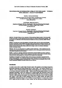
In vitro and in vivo interaction of UvrY with luxS promoter Purified UvrY interacts with luxS promoter without and with acetyl phosphate (Fig 2). The shift in the probe was shown by an arrow. The interaction of UvrY with luxS promoter is relatively weak compared to that with csrB promoter. Furthermore, interaction was also modulated by addition of negative and positive competitor DNA. Addition of positive competitor releases the binding whereas that of negative competitor tightens it. Since the interaction of uvrY with luxS promoter was relatively weak indicating there may be yet other factors associated with the interaction, we performed a in vivo chromatin immunoprecipitation of the luxS promoter fragment to determine the in vivo interaction using anti-uvrY antibody. Our result confirms the in-vivo binding of UvrY with luxS promoter (Fig 3). For the control reactions, we found the in vivo interaction of UvrY with csrB is relatively strong, while that with csrA was very weak, as expected from previous studies.
UvrY is required for cAMP-CRP repression of luxS-lacZ expression We further wanted to test any additional regulator might be involved in expression of luxSlacZ fusion. An important regulation occurs through the cyclic AMP-cyclic AMP Receptor Protein (cAMP-CRP) system. To further determine the role of UvrY in the glucose repression of luxS-lacZ fusion, we deleted adenylate cyclase encoding gene cyaA, which blockssynthesis of cAMP from ATP. Disruption of cya gene led to a constitutive expression of the luxS-lacZ fusion (Fig 4A). Addition of 1mM cAMP reduced the expression of luxS fusion to a basal level in the mutant. In case of ΔuvrYΔcya double mutant, the expression of the fusion is constitutive and addition of 1mM cAMP reduced the expression marginally (Fig 4B). The repression was faster in a Δcya mutant (30 min) compared to a ΔuvrYΔcya mutant (~90 min). Only addition
Fig 2. Electrophoretic mobility shift assay (EMSA) demonstrating interaction of UvrY with A) labeled csrB B) labeled luxS promoter. The promoter DNA was radiolabelled with P32. Purified UvrY was added at indicated concentrations without and with 20mM acetyl phosphate. The shift in probe was indicated by an arrow. Unlabeled DNA were added as negative and positive competitors shown by * and ¶ respectively. doi:10.1371/journal.pone.0157532.g002
PLOS ONE | DOI:10.1371/journal.pone.0157532 June 30, 2016
9 / 19
Regulation of luxS
Fig 3. Direct in-vivo binding of UvrY with luxS promoter by chromatin immunoprecipitation assay (ChIP). The occupancy of UvrY at the csrA, csrB and luxS promoters in E. coli was analyzed by ChIP using an anti-UvrY antibody. Purified DNA was PCR amplified using target specific primers. Genomic DNA and cell lysates were used as template for additional controls in PCR reaction. A 100 bp ladder marker was used for size comparison of amplicons. Expected sizes for all amplicons were between 300–400 bp. doi:10.1371/journal.pone.0157532.g003
of 5mM reduced the expression of the double mutant to a basal level. Similarly, deletion of crp resulted in constitutive expression of luxS and a ΔuvrYΔcrp mutant reduced the expression of the fusion marginally (Fig 5).
Effect of extracellular AI-2 accumulation and expression of luxS-lacZ fusion in csrA mutant In contrast to uvrY mutant, mutation in the csrA leads to elevated levels of AI-2 relative to the wild type and complementation of csrA in the mutant reduced extracellular accumulation of AI-2 to the wild-type level (Fig 6A). The inverse correlation of AI-2 accumulation in the uvrY and csrA deficient strains suggested a regulation both at the level of transcription and post transcription of luxS and AI-2 uptake. The uptake of AI-2 was also evaluated in the uvrY and csrA mutants and indicated an inverse relationship in the lsr operon expression. Similarly in case of the csrA mutant, the expression of the fusion was significantly higher at the entry of stationary
PLOS ONE | DOI:10.1371/journal.pone.0157532 June 30, 2016
10 / 19
Regulation of luxS
Fig 4. UvrY influences cAMP-CRP catabolite repression of luxS. A) Expression of luxS::lacZ in SM1020 (ΔcyaA::kan luxS::lacZ) in the absence of cAMP and in the presence of 1 mM cAMP. (B) Expression of luxS:: lacZ in SM1021 (ΔuvrY::cam ΔcyaA::kan luxS::lacZ) in the absence of cAMP, 1mM cAMP, and 5 mM cAMP. The point of cAMP addition is marked with an arrow on the graphs. Mean of three experiments were plotted and a representative data point used for calculation of significance is shown in asterisk (p