BASIC RESEARCH
www.jasn.org
Integrin-linked Kinase Controls Renal Branching Morphogenesis via Dual Specificity Phosphatase 8 Joanna Smeeton,*†‡ Priya Dhir,* Di Hu,* Meghan M. Feeney,* Lin Chen,* and Norman D. Rosenblum*†§ *Program in Developmental and Stem Cell Biology, and §Division of Nephrology, The Hospital for Sick Children, Toronto, Ontario, Canada; and †Departments of Paediatrics, and ‡Laboratory Medicine and Pathobiology, University of Toronto, Toronto, Ontario, Canada
ABSTRACT Integrin-linked kinase (ILK) is an intracellular scaffold protein with critical cell-specific functions in the embryonic and mature mammalian kidney. Previously, we demonstrated a requirement for Ilk during ureteric branching and cell cycle regulation in collecting duct cells in vivo. Although in vitro data indicate that ILK controls p38 mitogen-activated protein kinase (p38MAPK) activity, the contribution of ILKp38MAPK signaling to branching morphogenesis in vivo is not defined. Here, we identified genes that are regulated by Ilk in ureteric cells using a whole-genome expression analysis of whole-kidney mRNA in mice with Ilk deficiency in the ureteric cell lineage. Six genes with expression in ureteric tip cells, including Wnt11, were downregulated, whereas the expression of dual-specificity phosphatase 8 (DUSP8) was upregulated. Phosphorylation of p38MAPK was decreased in kidney tissue with Ilk deficiency, but no significant decrease in the phosphorylation of other intracellular effectors previously shown to control renal morphogenesis was observed. Pharmacologic inhibition of p38MAPK activity in murine inner medullary collecting duct 3 (mIMCD3) cells decreased expression of Wnt11, Krt23, and Slo4c1. DUSP8 overexpression in mIMCD3 cells significantly inhibited p38MAPK activation and the expression of Wnt11 and Slo4c1. Adenovirus-mediated overexpression of DUSP8 in cultured embryonic murine kidneys decreased ureteric branching and p38MAPK activation. Together, these data demonstrate that Ilk controls branching morphogenesis by regulating the expression of DUSP8, which inhibits p38MAPK activity and decreases branching morphogenesis. J Am Soc Nephrol 27: ccc–ccc, 2015. doi: 10.1681/ASN.2015020139
Renal branching morphogenesis, defined as growth and branching of the ureteric bud (UB) and its derivatives, is essential to mammalian kidney development. The UB grows, branches, and differentiates to form the collecting ducts and pelvis of the mature kidney. Further, UB tips induce adjacent metanephric mesenchyme cells to undergo the process of nephrogenesis.1 Defects in renal branching morphogenesis cause congenital renal hypodysplasia, characterized by abnormal collecting-system morphology and function and low nephron number.2 The UB arises as a direct evagination of the intermediate mesoderm-derived Wolffian duct. Extracellular ligands and cell-surface receptors in multiple distinct molecular signaling pathways function during the induction and early patterning of the UB. In J Am Soc Nephrol 27: ccc–ccc, 2015
contrast, a limited number and variety of intracellular molecules that function within these signaling pathways to control ureteric branching have been identified. In vitro treatment of embryonic urogenital explants with pharmacologic inhibitors revealed distinct roles for extracellular signal-regulated kinase
Received February 6, 2015. Accepted August 11, 2015. Published online ahead of print. Publication date available at www.jasn.org. Correspondence: Dr. Norman D. Rosenblum, The Hospital for Sick Children, Peter Gilgan Centre for Research and Learning, 686 Bay Street, Toronto, ON M5G 0A4, Canada. Email: norman.
[email protected] Copyright © 2015 by the American Society of Nephrology
ISSN : 1046-6673/2705-ccc
1
BASIC RESEARCH
www.jasn.org
(ERK), protein kinase B (AKT), and p38 mitogen-activated protein kinase (p38MAPK) during renal development.3 Furthermore, ERK and AKT can be activated downstream of the tyrosine kinase receptor, RET, in the UB tip domain, suggesting that intracellular kinase signaling is functionally important during ureteric branching.4,5 Integrin-linked kinase (ILK) is a scaffold protein that was initially identified through its ability to interact with the cytoplasmic domain of b-integrins.6 Investigation of ILK function has revealed a requirement for ILK upstream of intracellular kinases AKT,7 glycogen synthase kinase 3b,8 and p38MAPK.9 While each of AKT, glycogen synthase kinase 3b, and p38MAPK have been shown to play a role in controlling renal branching morphogenesis, their role downstream of ILK in the embryonic kidney has not been defined. Nor is it predicted by their functions in nonrenal tissues because their regulation by ILK is context-dependent.10 Previously, we demonstrated that Ilk is required for renal branching morphogenesis in vivo11 and controls UB branching via p38MAPK in vitro.9,12 However, the role of p38MAPK in vivo and the genes that act downstream of Ilk during the early stages of branching morphogenesis have not been previously defined. In this study we characterize the intracellular signaling pathways and UB transcriptome in the early embryonic Ilkdeficient kidney to determine the primary role of Ilk in the regulation of renal branching. Our results demonstrate that Ilk regulates the expression of a distinct subset of UB branching genes through both p38MAPK-independent and -dependent pathways. Our data also demonstrate upregulation of a phosphatase, dual-specificity phosphatase 8 (DUSP8), not previously implicated in kidney development. We demonstrate a functional role for DUSP8 in attenuating activation of p38MAPK and p38MAPK-dependent genes and inhibiting branching morphogenesis. Together, these results describe a novel DUSP8-mediated mechanism by which ILK controls renal branching morphogenesis.
RESULTS
Mice with conditional deletion of Ilk in the developing UB (Ilk2/2UB) have decreased UB branching at E12.5.11 However, the underlying molecular mechanisms are unknown. We investigated these mechanisms using whole-genome–based analysis of gene expression in intact kidney tissue isolated at E12.5 (n=6 kidneys per sample, three biologic replicates per genotype). At the time of dissection, the presence of a branching phenotype in Ilk-deficient kidneys was documented using green fluorescent protein (GFP) expression driven by the Hoxb7 promoter (Figure 1). Gene expression changes were investigated through microarray analysis using Affymetrix GeneChip Mouse 430 2.0 arrays. Expression levels were normalized to wild-type (WT) and then analyzed for differential expression between Ilk 2 /2 UB and WT samples using the Bioconductor limma package in R statistical program.13 2
Journal of the American Society of Nephrology
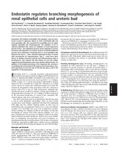
Figure 1. E12.5 Ilk2/2UB kidneys demonstrate reduced UB branching and abnormal gene regulation. (A,B) GFP branching pattern of (A) GFP+ heterozygote and (B) Ilk2/2UB kidneys confirmed the branching defect in E12.5 Ilk2/2UB kidneys selected for microarray analysis. (C) Gene expression differs between WT and Ilk2/2UB mutant (MUT) kidneys with similar expression patterns clustering within the WT and mutant samples. Expression values of differentially regulated genes (P,0.003) are shown in this heatmap, with red indicating lower expression and yellow higher expression.
To determine changes in expression levels for individual genes, the top table of genes based on the t-statistic ranking was generated. Using a statistical cutoff of P,0.003 to identify changes in the expression of individual genes, we identified 131 downregulated probe sets and 96 upregulated (Figure 1C, Supplemental Tables 2 and 3). The magnitude of changes in gene expression, particularly for downregulated genes, was twofold or lower. This may be due to the fact that UB gene expression was assayed in whole tissue in which UB cells constitute a minority of cells. While a gene expression analysis in isolated UB cells may have generated gene expression changes of higher magnitude, the quality of RNA isolated from flow-sorted UB cells derived from mutant mice precluded a whole-genome gene expression analysis. Notwithstanding these considerations, consistent with genetic deletion of Ilk, the most statistically significant gene expression change was a downregulation of Ilk in Ilk2/2UB kidneys. J Am Soc Nephrol 27: ccc–ccc, 2015
www.jasn.org
Gene Ontology Analysis Suggests Abnormal Regulation of Tube Morphogenesis Genes
To define the global state of gene expression in an Ilk-deficient state, we performed pathway analysis to identify gene ontology (GO) biologic processes that are enriched within the downregulated gene set. Sixty-one biologic process GO terms were statistically enriched in the downregulated gene list (Supplemental Table 4). Within the downregulated gene set, GO terms involved in epithelial tube development as well as the regulation of organ morphogenesis were enriched (Table 1). These terms are highly consistent with the mutant phenotype and a putative role for Ilk in controlling the expression of genes involved in branching morphogenesis and tube morphogenesis. A Subset of Ilk-Dependent Genes have a UB-Specific Expression Pattern
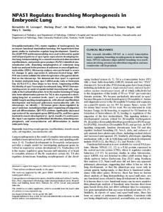
Because knockdown of Ilk was specific to the UB lineage, next we investigated genes expressed in the ureteric lineage and which function in ureteric branching. Accordingly, the whole-kidney gene data set was filtered for genes that are known to be expressed in the UB by crossreferencing the downregulated genes with the microarray and in situ hybridization expression data available in GUDMAP (www.gudmap. org).14 This procedure resulted in reduction of the 131 downregulated genes to 14 genes with confirmed UB-specific expression, including Wnt11, a UB tip–specific gene essential for UB development15 (Table 2). While a subset of these genes (Wnt11, Sox8, CXCR4, Myb, Etv5, Etv4, and Spry1) are components of the RET signaling pathway or are RET signaling targets,16 expression of Ret mRNA was not significantly altered in Ilk2/2UB kidneys (Figure 2, B and C). Expression of RETwas further investigated at the level of protein activity by assaying the levels of phosphorylated RET-1062, an isoform critical for RET activity.17 Results demonstrated that levels of phosphorylated RET 1062, assayed by Western blot analysis of protein lysates generated from E12.5 kidney tissue, were not significantly different between Ilk2/2UB and control mice (Supplemental Figure 1, A and B). The expression of Ilk-dependent UB genes was validated by quantitative RT-PCR (qRT-PCR). As shown in Figure 2A, differential expression reached
BASIC RESEARCH
statistical significance for six genes by qRT-PCR (n=6/genotype, two technical replicates) and these were further confirmed by in situ hybridization in tissue sections at E12.5 (Figure 2). In contrast to UB tip genes Ret and Sox9, the mRNA expression of which was not significantly altered, in situ hybridization analysis confirmed decreased expression of Sox8, Wnt11, Myb, CXCR4, Krt23, and Slco4c1 in the UB of E12.5 Ilk2/2UB kidneys (Figure 2). Ilk Deficiency Causes a Specific Decrease in the Activation of p38MAPK
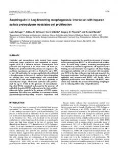
Because previous studies in nonrenal tissues have implicated ILK in the control of kinase activity,7,8 we investigated the state of kinase signaling in E13.5 Ilk2/2UB embryonic kidney tissue protein lysates. Consistent with our prior results in vitro,12 phosphorylation of p38MAPK, an indicator of p38MAPK activation, was significantly decreased by 61% in Ilk2/2UB embryonic kidney tissue (Figure 3, A and B). In contrast, we observed no statistically significant difference in the expression of the phosphorylated forms of AKT and ERK in Ilk2/2UB embryonic kidney tissue as compared with WT (Figure 3, A and B), nor was there a significant difference in the expression of the functionally active form of b-catenin protein, in which residues Ser33, Ser37, and Thr41 are not phosphorylated (Figure 3, A and B). Consistent with the analysis of protein expression, expression of the TCF/LEF-LacZ reporter allele, a surrogate measure of canonical WNT target gene activation, was comparable in Ilk2/2UB and WT kidney tissue (Figure 3C). p38MAPK Activation is Required for the Expression of a Subset of Ilk-Dependent Genes
We investigated the functional role of p38MAPK in regulating expression of Ilk-dependent genes in the UB by inhibiting p38MAPK function. Inhibition of p38MAPK was achieved through treatment of explanted kidneys with commercially available p38MAPK inhibitors SB203580 and SB202190. Following two days of treatment with SB203580 or SB202190, cultured E12.5 embryonic kidneys displayed abnormal UB branch morphology (Figure 4, A–C) and 40% or 66% fewer branches, respectively, compared with vehicle-treated littermate kidneys (Figure 4D). Next, we determined the effect
Table 1. Selected GO terms enriched in genes decreased in Ilk2/2UB kidneys GO ID
Name
P Value
Genes in Test Set UNCX|HES1|GCNT3|MYO7A|POU3F4|SOX8|GJA1|FGF9|FG F1|CXCR4|BMP2|LAMA1|BCL11B|JAG1|ATIC|HIF1A|SOST DC1|WNT11|CA2|ETV4|ETV5|ALDH1A1|FAM20C|SPRY1|S PRY2|WNT8B HES1|CAPRIN1|SOX8|GJA1|NRCAM|CHRNA7|FGF9|FGF1|N PNT|CXCR4|BTC|NELL1|LRP8|BMP2|KITLG|JAG1|MYB|HIF 1A|CA2|CDH4|ETV5|FAM20C|SPRY1|ILK HES1|SOX8|GJA1|FGF1|NPNT|CXCR4|BMP2|LAMA1|JAG1| HIF1A|WNT11|CA2|ETV4|ETV5|ALDH1A1|FREM2|ILK HES1|SOX8|GJA1|FGF9|FGF1|NPNT|CXCR4|ARG2|BMP2|LA MA1|JAG1|HIF1A|WNT11|ETV4|ETV5|SPRY1|SPRY2|ILK
GO:0009887
Organ morphogenesis
6.22E-08
GO:0051094
Positive regulation of developmental process
1.66E-07
GO:0002009
Morphogenesis of an epithelium
1.39E-06
GO:0035295
Tube development
2.25E-06
J Am Soc Nephrol 27: ccc–ccc, 2015
Ilk Controls Ureteric Branching via DUSP8
3
BASIC RESEARCH
www.jasn.org
Table 2. Selected UB-expressed mRNA transcripts downregulated in Ilk2/2UB kidneys (P,0.003) Probe ID
Gene Name
1435438_at 1437870_at 1418213_at 1448710_at 1450772_at 1420998_at 1435303_at 1422165_at 1423232_at 1424367_a_at 1450194_a_at 1415874_at 1418153_at 1415855_at
SRY-box containing gene 8 Solute carrier organic anion transporter family, member 4C1 Keratin 23 Chemokine (C-X-C motif) receptor 4 Wingless-related MMTV integration site 11 Ets variant gene 5 TAF4B RNA polymerase II, TATA box binding protein-associated factor POU domain, class 3, transcription factor 4 Ets variant gene 4 (E1A enhancer binding protein, E1AF) Homer homolog 2 (Drosophila) Myeloblastosis oncogene Sprouty homolog 1 (Drosophila); similar to sprouty 1 Laminin, a 1 Kit ligand
of p38MAPK inhibition on the expression of the six Ilkdependent UB genes using qRT-PCT. While levels of Sox8, CXCR4, and Myb were not significantly affected by inhibition of p38MAPK in kidney explant culture (Figure 4E), expression of Wnt11, Krt23, and Slco4c1 were each significantly decreased (Figure 4F). These data indicate that Ilk acts via p38MAPKdependent and -independent mechanisms to regulate genes expressed in the developing UB. Overexpression of DUSP8 Decreases Both p38MAPK Activation and Expression of UB Tip–Specific Genes
GO analysis of the complete transcriptome identified altered regulation of MAPK phosphatases in Ilk 2 /2 UB kidneys (Table 3). At the individual gene level, the p38MAPK phosphatase, Dusp8, was among the most significantly upregulated genes in Ilk2/2UB kidneys (Table 4). We confirmed upregulation of DUSP8 in Ilk2/2UB embryonic kidneys by qRT-PCR analysis (n=6/genotype, two technical replicates; Figure 5A). While our published work demonstrated that ILK activates p38MAPK, the mechanism underlying their interaction is not understood. Accordingly, we investigated the functional contribution of DUSP8 to p38MAPK activity in the UB lineage using inner medullary collecting duct cells (mIMCD3) in which we varied DUSP8 expression and stimulated p38MAPK signaling using EGF12 (Figure 5B). mIMCD3 cells respond to EGF within 15 minutes with a 52.2% increase in p38MAPK phosphorylation (Figure 5, C and D). In contrast, no increase in p38MAPK phosphorylation was observed in DUSP8-overexpressing mIMCD3 cells. While a further increase in p38MAPK was observed by 30 minutes following administration of EGF in mIMCD3 cells, no change in p38MAPK phosphorylation was observed in DUSP8-overexpressing mIMCD3 cells. Next, we investigated whether DUSP8 regulates the genes whose expression is regulated by ILK. Expression of p38MAPKregulated genes (Wnt11, Krt23, and Slco4c1) and p38MAPKindependent genes (Sox8, CXCR4, and Myb) was analyzed after EGF-stimulation of mIMCD3 cells and DUSP8-overexpressing 4
Journal of the American Society of Nephrology
Gene Symbol Sox8 Slco4c1 Krt23 Cxcr4 Wnt11 Etv5 Taf4b Pou3f4 Etv4 Homer2 Myb Spry1 Lama1 Kitl
Fold Change
P Value
22.037065358 21.963658776 21.835321044 21.613128354 21.572346139 21.459696529 21.413147086 21.379100185 21.368048482 21.344712561 21.307002458 21.304973111 21.291465414 21.283595843
8.71E-06 2.48E-05 7.02E-04 4.71E-05 3.12E-04 3.89E-04 1.05E-03 2.44E-03 8.53E-04 1.14E-03 6.57E-04 4.38E-04 1.62E-03 1.33E-03
mIMCD3 cells. Our data demonstrate that overexpression of DUSP8 results in a significant decrease in the expression of Wnt11 and Slco4c1 mRNA (Figure 5E). Krt23 was also decreased, but the change in expression was not statistically significant. mRNA levels of Myb and Sox8 were unchanged. Because CXCR4 mRNA was undetected in both mIMCD3and DUSP8-overexpressing mIMCD3 cells, we could not evaluate its regulation by DUSP8. Together, these data show that Wnt11 and Slco4c1, both of which are regulated by p38MAPK, are also regulated by DUSP8. Adenovirus-Mediated DUSP8 Overexpression in Embryonic Kidney Explants Decreases UB Branching
We investigated the functional contribution of DUSP8 in renal branching morphogenesis in intact kidney tissue. E12.5 WT kidney explants were treated with either GFP-expressing recombinant adenovirus (Ad-Gfp) or adenovirus carrying Gfp and Dusp8 (Ad-Gfp-Dusp8) for 5 days at 107 plaqueforming units/ml. Transduction efficiency of Dusp8 transcript was analyzed in three independent experiments by qRT-PCR analysis (n=9/group, three technical replicates; Figure 6A). A minus-reverse transcription (RT) negativecontrol sample was included in qRT-PCR experiments to establish the effective removal of adenoviral DNA in the RNA samples. The mRNA of Dusp8 was 68-fold more abundant in the Ad-Gfp-Dusp8–treated explants compared with control explants. Levels of DUSP8 protein were remarkably higher in Ad-Gfp-Dusp8–treated explants compared with control explants (Figure 6B). Next, we measured the effect of DUSP8 overexpression on ureteric branching. Following 5 days of treatment with Ad-Gfp-Dusp8, ureteric branches were analyzed by whole-mount immunofluorescence analysis of cytokeratin expression (Figure 6, C and D). Quantification of ureteric tips demonstrated a significant branching phenotype in the DUSP8-overexpressing kidneys, with an average of 45 UB branch tips compared with the 74 branch tips present in the control explants (n=14/group) (Figure 6E). J Am Soc Nephrol 27: ccc–ccc, 2015
www.jasn.org
BASIC RESEARCH
Figure 2. Validation of Ilk-dependent genes confirmed differential expression in the UB. (A) Results of qRT-PCR analysis of UBexpressed genes downregulated in Ilk2/2UB kidneys. Significantly decreased genes Ilk, Sox8, Wnt11, CXCR4, Slco4c1, Myb, and Krt23 are shown (P,0.05). (B) In situ hybridization analysis of gene expression in tissue sections demonstrated normal expression of (B and C) Ret and (R and S) Sox9 in UB tips. (D–Q) Decreased UB-specific expression of Ilk-target genes was confirmed. Arrows indicate UBs.
In parallel experiments we investigated the effect of DUSP8 overexpression on p38MAPK activity in embryonic kidney explants. Experiments were designed in a manner parallel to those using mIMCD3 cells, using EGF to stimulate p38MAPK activity (Figure 5). Three independent experiments were performed, each consisting of three explants isolated from J Am Soc Nephrol 27: ccc–ccc, 2015
different mice and treated individually and then pooled to generate sufficient protein for Western blot analysis. The levels of phosphorylated p38MAPK, controlled for total p38MAPK, in each treatment group were compared with Ad-GFP plus EGF (no DUSP8). Treatment of explants with Ad-GFPDUSP8 or Ad-GFP did not significantly change p38MAPK Ilk Controls Ureteric Branching via DUSP8
5
BASIC RESEARCH
www.jasn.org
DISCUSSION
A requirement for Ilk expression in ureteric cells during branching morphogenesis has been demonstrated in the murine kidney.11 Yet, the molecular mechanisms that underlie its actions have not been previously defined in vivo. Our previous analysis of intracellular signaling downstream of ILK in cultured collecting duct cells with gain or loss of ILK expression suggested that ILK controls tubule morphogenesis via p38MAPK.9 In this study, we demonstrate that Ilk controls p38MAPK-dependent expression of genes involved in renal branching morphogenesis by regulating the expression of DUSP8, which inhibits p38MAPK activity. We first identified genes that are regulated by Ilk in ureteric cells using a whole-genome mRNA expression analysis of whole-kidney mRNA in mice with Ilk deficiency targeted to the UB. The expression of six genes with expression in ureteric tip cells – Wnt11, Krt23, Slo4c1, Myb, Sox8, and Cxcr4 – was downregulated in Ilk-deficient tissue. Next, we demonstrated that kidney tissue with Ilk deficiency is characterized by decreased phosphorylated p38MAPK but no significant decrease in the phosphorylation of other intracellular effectors (ERK, AKT, or b-catenin) previously shown to control renal morphogenesis. We then showed that inhibition of p38MAPK activity controls the expression of Wnt11, Krt23, and Slo4c1. Upregulation of DUSP8, a dualspecificity phosphatase, in Ilk-deficient kidney tissue suggested a mechanism by which ILK controls p38MAPK and ureteric tip cell gene expression. Our data show that DUSP8 overexpression in murine-collecting duct cells inhibited p38MAPK activation Figure 3. Embryonic Ilk2/2UB kidneys have decreased activation of p38MAPK. (A) and expression of Wnt11 and Slo4c1 and deWestern blot analysis of E13.5 kidney lysates for expression of phosphorylated (phoscreased ureteric branching and p38MAPK pho) p38MAPK, p38MAPK, phospho-ERK, phospho-AKT, pS33/S37/741 b-catenin, and activation in intact embryonic kidney tissue. total b-catenin. (B) Quantification of protein bands relative to total protein demon2/2UB kidneys Our data support a model of ILK signaling strated significantly decreased expression of phospho-p38MAPK in Ilk (P,0.001). (C) TCF-LacZ expression in the UB lineage is unchanged in Ilk2/2UB kidneys. during UB branching (Figure 7). Our model suggests that ILK acts upstream of p38MAPK to regulate Wnt11, Krt23, and Slco4c1 expression in the UB tip to stimulate UB branching. activation compared with Ad-GFP plus EGF. In contrast, treatment with Ad-GFP-DUSP8 plus EGF decreased ILK also regulates expression of Sox8, CXCR4, and Myb in the UB. Our model further proposes that ILK negatively regulates p38MAPK activation by 62% (P,0.02) compared with the the expression of DUSP8, a protein phosphatase, which can act Ad-GFP plus EGF control (Figure 6F). Together, these data show that DUSP8 decreases branching morphogenesis and to modulate p38MAPK activation and downstream target gene expression. p38MAPK activation in intact murine kidney tissue. 6
Journal of the American Society of Nephrology
J Am Soc Nephrol 27: ccc–ccc, 2015
www.jasn.org
BASIC RESEARCH
Figure 4. p38MAPK inhibition in kidney culture causes decreased branching and decreased expression of a subset of Ilk-dependent genes. (A–C) E12.5 Hoxb7Cre-EGFP kidneys were cultured in the presence of DMSO, SB203580, or SB202190 for 48 hours. Branching was visualized by GFP expression in the UB lineage. Insets show high magnification of UB tip regions. (D) Quantification demonstrated decreased tip number in p38MAPK-inhibitor–treated kidneys. (E) qRT-PCR analysis of gene expression demonstrated that Sox8, Myb, and CXCR4 were not significantly altered following inhibition of p38MAPK activity (P.0.05). (F) qRT-PCR analysis demonstrated that expression of Wnt11, Krt23, and Slco4c1 were decreased following inhibition of p38MAPK activity in kidney culture (P,0.05).
Ilk-Dependent Genes and Control of Renal Branching Morphogenesis
The expression of genes (Wnt11, Cxcr4, and Sox9) expressed in the ureteric tip and with demonstrated functions in ureteric branching are decreased in Ilk2/2UB mice. Wnt11-deficient mice are J Am Soc Nephrol 27: ccc–ccc, 2015
characterized by renal hypoplasia and decreased ureteric branching.15 This phenotype is similar to that observed in Ilk2/2UB mice and suggests that Wnt11 deficiency in the absence of Ilk is critical to the genesis of renal hypoplasia. We also observed decreased expression of Cxcr4, a chemokine encoding gene that is highly Ilk Controls Ureteric Branching via DUSP8
7
BASIC RESEARCH
www.jasn.org
Table 3. Selected GO terms differentially regulated in Ilk2/2UB kidneys P Value
Genes in Test Set
GO:0008009
Chemokine activity
1.36E-03
GO:0019838
Growth factor binding
7.10E-03
GO:0033549
MAP kinase phosphatase activity MAP kinase activity
0.03
Ccl1|Ccl11|Ccl12|Ccl17|Ccl19|Ccl2|Ccl20|Ccl21a|Ccl21bCcl21c| Gm10591|Gm13304|Gm1987|LOC100041593|Ccl22|Ccl24|C cl25|Ccl27a|Ccl27b|Ccl27a|Ccl27b|Gm13306|Ccl28|Ccl3|Ccl4| Ccl5|Ccl6|Ccl7|Ccl8|Ccl9|Cklf|Cx3cl1|Cxcl1|Cxcl10|Cxcl11|Cxc l12|Cxcl13|Cxcl14|Cxcl15|Cxcl16|Cxcl2|Cxcl3|Cxcl5|Cxcl9|Elav l1|Pf4|Ppbp|Xcl1| A2m|Acvr1|Acvr1b|Acvr2a|Acvr2b|Acvrl1|Api5|Bmpr2|Cd36|Cep 57|Col1a1|Col1a2|Col2a1|Col3a1|Col4a1|Col5a1|Col6a1|Crim 1|Ctgf|Cyr61|Egfr|Eng|Erbb2|Esm1|Fgf2|Fgfbp1|Fgfbp3|Fgfr1| Fgfr2|Fgfr3|Fgfrl1|Fibp|Flt1|Flt4|Furin|Ghr|Ghrhr|Gm13305|Gm 2002|Il11ra1|Il11ra2|Gm614|Il2rg|Htra1|Htra3|Htra4|Igf1r|Igf2r| Igfals|Igfbp1|Igfbp2|Igfbp3|Igfbp4|Igfbp5|Igfbp6|Igfbp7|Igfbp l1|Il1rapl1|Il2ra|Il2rb|Il2rg|Il6ra|Il6st|Il9r|Insr|Kazald1|Kdr|Kl|Klb| Lifr|Ltbp1|Ltbp2|Ltbp3|Ltbp4|Ngfr|Nov|Nrp1|Ntf3|Ntrk2|Osmr| Pdap1|Pdgfa|Pdgfb|Pdgfra|Pdgfrb|Sdcbp|Smn1|Sort1|Tek|Tgf b3|Tgfbr1|Tgfbr3|Tnxb|Wisp1|Wisp2| 5530400B01Rik|Dusp8|Dusp1|Dusp10|Dusp14|Dusp16|Dusp18|Du sp2|Dusp21|Dusp3|Dusp4|Dusp5|Dusp6|Dusp7|Dusp8|Dusp9| Mapk1|Mapk10|Mapk11|Mapk12|Mapk13|Mapk14|Mapk15|Map k3|Mapk4|Mapk6|Mapk7|Mapk8|Mapk9|Nlk|
GO ID
GO:0004707
Name
0.03
expressed in the UB tip. CXCR42/2 mice are characterized by a mild degree of renal hypoplasia.18 Pharmacologic inhibition of CXCR4 in embryonic kidney explants inhibits branching morphogenesis.19 Moreover, the overall state of chemokine signaling is misregulated in Ilk2/2UB kidneys (Supplemental Table 5, Table 3). Together, these observations suggest that loss of Cxcr4 may also contribute to the hypoplastic phenotype in Ilk2/2UB mice. Sox8 was markedly decreased in Ilk2/2UB mice. Sox8 acts in combination with Sox9 and alone to control branching morphogenesis.20 However, the effects of Sox9 deficiency on branching morphogenesis are far less severe than those observed in mice with deficiency of both Sox8 and Sox9, suggesting that loss of Sox8 may act in combination with the loss of Wnt11 and Cscr4 to disrupt branching morphogenesis. We identified loss of expression of other genes in ureteric tip cells, but without known functions during branching morphogenesis, in Ilk2/2UB mice. Keratin 23 is an intermediate filament protein,21
which could play a structural role specific to the ureteric tip. The function of Slco4c1, a unique marker of the ureteric tip domain22 and a uremic toxin transporter protein,23 remains to be defined. Growth factor signaling via the RET receptor is crucial for renal branching morphogenesis. Ret expression and phosphorylation is not decreased in Ilk2/2UB kidney tissue. Interestingly, UB isolated from Ilk loss-of-function (K220A or K220M) mutant mice is unresponsive to the RET ligand, and GDNF24 and GDNF-responsive genes are downregulated in Ilk2/2UB kidneys. Phosphorylation of specific tyrosines in the cytoplasmic domain of RET causes phosphorylation of multiple distinct pathways downstream of RET.25 Because loss of Ilk results in alterations in a subset of GDNF-dependent genes, including Myb and CXCR4, it is possible that ILK may be acting downstream of a distinct phosphorylation site of RET to regulate a subset of signals downstream of RET.
Table 4. Selected mRNA transcripts upregulated in Ilk2/2UB kidneys (P,0.003) Probe ID 1455625_at
1418930_at
1418714_at 1427975_at 1422029_at 1449195_s_at
8
Gene Name Predicted gene 4799; TAF10 RNA polymerase II, TATA box binding protein-associated factor Chemokine (C-X-C motif) ligand 10; similar to small inducible cytokine B10 precursor (CXCL10) (interferon-g-induced protein CRG-2) (g-IP10) (IP-10) (C7) Dual-specificity phosphatase 8 RAS-like, family 10, member A Chemokine (C-C motif) ligand 20 Chemokine (C-X-C motif) ligand 16
Journal of the American Society of Nephrology
Fold Change
P Value
Taf10
2.43524092
3.45E-06
Cxcl10
2.09356283
5.34E-06
Dusp8 Rasl10a Ccl20 Cxcl16
1.73978671 1.72559525 1.46633699 1.4264928
8.55E-05 4.78E-06 5.64E-05 5.05E-05
Gene Symbol
J Am Soc Nephrol 27: ccc–ccc, 2015
www.jasn.org
BASIC RESEARCH
Figure 5. DUSP8 overexpression results in altered p38MAPK phosphorylation and decreased UB gene expression. (A) Dusp8 overexpression in Ilk2/2UB kidneys was confirmed by qRT-PCR (P,0.05). (B) Following Dusp8 cDNA transfection and stable colony selection, qRT-PCR analysis demonstrated successful overexpression of Dusp8 in mIMCD3 cells (DUSP8 o/e) (P,0.05). (C and D) After 15 minutes of EGF treatment in culture, levels of phosphorylated (phospho) p38MAPK are comparable between WT and DUSP8-overexpressing cells. After 30 minutes of EGF treatment, phospho-p38MAPK levels in DUSP8-overexpressing mIMCD cells are significantly decreased compared with WT cells (P,0.001). (E) In an EGF-stimulated manner, DUSP8-overexpressing mIMCD cells have significantly decreased the expression of Wnt11 and Slco4c1 (*Wnt11, P,0.05; **Slco4c1, P,0.001). Inhibition of Krt23 and Myb in DUSP8overexpressing cells was observed, but not statistically significant.
DUSP8 is a Novel Modulator of p38MAPK Activity in the Murine Embryonic Kidney
We identified DUSP8 as a novel ILK target that modulates p38MAPK activity. To our knowledge, this is the first reported functional role of DUSP8 in regulating branching J Am Soc Nephrol 27: ccc–ccc, 2015
morphogenesis. In fact, the body of evidence related to the role of phosphatases during kidney development has previously been limited to proteins that interact with RET. The tyrosine phosphatase SHP2 promotes signaling downstream of RET; loss of Shp2 in the developing UB decreases both ERK Ilk Controls Ureteric Branching via DUSP8
9
BASIC RESEARCH
www.jasn.org
unchanged in contrast to a significant decrease in phosphorylation of p38MAPK. Three members of the DUSP family – DUSP8, DUSP10, and DUSP16 – have been identified in nonrenal tissues and shown to modulate p38MAPK activity.28–30 These observations are consistent with our data, which demonstrate that DUSP8 decreases p38MAPK phosphorylation and expression of genes that are modulated in their expression by p38MAPK in the embryonic kidney. Interestingly, attenuation of p38MAPK activation by DUSPs can lead to positive regulation of ERK phosphorylation and activity.31 Our finding that phosphorylation of ERK is unchanged suggests that abnormal ERK activity does not contribute to the phenotype in Ilk2/2UB kidneys. The regulation of DUSP8 expression is incompletely understood. Given our previous demonstration of a BMP7-ILK-p38MAPK signaling axis and our observation that DUSP8 modulates p38MAPK activation, an autoregulatory feedback loop to regulate its expression is likely. If so, our model (Figure 7) suggests that p38MAPK activation leads to expression of DUSP8, but ILK is required directly or indirectly to negatively modulate DUSP8 and maintain the levels of p38MAPK within a precise range to facilitate gene expression and branching. Loss of negFigure 6. Adenovirus-mediated DUSP8 overexpression in WT E12.5 embryonic kidney ative inhibition in Ilk2/2UB kidneys would explants cultured for 5 days results in impaired UB branching and decreased activation cause excessive DUSP8 expression leading of p38MAPK. (A) Overexpression of Dusp8 in explant cultures treated with Ad-GFPto decreased phosphorylated p38MAPK actiDusp8 compared with Ad-GFP control was confirmed by qRT-PCR (P,0.001). (B) vation in the UB, altered gene expression, and DUSP8 protein is expressed in explants treated with Ad-GFP-Dusp8 compared with explants treated with Ad-GFP. (C and D) Immunofluorescence analysis of cytokeratin decreased branching morphogenesis. In summary, we demonstrated that Ilk expression in whole-mount preparations of E12.5 kidney explants at 5 days postinfection demonstrate markedly decreased branching. (D) The number of tips was is crucial for the sustained activation of quantified from Ad-GFP and Ad-GFP-Dusp8 kidneys stained with cytokeratin. Differ- p38MAPK and downstream gene expression ences between the two treatment groups were statistically significant (n=14 kidneys/ in the developing ureteric cell in lineage. Degroup; P,0.001). (E and F) Western blot analysis of p38MAPK activation in EGF- creased branching morphogenesis in Ilk2/2UB stimulated Dusp8-overexpressing explants cultured for 48 hours shows significantly mice is due, in part, to increased expression decreased phosphorylation of p38MAPK in Ad-GFP-Dusp8 compared with Ad-GFP of DUSP8, which attenuates p38MAPK (n=3 groups, P,0.02) and relevant controls. signaling. Ilk is required in the UB for the expression of specific markers of the tip cell doactivation in UB tips as well as ureteric branching.26 The phosmain, as well as expression of renal branching genes crucial for phatase PTEN antagonizes PI3K activity downstream of the morphogenesis. RET receptor affecting branching morphogenesis through the regulation of the localization and generation of phosphoinositol 3–5 triphosphates.27 CONCISE METHODS The DUSP family of proteins are specific to MAPKs. DUSPs dephosphorylate the serine-threonine and tyrosine residues in Mice the kinase domain, thereby inactivating the kinases. In IlkMice with conditional ILK deficiency targeted to the ureteric lineage deficient kidney tissue, the effect of ILK deficiency was specific were generated using Hoxb7-Cre-EGFP32 and IlkloxP mice33 to generto p38MAPK because phosphorylation of ERK and AKT was ate Hoxb7-Cre-EGFP;Ilk-/loxP (Ilk2/2UB) progeny with a specific 10
Journal of the American Society of Nephrology
J Am Soc Nephrol 27: ccc–ccc, 2015
www.jasn.org
BASIC RESEARCH
Microarray Data Set Analysis The six samples were analyzed and compared using the Bioconductor limma package in the R statistical program. Briefly, the limma package was used to perform background correction, robust multiarray (GC-RMA) normalization, expression calculation, and comparative analysis using a contrast matrix between WT and Ilk2/2UB samples to determine log-fold change and differential expression for each probe. The limma package calculates the expression values for each probe and ranks the probe list by statistical significance.35 To investigate the contributions to the mutant phenotype of genes with common attributes, the relative enrichment of GO terms in the downregulated gene list was investigated using the ToppGene server (false discovery rate, P,0.05).36 The complete data set was analyzed by the program ErmineJ37 to identify globally altered molecular pathways.
qRT-PCR
Figure 7. Model of the role of ILK in UB branching. ILK acts upstream of p38MAPK to regulate Wnt11, Krt23, and Slco4c1 expression in the UB tip to stimulate UB branching. Wnt11 is required for maintenance of Gdnf expression in the adjacent metanephric mesenchyme. ILK also regulates expression of Sox8, CXCR4, and Myb in the UB. ILK negatively regulates the expression of the phosphatase DUSP8, which can act to modulate p38MAPK activation and downstream target gene expression.
deficiency of Ilk in the UB lineage. Ilk2/2UB mice were crossed with the TCF/LEF-b-galactosidase reporter strain.34 All mice were housed at the Toronto Centre for Phenogenomics according to the guidelines of the Canadian Council for Animal Care.
Global Gene Expression Analysis in Mouse Kidney Tissue
E12.5 embryos were dissected and kidneys removed. GFP fluorescence and branching pattern were used to identify mutant and WT kidneys prior to RNA stabilization in RNAlater (Invitrogen). Due to the absence of GFP expression in both heterozygote and WT littermates, the genotypes were confirmed by PCR prior to RNA isolation. Six kidneys were pooled in each biologic replicate prior to RNA isolation by Qiagen RNeasy Micro Kit according to the manufacturer’s instructions. Prior to microarray sample submission, total RNA quality and quantity was determined by Nanodrop analysis. RNA quality was confirmed by Agilent 2100 Bioanalyzer nano chip analysis prior to linear RNA amplification and microarray profiling on the Affymetrix GeneChip Mouse Genome 430 2.0 Array. All samples were of sufficient RNA quality (RNA integrity number .9) and 400 ng of each sample was amplified for array analysis using the Ambion IVT kit. Each sample was amplified, hybridized, and scanned according to the standard protocol by The Centre for Applied Genomics, The Hospital for Sick Children, Toronto, Canada. J Am Soc Nephrol 27: ccc–ccc, 2015
RNA was isolated using Qiagen RNeasy Micro Kit and cDNA was generated using First Strand cDNA Synthesis (Invitrogen) from total RNA. Real-time PCR was performed using the Applied Biosystems 7900 HT Fast RT-PCR system with a PCR reaction mix containing a cDNA sample, SYBR green, and gene-specific primers (Supplemental Table 1). Relative mRNA expression levels were determined using the standard curve method and individual expression values were normalized by comparison to b2-microglobulin. In embryonic kidney explants treated with adenovirus, relative mRNA expression levels were determined using the comparative CT method and individual expression values were normalized by comparison to glyceraldehyde 3-phosphate dehydrogenase.
In Situ mRNA Hybridization, Western Analysis, and Immunocytochemistry
Whole embryos were fixed in 4% paraformaldehyde in PBS for 16 hours at 4°C. In situ hybridization was performed as described,38 on paraffin-embedded sections (4 mm) using dioxygenin-labeled cDNA probes for Ret, Wnt11, Sox8, Sox9, Slco4c1, Krt23, Myb, and CXCR4. For Western blot analysis, kidneys or cultured cells were lysed in radioimmunoprecipitation assay buffer and subjected to immunoblotting as described.11 The following primary antibodies were used at a dilution of 1:1000: p38MAPK, 9212; P-p38MAPK, 9211; ERK, 4695; P-ERK, 9101; AKT, 9272; P-AKT, 9275; nonphospho (Active) b-catenin (Ser33/37/Thr41), 8814; b-catenin, 8480 (Cell Signaling Technology), DUSP8, NBP1–58302 (from Novus Biologicals), P-RET (Tyr-1062)-R, sc-20252-R (Santa Cruz Biotechnology), RET (C-19), sc-167 (Santa Cruz Biotechnology), and Actin, A5441 (SigmaAldrich). E12.5 urogenital ridges were stained for b-galactosidase expression as previously described.39
Cell Culture Both mIMCD3- and DUSP8-overexpressing mIMCD3 cells were grown in monolayer in DMEM-Ham’s F12 medium (DMEM-F12), supplemented with 10% FBS (Gibco), 1% penicillin-streptomycin in an incubator with 5% CO2 at 37°C. The medium for DUSP8overexpressing mIMCD3 cells was supplemented with 8 mg/ml G418. DUSP8 (NM_008748) was overexpressed in mIMCD3 cells using a TrueClone full-length cDNA clone from Origene (MC203407). Cells Ilk Controls Ureteric Branching via DUSP8
11
BASIC RESEARCH
www.jasn.org
were transfected using Turbofect reagent (R0531; Thermo Fisher Scientific), according to the manufacturer’s instructions, and stable clones were generated following G418 selection over one week. EGF stimulation experiments were performed in triplicate after 18 hours of serum starvation in culture using 10 ng/ml EGF (E4127; Sigma-Aldrich).
Cultured Embryonic Kidney Explants Kidneys were dissected from E12.5 Hoxb7-Cre-EGFP mice and grown in culture on filter membrane inserts for 2 days in the presence of DMSO vehicle control or chemical inhibitors SB203580 (20 mM, V1161; Promega) and SB202190 (10 mM, S7067; Sigma-Aldrich) (n=3 kidneys/treatment). For adenovirus treatment, embryonic kidneys dissected at E12.5 were grown in culture on filter membrane inserts for 5 days. Ad-Gfp and Ad-Gfp-Dusp8 were purchased from Vector Biolabs and added daily to culture medium at 107 plaque-forming units/ml for 5 days. For imaging of ureteric branches, explants were fixed for 1 hour in methanol at –20°C, permeabilized with 0.1% Triton X-100 for 10 minutes, blocked with Protein Block Serum Free (Dako) for 1 hour and incubated overnight with cytokeratin mAb (1:200; Sigma-Aldrich). After washes in PBS, staining was visualized by incubation with Alexa Fluor 488 goat anti-mouse antibody (1:2000 dilution; Invitrogen). The number of UB tips was quantified using the cell counter plugin in ImageJ. For assays of p38MAPK activation, 12.5 embryonic kidney explants were treated for 2 days and then analyzed.
Statistical Analyses Results were analyzed for statistical differences between two groups using the t test, on GraphPad Prism software (version 6.0). P,0.05 was considered statistically significant. Values are given as mean6SEM.
ACKNOWLEDGMENTS We thank Shoukat Dedhar for kindly providing IlkloxP mice and Carlton Bates for Hoxb7GFPCre mice. This work was supported by the Hospital for Sick Children Foundation Graduate Training Award (to J.S.) and by grants from the Canadian Institutes of Health Research and Canada Research Chairs Program (to N.D.R.).
DISCLOSURES None.
REFERENCES 1. Saxen L: Organogenesis of the kidney, Cambridge, Cambridge University Press, 1987 2. Cain JE, Di Giovanni V, Smeeton J, Rosenblum ND: Genetics of renal hypoplasia: insights into the mechanisms controlling nephron endowment. Pediatr Res 68: 91–98, 2010 3. Tang MJ, Cai Y, Tsai S-J, Wang Y-K, Dressler GR: Ureteric bud outgrowth in response to RET activation is mediated by phosphatidylinositol 3-kinase. Dev Biol 243: 128–136, 2002
12
Journal of the American Society of Nephrology
4. Jijiwa M, Fukuda T, Kawai K, Nakamura A, Kurokawa K, Murakumo Y, Ichihara M, Takahashi M: A targeting mutation of tyrosine 1062 in Ret causes a marked decrease of enteric neurons and renal hypoplasia. Mol Cell Biol 24: 8026–8036, 2004 5. Jain S, Encinas M, Johnson EM Jr, Milbrandt J: Critical and distinct roles for key RET tyrosine docking sites in renal development. Genes Dev 20: 321–333, 2006 6. Hannigan GE, Leung-Hagesteijn C, Fitz-Gibbon L, Coppolino MG, Radeva G, Filmus J, Bell JC, Dedhar S: Regulation of cell adhesion and anchorage-dependent growth by a new beta 1-integrin-linked protein kinase. Nature 379: 91–96, 1996 7. Persad S, Attwell S, Gray V, Delcommenne M, Troussard A, Sanghera J, Dedhar S: Inhibition of integrin-linked kinase (ILK) suppresses activation of protein kinase B/Akt and induces cell cycle arrest and apoptosis of PTEN-mutant prostate cancer cells. Proc Natl Acad Sci U S A 97: 3207– 3212, 2000 8. Delcommenne M, Tan C, Gray V, Rue L, Woodgett J, Dedhar S: Phosphoinositide-3-OH kinase-dependent regulation of glycogen synthase kinase 3 and protein kinase B/AKT by the integrin-linked kinase. Proc Natl Acad Sci U S A 95: 11211–11216, 1998 9. Leung-Hagesteijn C, Hu MC, Mahendra AS, Hartwig S, Klamut HJ, Rosenblum ND, Hannigan GE: Integrin-linked kinase mediates bone morphogenetic protein 7-dependent renal epithelial cell morphogenesis. Mol Cell Biol 25: 3648–3657, 2005 10. McDonald PC, Fielding AB, Dedhar S: Integrin-linked kinase–essential roles in physiology and cancer biology. J Cell Sci 121: 3121–3132, 2008 11. Smeeton J, Zhang X, Bulus N, Mernaugh G, Lange A, Karner CM, Carroll TJ, Fässler R, Pozzi A, Rosenblum ND, Zent R: Integrin-linked kinase regulates p38 MAPK-dependent cell cycle arrest in ureteric bud development. Development 137: 3233–3243, 2010 12. Hu MC, Wasserman D, Hartwig S, Rosenblum ND: p38MAPK acts in the BMP7-dependent stimulatory pathway during epithelial cell morphogenesis and is regulated by Smad1. J Biol Chem 279: 12051–12059, 2004 13. Smyth GK: Limma: linear models for microarray data. In: Bioinformatics and Computational Biology Solutions using R and Bioconductor, edited by Gentleman VCR, Dudoit S, Irizarry R, Huber W, New York, Springer, 2005, pp 397–420 14. Harding SD, Armit C, Armstrong J, Brennan J, Cheng Y, Haggarty B, Houghton D, Lloyd-MacGilp S, Pi X, Roochun Y, Sharghi M, Tindal C, McMahon AP, Gottesman B, Little MH, Georgas K, Aronow BJ, Potter SS, Brunskill EW, Southard-Smith EM, Mendelsohn C, Baldock RA, Davies JA, Davidson D: The GUDMAP database–an online resource for genitourinary research. Development 138: 2845–2853, 2011 15. Majumdar A, Vainio S, Kispert A, McMahon J, McMahon A: Wnt11 and Ret/Gdnf pathways cooperate in regulating ureteric branching during metanephric kidney development. Development 130: 3175–3185, 2003 16. Lu BC, Cebrian C, Chi X, Kuure S, Kuo R, Bates CM, Arber S, Hassell J, MacNeil L, Hoshi M, Jain S, Asai N, Takahashi M, Schmidt-Ott KM, Barasch J, D’Agati V, Costantini F: Etv4 and Etv5 are required downstream of GDNF and Ret for kidney branching morphogenesis. Nat Genet 41: 1295–1302, 2009 17. Wong A, Bogni S, Kotka P, de Graaff E, D’Agati V, Costantini F, Pachnis V: Phosphotyrosine 1062 is critical for the in vivo activity of the Ret9 receptor tyrosine kinase isoform. Mol Cell Biol 25: 9661–9673, 2005 18. Takabatake Y, Sugiyama T, Kohara H, Matsusaka T, Kurihara H, Koni PA, Nagasawa Y, Hamano T, Matsui I, Kawada N, Imai E, Nagasawa T, Rakugi H, Isaka Y: The CXCL12 (SDF-1)/CXCR4 axis is essential for the development of renal vasculature. J Am Soc Nephrol 20: 1714–1723, 2009 19. Ueland J, Yuan A, Marlier A, Gallagher AR, Karihaloo A: A novel role for the chemokine receptor Cxcr4 in kidney morphogenesis: an in vitro study. Dev Dyn 238: 1083–1091, 2009 20. Reginensi A, Clarkson M, Neirijnck Y, Lu B, Ohyama T, Groves AK, Sock E, Wegner M, Costantini F, Chaboissier MC, Schedl A: SOX9 controls epithelial branching by activating RET effector genes during kidney development. Hum Mol Genet 20: 1143–1153, 2011
J Am Soc Nephrol 27: ccc–ccc, 2015
www.jasn.org
21. Zhang S, Lin Y, Itäranta P, Yagi A, Vainio S: Expression of Sprouty genes 1, 2 and 4 during mouse organogenesis. Mech Dev 109: 367–370, 2001 22. Thiagarajan RD, Georgas KM, Rumballe BA, Lesieur E, Chiu HS, Taylor D, Tang DT, Grimmond SM, Little MH: Identification of anchor genes during kidney development defines ontological relationships, molecular subcompartments and regulatory pathways. PLoS One 6: e17286, 2011 23. Toyohara T, Suzuki T, Morimoto R, Akiyama Y, Souma T, Shiwaku HO, Takeuchi Y, Mishima E, Abe M, Tanemoto M, Masuda S, Kawano H, Maemura K, Nakayama M, Sato H, Mikkaichi T, Yamaguchi H, Fukui S, Fukumoto Y, Shimokawa H, Inui K, Terasaki T, Goto J, Ito S, Hishinuma T, Rubera I, Tauc M, Fujii-Kuriyama Y, Yabuuchi H, Moriyama Y, Soga T, Abe T: SLCO4C1 transporter eliminates uremic toxins and attenuates hypertension and renal inflammation. J Am Soc Nephrol 20: 2546– 2555, 2009 24. Lange A, Wickström SA, Jakobson M, Zent R, Sainio K, Fässler R: Integrin-linked kinase is an adaptor with essential functions during mouse development. Nature 461: 1002–1006, 2009 25. Kawamoto Y, Takeda K, Okuno Y, Yamakawa Y, Ito Y, Taguchi R, Kato M, Suzuki H, Takahashi M, Nakashima I: Identification of RET autophosphorylation sites by mass spectrometry. J Biol Chem 279: 14213– 14224, 2004 26. Willecke R, Heuberger J, Grossmann K, Michos O, Schmidt-Ott K, Walentin K, Costantini F, Birchmeier W: The tyrosine phosphatase Shp2 acts downstream of GDNF/Ret in branching morphogenesis of the developing mouse kidney. Dev Biol 360: 310–317, 2011 27. Kim D, Dressler GR: PTEN modulates GDNF/RET mediated chemotaxis and branching morphogenesis in the developing kidney. Dev Biol 307: 290–299, 2007 28. Muda M, Theodosiou A, Rodrigues N, Boschert U, Camps M, Gillieron C, Davies K, Ashworth A, Arkinstall S: The dual specificity phosphatases M3/6 and MKP-3 are highly selective for inactivation of distinct mitogen-activated protein kinases. J Biol Chem 271: 27205–27208, 1996 29. Masuda K, Shima H, Watanabe M, Kikuchi K: MKP-7, a novel mitogenactivated protein kinase phosphatase, functions as a shuttle protein. J Biol Chem 276: 39002–39011, 2001
J Am Soc Nephrol 27: ccc–ccc, 2015
BASIC RESEARCH
30. Tanoue T, Yamamoto T, Maeda R, Nishida E: A Novel MAPK phosphatase MKP-7 acts preferentially on JNK/SAPK and p38 alpha and beta MAPKs. J Biol Chem 276: 26629–26639, 2001 31. Finch AR, Caunt CJ, Perrett RM, Tsaneva-Atanasova K, McArdle CA: Dual specificity phosphatases 10 and 16 are positive regulators of EGFstimulated ERK activity: indirect regulation of ERK signals by JNK/p38 selective MAPK phosphatases. Cell Signal 24: 1002–1011, 2012 32. Zhao H, Kegg H, Grady S, Truong HT, Robinson ML, Baum M, Bates CM: Role of fibroblast growth factor receptors 1 and 2 in the ureteric bud. Dev Biol 276: 403–415, 2004 33. Terpstra L, Prud’homme J, Arabian A, Takeda S, Karsenty G, Dedhar S, St-Arnaud R: Reduced chondrocyte proliferation and chondrodysplasia in mice lacking the integrin-linked kinase in chondrocytes. J Cell Biol 162: 139–148, 2003 34. Mohamed OA, Clarke HJ, Dufort D: Beta-catenin signaling marks the prospective site of primitive streak formation in the mouse embryo. Dev Dyn 231: 416–424, 2004 35. Smyth GK: Linear models and empirical bayes methods for assessing differential expression in microarray experiments. Stat Appl Genet Mol Biol 3: 1–25, 2004 36. Chen J, Bardes EE, Aronow BJ, Jegga AG: ToppGene Suite for gene list enrichment analysis and candidate gene prioritization. Nucleic Acids Res 37: W305–311, 2009 37. Lee HK, Braynen W, Keshav K, Pavlidis P: ErmineJ: tool for functional analysis of gene expression data sets. BMC Bioinformatics 6: 269, 2005 38. Bridgewater D, Di Giovanni V, Cain JE, Cox B, Jakobson M, Sainio K, Rosenblum ND: b-catenin causes renal dysplasia via upregulation of Tgfb2 and Dkk1. J Am Soc Nephrol 22: 718–731, 2011 39. Bridgewater D, Cox B, Cain J, Lau A, Athaide V, Gill PS, Kuure S, Sainio K, Rosenblum ND: Canonical WNT/beta-catenin signaling is required for ureteric branching. Dev Biol 317: 83–94, 2008
This article contains supplemental material online at http://jasn.asnjournals. org/lookup/suppl/doi:10.1681/ASN.2015020139/-/DCSupplemental.
Ilk Controls Ureteric Branching via DUSP8
13